Abstract
Bouveret’s syndrome is a rare cause of intestinal obstruction caused by gallstones and is usually seen in older patients with poor medical status. The surgical treatment for these patients is controversial. The authors present a case of a 73-year-old woman who presented with coffee ground vomiting. An upper gastrointestinal endoscopy showed a big gallstone obstructing the duodenal bulb and a CT scan showed a cholecystoduodenal fistula. The stone could not be removed or crushed endoscopically and a laparotomy was undertaken to relieve the obstruction. The stone was removed by gastrotomy and a delayed cholecystectomy was not offered due to her co-morbid conditions. She presented 18 months later with pancreatitis and has now been offered an elective cholecystectomy.
Background
Gall stones are completely asymptomatic in majority of patients.1 Patients with mild symptoms are at a higher risk of developing severe but less frequent complication. Biliary fistula is a rare complication that is frequently preceded by an episode of acute cholecystitis.2 It is mostly encountered in the duodenum although it can occur anywhere in the gastrointestinal (GI) tract. In rare cases, the stones may get anchored at the duodenum and produce symptoms of gastric outlet obstruction as first described by Bouveret in 1896.3 The management of patients with Bouveret’s syndrome is controversial. We present a case of Bouveret’s syndrome which was managed by gastrotomy and presented 18 months later with acute pancreatitis.
Case presentation
A 73-year-old woman presented to the emergency department with coffee ground vomiting. She was known to have chronic kidney disease, ischaemic heart disease and bronchiectasis. On examination she was haemodynamically stable, abdomen was mildly distended with epigastric tenderness.
Investigations
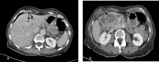
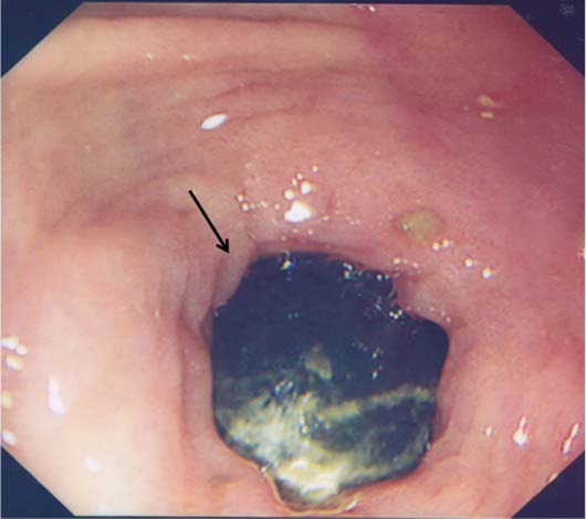
Laboratory investigations showed a haemoglobin of 14 g/dl, white cell count of 18.8×10⁹/l, normal amylase and liver function tests (LFTs). A CT scan showed pneumobilia and a cholecystoduodenal fistula (figure 1). An upper GI endoscopy was performed the subsequent day which showed panbulbar erosion with no active bleed and a 40 mm solitary black stone obstructing the duodenal bulb (figure 2).
Figure 1.
CT scan: Oral contrast enhanced CT scan showing (A) pneumobilia (B) gallstone fistulating into the duodenum.
Figure 2.
Upper GI endoscopy: endoscopic picture showing gallstone in the duodenal bulb.
Treatment
During endoscopy, the stone was extracted into the stomach but could not be crushed. Two days later an endoscopic lithotripsy was attempted. Due to its size, the stone could not be engaged with the keymed lithotripter or a web basket and the procedure was abandoned. A decision was made for a laparotomy but due to her co-morbid conditions, a simple procedure was undertaken to relieve the obstruction. A gastrotomy was performed and a 6×5 cm stone was removed from the stomach.
Outcome and follow-up
She had an uncomplicated postoperative recovery, her LFTs remained normal and magnetic resonance cholangio-pancreatography (MRCP) did not reveal any stones in the common bile duct. Due to her co-morbidities, an elective cholecystectomy was not offered. She presented 18 months later with acute pancreatitis and deranged LFTs. MRCP on this occasion showed multiple gallstones but CBD could not be visualised. In view of her deranged LFTs, an endoscopic retrograde cholangio-pancreatography was performed which showed a dilated CBD of 11 mm but no obvious stones were seen. CT scan and barium meal did not reveal a fistula between the gall bladder and the bowel. She has now been offered an elective cholecystectomy.
Discussion
Patients with Bouveret’s syndrome usually present with symptoms of gall stone obstruction, although presentation with other complications of gallstone disease or upper GI bleeding has also been reported.4
The diagnosis is often delayed because routine investigations generally fail to identify the exact cause of bowel obstruction. Plain abdominal x-ray is diagnostic in 23% cases and may demonstrate pneumobilia and an ectopic gallstone.5 Abdominal ultrasound is useful to confirm the presence of cholelithiasis and may also identify the fistula. When compared to the plain radiograph and abdominal ultrasound, CT scan has proven to be a more valuable technique in the diagnosis of mechanical bowel obstruction, pneumobilia and ectopic gall stone.6
Since this syndrome is usually seen in older patients with poor medical status, a non-surgical approach such as endoscopic removal is usually the first line of treatment. This may be performed by simple endoscopic lithotomy with snare or specific stone baskets, or laser lithotripsy.7 There is no consensus on the choice of surgical procedures. Possible strategies are a one stage approach with duodenotomy or gastrotomy, cholecystectomy and repair of the fistula at once, or a two stage approach with an emergency surgery to remove the obstructing gallstone and a delayed cholecystectomy, or a duodenotomy or a gastrotomy alone.
Our patient was considered to be a high perioperative risk and a delayed cholecystectomy was not offered. A review of series reported an 8.2% risk of developing recurrent gall stone ileus in patients who survived enterolithotomy alone with an associated mortality of 12–20%.8 The risks of the surgical procedure and the potential further complications from gallstones needs to be carefully weighed before undertaking a definitive treatment in these patients.
Learning points.
-
▶
Bouveret’s syndrome should be considered in patients with gallstones who present with intestinal obstruction.
-
▶
Patients with biliary fistulae can present with recurrent gallstone related complications.
-
▶
Cholecytectomy should be considered in patients presenting with Bouveret’s syndrome.
Footnotes
Competing interests None.
Patient consent Obtained.
References
- 1.Haris HW. Biliary system. In: Norton JA, Bollinger RR, Chang AE, et al. (eds.) Surgery Basic Science and Clinical evidence. New York, NY: Springer Veralg; 2001:553–84 [Google Scholar]
- 2.Abou-Saif A, Al-Kawas FH. Complications of gallstone disease: Mirizzi syndrome, cholecystocholedochal fistula, and gallstone ileus. Am J Gastroenterol 2002;97:249–54 [DOI] [PubMed] [Google Scholar]
- 3.Bouveret L. Stenose du pylore adherant a la vesicule. Rev Med (Paris) 1896;16:1–16 [Google Scholar]
- 4.Salah-Eldin AA, Ibrahim MA, Alapati R, et al. The Bouveret syndrome: an unusual cause of hematemesis. Henry Ford Hosp Med J 1990;38:52–4 [PubMed] [Google Scholar]
- 5.Lawther RE, Diamond T. Bouveret’s syndrome: gallstone ileus causing gastric outlet obstruction. Ulster Med J 2000;69:69–70 [PMC free article] [PubMed] [Google Scholar]
- 6.Liew V, Layani L, Speakman D. Bouveret’s syndrome in Melbourne. ANZ J Surg 2002;72:161–3 [DOI] [PubMed] [Google Scholar]
- 7.Schweiger F, Shinder R. Duodenal obstruction by a gallstone (Bouveret’s syndrome) managed by endoscopic stone extraction: a case report and review. Can J Gastrenterol 1997;11:493–6 [DOI] [PubMed] [Google Scholar]
- 8.Doogue MP, Choong CK, Frizelle FA. Recurrent gallstone ileus: underestimated. Aust N Z J Surg 1998;68:755–6 [DOI] [PubMed] [Google Scholar]


