Abstract
Traumatic hip dislocations in children are uncommon, yet even trivial injuries may result in dislocation. Avascular necrosis (AVN) occurs in 3–6% of dislocations if reduction is performed within the first 4 h, however, the incidence rises to 66% if the reduction is performed 24 h after dislocation. Awareness and early identification is therefore critical to long term prognosis. The authors report a case of a relatively trivial trauma resulting in hip dislocation in a 5-year-old boy. This case is useful in highlighting that (a) hip dislocation may occur in children with relatively trivial mechanisms, (b) knee pain often indicates an underlying hip pathology and (c) traumatic AVN follows a similar course to Perthes disease and therefore may be management should be tailored in a similar manner to achieve containment within the acetabulum.
Background
This case report underlines two important clinical priniciples.
It emphasises the importance of investigating knee pain in children, and that even significant hip injuries may present solely as ‘knee pain’.
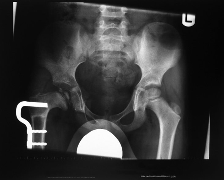
Avascular necrosis (AVN) following trauma in younger children behaves like Perthes disease. There is a period of sclerosis and then fragmentation followed by healing. During the fragmentation phase the cartilaginous femoral head, unsupported by the bony femoral nucleus, becomes plastic and will deform. Approaching the problem as Perthes disease would be approached in a child of similar age using a containment procedure seems to offer the same outcome following traumatic AVN as it does following AVN as a result of Perthes disease.
Case presentation
A 5-year-old boy attended the emergency department following a football injury whereby he slipped while attempting to kick the ball. He immediately had right knee pain and was unable to walk. Examination and radiographs of the knee were normal. A knee sprain was diagnosed and the patient was discharged with analgesia.
The patient represented 2 days later with increasing knee pain. Examination noted a flexion, adduction and internal rotation deformity at the right hip and shortening of leg by approximately 2 cm. All movements of the right hip were painful, knee examination was unremarkable and there was no neurovascular deficit.
Investigations
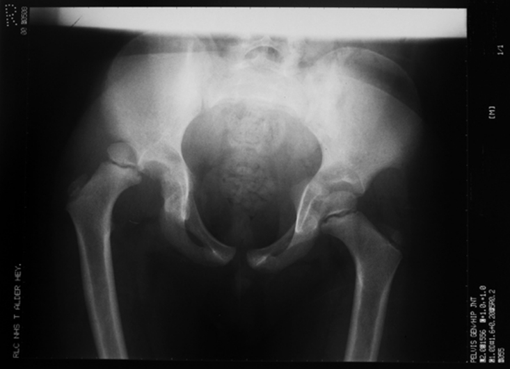
Radiographs revealed a posterior dislocation of the right hip (figure 1).
Figure 1.
Radiograph illustrating a posterior dislocation of the right hip identified 2 days following injury.
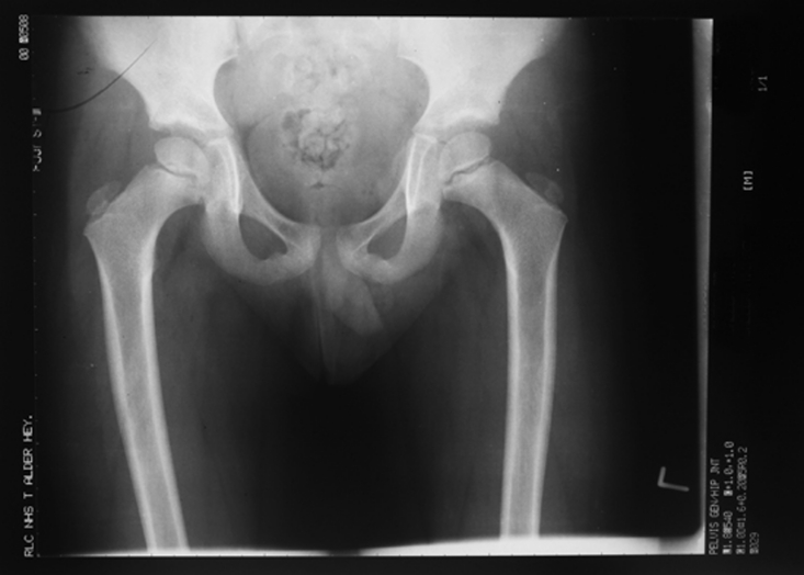
Closed reduction of the right hip under general anaesthesia was performed on the same day (figure 2). A postoperative CT scan confirmed concentric reduction and no associated fractures. The postoperative regimen encompassed 2 weeks of non-weight bearing, 6 weeks of partial weight bearing following which full weight bearing was allowed.
Figure 2.
Postreduction radiograph.
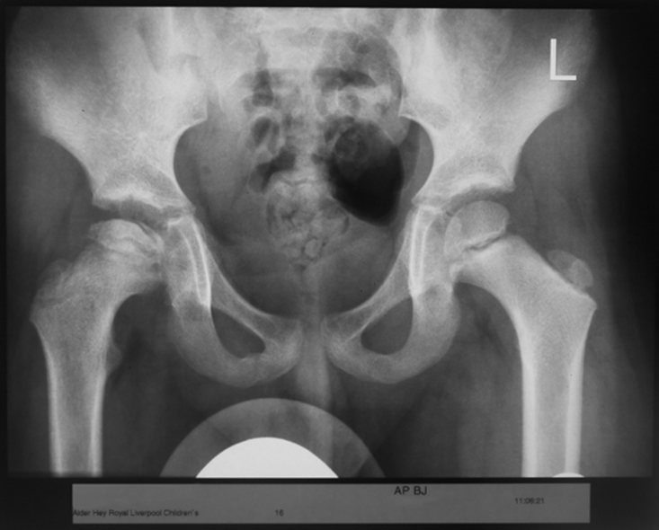
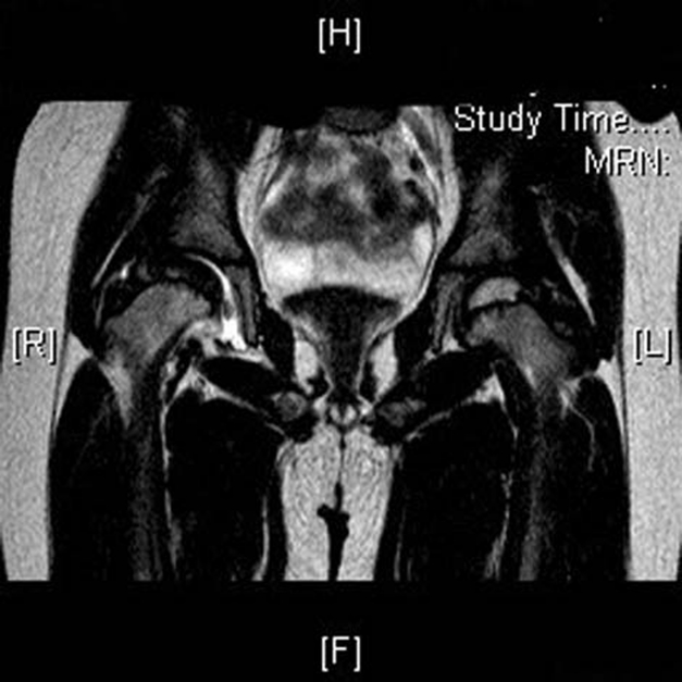
Six months postreduction, the patient presented with insidious onset right groin pain. Examination revealed a Trendelenberg gait with restricted movements of the right hip. There was a complete loss of abduction and restricted rotation. Radiographs identified the femoral epiphysis to be sclerotic with early fragmentation, similar to the changes of Perthes disease (figures 3 and 4).
Figure 3.
Radiograph at 6 months postreduction. The right femoral epiphysis is seen to be sclerotic and flattened with evidence early fragmentation. This is typical of changes seen in avascular necrosis.
Figure 4.
MRI demonstrating avascularity of the right femoral epiphysis.
Treatment
Given the restriction in movement it was identified that passive containment was not being achieved and therefore active containment was required. A varus osteotomy, incorporating 20° of varusisation, with blade plate fixation was therefore performed (figure 5).
Figure 5.
Radiograph at 1 year postreduction. This demonstrates the blade plate in-situ following a right femoral varus osteotomy. The epiphysis is well contained within the acetabulum and is late in the fragmentation stage of AVN. >50% collapse of the lateral pillar (equivalent to a Herring C hip).
Outcome and follow-up
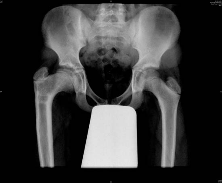
Nine years after corrective osteotomy, the patient is actively participating in sports. His only complaint is a limp following lengthy sporting activity. He has a full and painless range of movement in all planes except a slight restriction of internal rotation of 10° compared to the contralateral hip. There is no clinically significant limb length discrepancy. Radiograph (figure 6) shows a concentrically located, well rounded and remodeled femoral head.
Figure 6.
Radiograph 9 years postosteotomy. The femoral head is spherical, with a congruent acetabulum. Slight coxa vara and coxa magna remains.
Discussion
This case demonstrates a well recognised pitfall. Children presenting with knee pain may be experiencing referred hip pain and the hips must be examined. The case further highlights that children suffer traumatic hip dislocations even with seemingly trivial trauma.1–5
Early identification of hip dislocation is crucial to the long term prognosis. Funk in 1962 studied different factors influencing the outcome after traumatic hip dislocation in children. He stated that the fate of the femoral head seems to be determined by the delay in reduction of the dislocated hip. He concluded that permanent hip changes should be expected when reduction is delayed for more than 24 h.2 Similar more recent reports support this notion that earlier reduction results in a better outcome.3 6
Barquet7 in 1982 conducted a retrospective review of 145 cases of traumatic dislocation of hip in childhood and attempted to outline the natural history of this complication. He concluded that in children younger than 12 years the changes of AVN following traumatic hip dislocation are similar to Perthes disease while in older children these changes resembled AVN of femoral head in adults.7
AVN that follows trauma results from a tear and resultant division of the vasculature around the hip,8 yet the mechanism which underpins Perthes disease of the hip is unknown. The radiological appearance in both is similar with initial flattening and sclerosis of the epiphysis, progressing to fragmentation with later reossification.
Containment is necessary to support the demineralised and therefore malleable femoral head within the acetabulum.9 This achieves a femoral head which moulds to the shape of the acetabulum and sustains this shape as revascularistation and remineralisation occurs along with the return of the structural integrity.
This case emphasises the importance of investigating knee pain in children, and that even significant hip injuries may present solely as ‘knee pain’. It similarly demonstrates that ‘containment’ procedures may achieve successful results in AVN in childhood, even in those hips with a poor prognosis. We advocate treating traumatic AVN of the hip as one would treat Perthes disease – with the necessary interventions to achieve ‘containment’.
Learning points.
-
▶
Children presenting with knee pain may be experiencing referred hip pain and the hips must be examined.
-
▶
The case further highlights that children suffer traumatic hip dislocations even with seemingly trivial trauma.
-
▶
‘Containment’ procedures may achieve successful results in AVN in childhood, even in those hips with a poor prognosis. We advocate treating traumatic AVN of the hip as one would treat Perthes disease – with the necessary interventions to achieve ‘containment’.
Footnotes
Competing interests None.
Patient consent Not obtained.
References
- 1.Barquet A. Traumatic Hip Dislocation in Childhood. Vol 1 Berlin, Heidelberg, New York: Springer; 1987 [Google Scholar]
- 2.Funk FJ. Traumatic dislocation of hip in children: factors influencing prognosis and treatment. J Bone Joint Surg 1962;44-A:1135–45 [Google Scholar]
- 3.Herrera-Soto JA, Price CT. Traumatic hip dislocations in children and adolescents: pitfalls and complications. J Am Acad Orthop Surg 2009;17:15–21 [DOI] [PubMed] [Google Scholar]
- 4.Offierski CM. Traumatic dislocation of the hip in children. J Bone Joint Surg Br 1981;63-B:194–7 [DOI] [PubMed] [Google Scholar]
- 5.Vontobel BJ, Hocevar Z, Jakob RP. Avascular necrosis following traumatic hip dislocation in an 8-year-old boy. Arch Orthop Trauma Surg 1994;113:83–5 [DOI] [PubMed] [Google Scholar]
- 6.Gürkan V, Dursun M, Orhun H, et al. Evaluation of pediatric patients with traumatic hip dislocation. Acta Orthop Traumatol Turc 2006;40:392–5 [PubMed] [Google Scholar]
- 7.Barquet A. Natural history of avascular necrosis following traumatic hip dislocation in childhood: a review of 145 cases. Acta Orthop Scand 1982;53:815–20 [DOI] [PubMed] [Google Scholar]
- 8.Somerville EW. Perthes’ disease of the hip. J Bone Joint Surg Br 1971;53:639–49 [PubMed] [Google Scholar]
- 9.Lloyd-Roberts GC, Catterall A, Salamon PB. A controlled study of the indications for and the results of femoral osteotomy in Perthes’ disease. J Bone Joint Surg Br 1976;58:31–6 [DOI] [PubMed] [Google Scholar]


