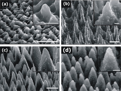
Figure 1.
Side-view scanning electron microscope image (45°) of Si surfaces structured by 800 nm, 180 fs irradiation at different laser fluences. (a) 0.37 J∕cm2, (b) 0.78 J∕cm2, (c) 1.56 J∕cm2, and (d) 2.47 J∕cm2 (scale bar of 5 μm). Higher magnifications of the obtained structures are shown in the corresponding insets (scale bar of 1 μm).

