Abstract
The analysis of the sagittal balance is important for the understanding of the lumbopelvic biomechanics. Results from previous studies documented the correlation between sacro-pelvic orientation and lumbar lordosis and a uniqueness of spino-pelvic alignment in an individual person. This study was subjected to determine the lumbopelvic orientation using pelvic radius measurement technique. The standing lateral radiographs in a standardized standing position were taken from 100 healthy volunteers. The measurements which included hip axis (HA), pelvic radius (PR), pelvic angle (PA), pelvic morphology (PR-S1), sacral translation distance (HA-S1), total lumbosacral lordosis (T12-S1), total lumbopelvic lordosis (PR-T12) and regional lumbopelvic lordosis angles (PR-L2, PR-L4 and PR-L5) were carried out with two independent observers. The relationships between the parameters were as follows. PR-S1 demonstrated positive correlation to regional lumbopelvic lordosis and revealed negative correlation to T12-S1. PA showed negative correlation to PR-S1 and regional lumbopelvic lordosis, but revealed positive correlation to HA-S1. T12-S1 was significantly increased when PR-S1 was lesser than average (35°–45°) and was significantly decreased when PR-S1 was above the average. PR-L4 and PR-L5 were significantly reduced when PR-S1 was smaller than average and only PR-L5 was significantly increased when PR-S1 was above the average. In conclusion, this present study supports that lumbar spine and pelvis work together in order to maintain lumbopelvic balance.
Keywords: Pelvic radius technique, Pelvic morphology, Lumbosacral lordosis, Sagittal balance
Introduction
The analysis of the sagittal balance is important for the understanding of the lumbopelvic biomechanics. However, there is only little information on these subjects. The problem refers to the great variability in spinal sagittal alignment among the normal populations [1, 2].
The sagittal alignment of the lumbopelvic region can be determined by pelvic and spinal parameters which have established the consistent correlations in normal population studies [1, 3, 4]. Furthermore, previous studies revealed that the sagittal orientation of the pelvis remained constant after cessation of growth [5–7]. These reflect that the sagittal orientation of the lumbar spine is influenced by the shape and orientation of sacro-pelvis which is individual for each person. The shape and orientation of sacro-pelvis can be measured with some fundamental parameters, pelvic incidence described by During [8] and Duval-Beaupere [9], and pelvic radius measurement described by Jackson [10, 11]. These two parameters measure pelvic morphology by using the approximated center of the hip on the lateral radiograph and draw a line to a specific point at the sacral endplate, to the middle of endplate for pelvic incidence measurement and to the posterior superior corner of endplate for pelvic radius measurement.
Studies of Legaye [3] and Jackson [11] confirmed that the degree of lumbar lordosis involves by the magnitude of the sacral inclination to maintain the sagittal balance of the entire spine. However, as mentioned previously, there is great variability in the sagittal alignment among the individuals. Thus, the classification of the lumbopelvic alignments should be obtained for clearer understanding on its effect to the lumbar spinal diseases. The classification of normal variation in sagittal orientation of lumbosacral spine based on the pelvic incidence has been described by Roussouly [12]. In the study, the difference in sagittal orientation of the lumbar spine was observed and the correlation between pelvic orientation and the degree of lumbar lordosis have been described. However, the pelvic radius measurement has been reported to have the higher correlation coefficient between pelvic orientation and the details of lumbar sagittal orientation with very high intra- and interobserver reliability [11, 13]. Thus, our hypothesis is that pelvic radius measurement technique should be a better represented parameter to obtain the orientation of pelvis and is useful for the analysis of the lumbar sagittal alignment. However, to the best of our knowledge, classification of lumbar lordosis based on the pelvic radius measurement has not yet been studied. Therefore, the aim of this study was to determine the lumbar sagittal alignment and pelvic orientation using pelvic radius measurement technique.
Materials and methods
One hundred (100) asymptomatic Thai volunteers were studied. The average age of the subjects was 33.3 ± 6.8 (21–50 years); there were 70 males and 30 females. The mean body weight was 60.0 ± 7.9 (40–82 kg) and mean body height was 146.6 ± 7.2 (145–178 cm). The inclusion criteria were as follows: (1) age 20–60 years, (2) no prior spine surgery and (3) no history of low back pain, except for occasional episodes, for at least 6 months before their participation in this study. The exclusion criteria were as follows: (1) definite diagnosis of lumbar spinal pathology such as spinal stenosis, spondylolisthesis, degenerative disc disease or inflammatory spinal diseases, (2) definite clinical spine deformities from physical examination or X-ray, (3) definite diagnosis of diabetic mellitus, hypertension, rickets, osteoporosis, and (4) history of smoking. Volunteers were informed of risks and benefits of participation in the study and signed the consent forms. This study was reviewed and had been approved by the hospital ethical research committees.
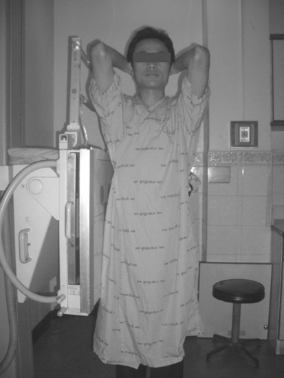
All subjects had standing lateral radiograph of the spine and pelvis by the same radio-technologist using the same radiographic equipments. The X-ray cassettes were set into a standard upright cassette adjustment devices then adjusted for a 72-inch-long focal film distance from the radiation source. The volunteers were carefully positioned with their right side against the cassette. They were asked to stand up straight, but relaxed, with the knees extended. The arms were brought upward with hands hold together behind the neck (Fig. 1).
Fig. 1.
Positioning technique for standing lateral radiograph of the spine and pelvis. The radiographs were taken with 72-inch-long distance from the X-ray source
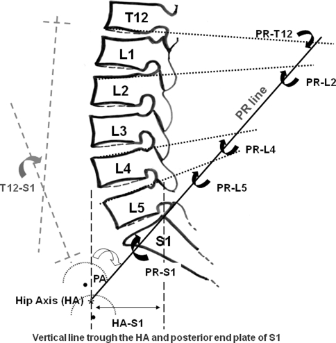
Radiographic measurements were made on the standing radiographs using pelvic radius measurement technique [11]. The hip center was determined by using mose template. The hip axis (HA) was then located midway between the two hip centers, and a line was drawn to the postero-superior corner of sacral end plate for the pelvic radius (PR); its length was measured in millimeters. Radiographic parameters were measured as follows: (1) anatomic PR-S1 angle, (2) total lumbosacral lordosis angle (T12-S1 angle) and total lumbopelvic lordosis angle (PR-T12 angle), (3) regional lumbopelvic lordosis angles (PR-L2, PR-L4 and PR-L5 angles), (4) sacral translation distance (HA-S1) and (5) pelvic angle (PA). The list of the nomenclature for the parameters that were measured with their abbreviation and description is outlined in Table 1. Figure 2 demonstrates the measurement methods. All radiographs were measured twice by two independent orthopedic trainees working in our department under supervision by the authors (PC and SW). The interobserver reliability was calculated.
Table 1.
Nomenclature for parameters measured on standing lateral radiographs
| Measurement | Abbreviation | Description |
|---|---|---|
| Hip axis | HA | Midpoint between approximate centers of both femoral heads. The other parameters measures in the study are measured from this point |
| Pelvic radius | PR | A distance from the HA to the posterior superior corner of S1 |
| Pelvic radius line | PR Line | A line connecting the HA and the posterior superior corner of S1 |
| Pelvic morphology | PR-S1 | Angular measurement between the PR line and a tangent line along the S1 endplate |
| Pelvic angle | PA | Angular measurement between the PR line and a vertical line draw through the HA |
| Sacral translation | HA-S1 | Horizontal distance between the vertical line troughs HA |
| Total lumbosacral lordosis | T12-S1 | Angular measurement between inferior endplate of T12 vertebral body and superior endplate of S1 |
| Total lumbopelvic lordosis | PR-T12 | Angular measurement from the PR line and a tangent line along the inferior endplate of T12 vertebral body |
| Regional lumbopelvic lordosis | PR-L2, PR-L4, PR-L5 | Angular measurement from the PR line and a tangent lines along the superior endplate of L2, L4 and L5, respectively |
Fig. 2.
Line drawing showing pelvic radius (PR line) and the pelvic radius measurement technique used in this study. List of the nomenclature with their abbreviation and description is outlined in Table 1. Black dashed lines demonstrate the vertical line trough HA and posterior superior corner of S1. Gray dashed lines show T12-S1 measurement. Arrows indicate the angles of representation
Statistical analysis
In attempt to classify the sacral orientation, mean and frequency of each parameter were analyzed. Correlations between two parameters were determined using Pearson correlation coefficient. The analysis of variances (ANOVAs) with Turkey–Kramer multiple comparisons procedure were used to compare the mean of radiographic parameters of each classification of pelvic alignment. Interobserver reliabilities were calculated using Pearson’s liner regression correlation coefficients. The statistically significant difference was considered for a P value less than 0.05.
Results
Spino-pelvic alignment parameters
The measurement values for all parameters measured by both observers were demonstrated as a perfect interobserver reliability. The correlation coefficients ranged from 0.9687 to 0.9952 with P value < 0.0001. Therefore, only the data from the first observer were reported (Table 2). The mean of PR distance was 123 mm and HA-S1 distance was 40.8 mm. The PR-S1 angle was lesser than PA angle. The maximum lumbopelvic lordosis angle was PR-T12 and then PR-L2, PR-L4, respectively. The PR-L5 angle revealed minimal lumbopelvic lordosis angle. The total lumbosacral lordosis angle (T12-S1) was less than the total lumbopelvic lordosis angle (PR-T12).
Table 2.
Means, SD and ranges of the measurements for spino-pelvic alignments
| Measurementa | Mean (range) | SD | Correlation* |
|---|---|---|---|
| PR (mm) | 123 (104–144) | 8.2 | 0.9831 |
| PR-S1 | 37.4 (18–63) | 9.5 | 0.9908 |
| PA | 19.5 (7–36) | 5.5 | 0.9815 |
| HA-S1 (mm) | 40.8 (14–68) | 11.0 | 0.9951 |
| PR-T12 | 92.4 (75–114) | 8.2 | 0.9687 |
| T12-S1 | 54.7 (30–76) | 9.9 | 0.9921 |
| PR-L2 | 86.7 (70–110) | 7.9 | 0.9891 |
| PR-L4 | 71.4 (48–89) | 9.0 | 0.9904 |
| PR-L5 | 57.7 (30–82) | 10.5 | 0.9952 |
The correlation between pelvic morphology parameters
The correlations between parameters describing the pelvic orientation and morphology are list in Table 3. PR distance did not show any strong correlation with the other parameters. PA angle, on the other hand, demonstrated perfect correlation with the HA-S1 distance and revealed inverted correlation to PR-S1 angle.
Table 3.
Correlation among pelvic and lumbar alignment parameters (Pearson correlation)
| Parameter variablesa | PR | PR-S1 | PA |
|---|---|---|---|
| Pelvic alignments | |||
| PR-S1 | 0.2387 (0.0168) | < > | −0.4882 (<0.0001) |
| PA | −0.07519 (0.4572) | < > | < > |
| HA-S1 | 0.1538 (0.1267) | −0.4214 (<0.0001) | 0.9411 (<0.0001) |
| Lumbar alignments | |||
| PR-T12 | 0.08041 (0.4264) | 0.2908 (0.0033) | −0.6296 (<0.0001) |
| T12-S1 | −0.1642 (0.0125) | −0.6567 (<0.0001) | −0.06103 (0.5464) |
| PR-L2 | 0.03744 (0.7116) | 0.5057 (<0.0001) | −0.7412 (<0.0001) |
| PR-L4 | 0.08082 (0.4241) | 0.7402 (<0.0001) | −0.7297 (0.0001) |
| PR-L5 | 0.1238 (0.2198) | 0.8275 (<0.0001) | −0.6783 (0.0001) |
The correlation between pelvic parameters and lumbar lordosis
Table 3 shows the correlations between pelvic morphology parameters and lumbar lordotic parameters. Similar to pelvic parameter correlations, PR distance did not reveal any strong correlation with the parameters describing the lumbar lordosis. On the other hand, PA angle showed inverse correlation to the lumbopelvic lordosis angle (PR-T12, PR-L2, PR-L4 and PR-L5), but not total lumbosacral lordosis angle (T12-S1). PR-S1 angle demonstrated invert correlation with T12-S1 and showed positive correlation with regional lumbopelvic lordosis (PR-L2, PR-L4 and PR-L5), but not total lumbopelvic lordosis (PR-T12).
The changed in lumbopelvic alignments in the relation to the changed of PR-S1
For better understanding of the change in lumbopelvic alignment in the relation to pelvic morphology, the PR-S1 angle was categorized into three types, average, lower than the average and higher than the average. Based on the distribution of PR-S1 angle in studied subjects, the median of PR-S1 angle was 38° (10–63°). With this value, we defined the average value of PR-S1 angle to 35–45°; the low value was less than 35° and the high value was more than 45°. Then, the parameters of lumbopelvic alignment were regrouped based on the PR-S1 angle and calculated for the mean and analyzed for the difference. Table 4 shows the mean of lumbopelvic parameters subjected by the PR-S1 angle. T12-S1 angle was significantly increased according to the decrease of PR-S1 angle. PR-L2 remained unchanged according to the change in PR-S1 angle. PR-L4 was decreased in cases of PR-S1 lower than the average, but unchanged when PR-S1 angle was higher than the average. The PR-L5 angle revealed a significant decrease when PR-S1 was lower than the average and a significant increase when PR-S1 was higher than the average (Table 4).
Table 4.
The characteristics (classification) of the lumbar lordosis according to the PR-S1 angle
| Parametersa |
Low PR-S1 | Average PR-S1b | High PR-S1 |
|---|---|---|---|
| PR-S1 < 35° | PR-S1 ~ 35–45° | PR-S1 > 45° | |
| T12-S1 | 61.6 ± 8.0** | 53.9 ± 7.7 | 45.9 ± 8.5* |
| PR-L2 | 82.1 ± 7.4 | 87.8 ± 6.8 | 91.5 ± 7.1 |
| PR-L4 | 64.1 ± 8.4* | 73.2 ± 6.1 | 78.9 ± 6.2 |
| PR-L5 | 48.5 ± 9.7* | 59.3 ± 6.4 | 68.0 ± 5.7** |
Discussion
Pelvic morphology has been shown to affect the standing lumbosacral lordosis in adult [3, 11]. However, there are many studies that demonstrated the lumbopelvic alignment and pelvic morphology, but the change in the lumbopelvic alignment by means of the difference in orientation of sacro-pelvic is still not clear. In this study, we demonstrated the change in lumbopelvic lordosis subjected by the difference in pelvic angle (PA) and PR-S1 angle.
The correlations of the sacro-pelvis orientation, described by the pelvic incidence or pelvic radius measurement, with the degree of lumbar lordosis have been reported in many literatures. Nevertheless, the pelvic incidence, when compared to the pelvic radius measurement, has been reported with lower correlation to the degree of lumbar lordosis and do not explain the degree of correlation upon the lumbar spine orientations [9–11]. Further, with the pelvic radius measurement, the correlations between the parameters describe the sacro-pelvic alignment and the lumbar spine orientations have been established [14]. Therefore, the set of parameters which illustrate the details of lumbar spine and the sacro-pelvic orientations by means of the pelvic radius measurement should be a better represented parameter to clarify the orientation of pelvis and it association to the lumbar sagittal alignment in order to understand the complex relationship between the lumbar spinal orientations and the pelvic morphology.
Spino-pelvic alignment and correlation with lumbopelvic parameters
In this study, the results of pelvic parameters and lumbopelvic parameters were different compared to the results from Jackson et al. [14] In our results, the PR distance was 123 mm, less than 136.8 mm. The PA angle in our study was 19.5°, the PR-S1 angle was 37.4° and HA-S1 distance was 40.8 mm, all values were larger than those of Jackson’s study. This may be caused by the difference in sacro-pelvic anatomy in the two populations. However, the alignment correlations between spino-pelvic alignment and lumbopelvic parameter were almost the same. The correlation results between pelvic parameters revealed a positive correlation between PA angle and HA-S1 distance (r = 0.9411, P < 0.0001), and demonstrated a negative correlation with PR-S1 angle (r = −0.4882, P < 0.0001). The results showed that when PA angle was decreased the sacral was closer to the hip axis, and the sacrum had a trend to be more in vertical orientation similar to the observation by Roussouly et al. [12].
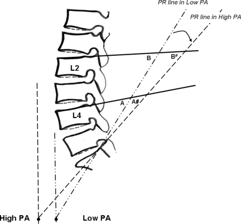
The previous studies on pelvic morphology using pelvic incidence have demonstrated high correlation between the sacral slope and lumbar lordosis [2, 3]. In our analysis, however, we used a different technique to describe the pelvic alignment. The result showed that PR distance did not reveal the correlation with other parameters. However, PA angle revealed a strong correlation with the HA-S1 and PR-S1 angles and lumbopelvic parameters (PR-T12, PR-L2, PR-L4 and PR-L5), but not with total lumbar lordosis (T12-S1). Unfortunately, PA angle cannot be used to predict the lumbar sagittal alignment, because the results observed in our study should be caused by the rearward projection of the PR line (Fig. 3). PR-S1 angle, on the other hand, revealed a correlation with regional lumbopelvic lordosis (PR-L2, PR-L4 and PR-L5) and total lumbar lordosis (T12-S1). Thus, PR-S1 angle may be suitable for the prediction of lumbar lordosis, while PA angle can be used to describe the pelvic orientation.
Fig. 3.
Drawing demonstrates the relationship between PA angle and pelvic radius measurements on lumbar spines (PR-L2, PR-L4 and PR-L5). The black dots represent the HA in high and low PA angles. The PR line in low PA angle is indicated by dash dotted lines and the dashed line is the High PA angle. The PR line was rearward projected as the PA angle was higher as indicated by curve arrow. The regional lumbar lordosis angles were then measured and decrease value even without the change of lumbar lordosis (PR-L4: A > A# and PR-L2: B > B#)
Gardocki et al. [15] described the association between pelvic morphology using the same technique as in our study with lumbar lordosis. The result showed that HA-S1 was negatively correlated with total segmental lumbar lordosis (L1-S1, as described in his report) and lumbopelvic lordosis (PR-T12 to PR L5, as described in his report). He concluded from this finding that the pelvic rotated posteriorly around the hip (HA-S1 increased) when the lumbar lordosis was decreased. However, our results revealed a negative correlation between PR-S1 and T12-S1 (r = −0.6567, P < 0.0001), and demonstrated a negative correlation with HA-S1 (r = −0.4214, P < 0.0001). Therefore, from our results, we could conclude that the change in sacral orientation (PR-S1) was negatively correlated with lumbar lordosis. Therefore, as lumbar lordosis decreases the sacrum has a trend to be vertical and, on the contrary, it tends to be horizontal as the lumbar lordosis increases. This phenomenon is not a result by the rotation of the pelvis around the hip, because the correlation to HA-S1 is negative. However, we believe in the concept that the lumbar spine and pelvis work together in order to maintain lumbopelvic balance. Thus, this finding should be explained by the adaptation of lumbar spine to facilitate balance for the pelvis.
The classification of lumbar sagittal alignment in relation to the PR-S1
The association between the alignment of the pelvis and the lumbar spine is an important determinant of sagittal balance. In this study, we categorized the pattern of lumbopelvic lordosis according to the PR-S1 angle into three types, high, average and low type, to clarify the relationship between patterns of lumbar sagittal alignment and pelvic morphology. Roussouly et al. [12] classified the lumbar sagittal orientation into four types. Based on his description, type 1 and type 2 lordosis were associated with sacral slope less than 35°. In both type 1 and type 2 lordosis, the lumbar lordosis angle was decreased at the lower lumbar level, but remained constant in upper lumbar level in type 1, and decreased in type 2. Type 3 lordosis was observed for the sacral slope between 35° and 45° and the lower lumbar lordosis was increasing. For type 4 lordosis, the sacral slope was greater than 45° when the lower and upper lumbar lordosis were prominent and spine became hyperlordosis. The results were compared to our study. In this study, however, we used the PR-S1 to classify the pattern of lumbar lordosis. Comparing to the pelvic incidence (PI) the sacral orientations when described by PR-S1 were as follows: if the sacral inclination was increased as described by increased PI the PR-S1 was reduced, and, on the other hand, the PR-S1 was increased when PI was reduced as the sacral became vertical orientation.
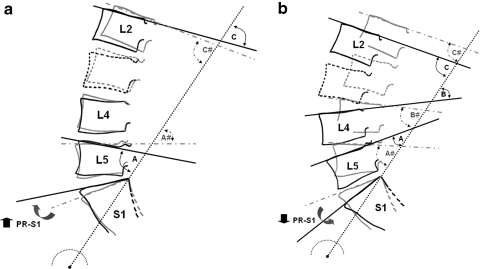
In our results, the total lumbar lordosis (T12-S1) was increased when PR-S1 angle was lower than the average and decreased as PR-S1 angle was higher than the average. The change in lumbar spines orientation appeared at the lower lumbar level, as same as the observation by Roussouly. We believed that, in our definition, type 3 lordosis was the lumbar lordosis in an average PR-S1. In high PR-S1 group, PR-L5 angle was significantly increased. As a result the lumbar lordosis was decreased at the L4–L5 level, and the spine was relatively flat as demonstrated in type 2 lordosis (Fig. 4a). In low PR-S1 group, PR-L4 and PR-L5 were significantly decreased then the lower lumbar lordosis was increased as appeared in type 4 lordosis. Although, in our finding the PR-L2 remained unchanged, which means that the lumbar spine appeared to move forward (Fig. 4b). However, we could not demonstrated type 1 lordosis from our study.
Fig. 4.
The drawings demonstrate the lumbar sagittal alignment which was altered by the change of PR-S1 angle (indicated by black arrows), high PR-S1 angle (a) and low PR-S1 angle (b). Gray line shows the lumbar alignment in average PR-S1. See text for the details. The PR-L2 (C and C#) remained unchanged. In high PR-S1 (a) PR-L5 (A) was higher than that of the average PR-S1 (A#). The lumbar spine was then flat. In low PR-S1 (b) PR-L5 (A) and PR-L4 (B) was less than those of the average PR-S1 (A# and B#, respectively). Lumbar lordosis was increased at lower lumbar segments and the lumbar spine appeared to move forward
There are some limitations in our study. First, we focus only on the parameters that illustrate the lumbar spine orientation that described in the pelvic radius measurement, so some important parameters may be missed for the correlation with the sacro-pelvic orientations. For example, the length of lumbar lordosis, that describe the number of vertebrae included in the lordosis, it may be possible to determine when the lordotic curvature is changing into kyphosis in the relation with sacro-pelvic orientation. This is probably one reason why we did not find the type 1 of Roussouly in high PR-S1 group. Second, in our present study, we concentrated only on the lumbopelvic orientation that represents only one part of the global sagittal balances. The future study should be focussed to answer the question about the alteration on the global sagittal alignment with the change in lumbopelvic orientation.
Conclusion
The results from this study support the concept that the lumbar spine and pelvis work together in order to maintain lumbopelvic balance. Additionally, the classification on the lumbar sagittal orientation using pelvic radius technique by means of PR-S1 was accomplished. However, additional studies should be conducted to clarify its relations to the lumbar spinal disorders. Furthermore, it is necessary to prospectively follow these populations to determine if the native sagittal alignment influences the development of lumbar spinal pathologies.
Conflict of interest
None.
References
- 1.Kobayashi T, Atsuta Y, Matsuno T, Takeda N. A longitudinal study of congruent sagittal spinal alignment in as adult cohort. Spine. 2004;29:671–676. doi: 10.1097/01.BRS.0000115127.51758.A2. [DOI] [PubMed] [Google Scholar]
- 2.Barrey C, Jund J, Noseda O, Roussouly P. Sagittal balance of the pelvis-spine complex and lumbar degenerative diseases. A comparative study about 85 cases. Eur Spine J. 2007;16:1459–1467. doi: 10.1007/s00586-006-0294-6. [DOI] [PMC free article] [PubMed] [Google Scholar]
- 3.Legaye J, Duval-Beaupère G, Hecquet J, Marty C. Pelvic incidence: a fundamental pelvic parameter for the three dimensional regulation of spinal sagittal curves. Eur Spine J. 1998;7:99–103. doi: 10.1007/s005860050038. [DOI] [PMC free article] [PubMed] [Google Scholar]
- 4.Vaz G, Roussouly P, Berthonnaud E, Dimnet J. Sagittal morphology and equilibrium of pelvis and spine. Eur Spine J. 2002;11:80–87. doi: 10.1007/s005860000224. [DOI] [PMC free article] [PubMed] [Google Scholar]
- 5.Mac-Thiong JM, Berthonnaud E, Dimar JR, Betz RR, Labelle H. Sagittal Alignment of the Spine and Pelvis During Growth. Spine. 2004;29:1642–1647. doi: 10.1097/01.BRS.0000132312.78469.7B. [DOI] [PubMed] [Google Scholar]
- 6.Mangione P, Gomez D, Sénégas J. Study of the course of the incidence angle during growth. Eur Spine J. 1997;6:163–167. doi: 10.1007/BF01301430. [DOI] [PMC free article] [PubMed] [Google Scholar]
- 7.Marty C, Boisaubert B, Descamps H, et al. The sagittal anatomy of the sacrum among young adults, infants, and spondylolisthesis patients. Eur Spine J. 2002;11:119–125. doi: 10.1007/s00586-001-0349-7. [DOI] [PMC free article] [PubMed] [Google Scholar]
- 8.During J, Goudfrooij H, Keessen W, Beker TW, Crowe A Towards standards for posture. Postural characteristics of the lower back system in normal and pathologic conditions. Spine. 1985;10:83–87. doi: 10.1097/00007632-198501000-00013. [DOI] [PubMed] [Google Scholar]
- 9.Duval-Beaupere G, Legaye J. Composante sagittale de la satatique rachidienne (in French) Rev Rhum. 2004;71:105–119. doi: 10.1016/j.rhum.2003.09.018. [DOI] [Google Scholar]
- 10.Jackson RP. Spinal balance, lumbopelvic alignments around the “hip axis” and positioning for surgery. Spine: State Art Rev. 1997;11:33–58. [Google Scholar]
- 11.Jackson RP, Peterson MD, McManus AC, Hales C. Compensatory spinopelvic balance over the hip axis and better reliability in measuring lateral radiographs of adult volunteers and patients. Spine. 1998;23:1750–1767. doi: 10.1097/00007632-199808150-00008. [DOI] [PubMed] [Google Scholar]
- 12.Roussouly P, Gollogly S, Berthonnaud E, Dimnet J. Classification of the normal variation in the sagittal alignment of the human lumbar spine and pelvis in the standing position. Spine. 2005;30:346–353. doi: 10.1097/01.brs.0000152379.54463.65. [DOI] [PubMed] [Google Scholar]
- 13.Jackson RP, Kanemura T, Kawakami N, Hales C. Lumbopelvic lordosis and pelvic balance on repeated standing lateral radiographs of adult volunteers and untreated patients with constant low back pain. Spine. 2000;25:575–586. doi: 10.1097/00007632-200003010-00008. [DOI] [PubMed] [Google Scholar]
- 14.Jackson RP, Hales C. Congruent spinopelvic alignment on standing lateral radiographs of adult volunteers. Spine. 2000;25:2208–2215. doi: 10.1097/00007632-200011010-00014. [DOI] [PubMed] [Google Scholar]
- 15.Gardocki RJ, Watkins RG, Williams LA. Measurements of lumbopelvic lordosis using the pelvic radius technique as it correlates with sagittal spinal balance and sacral translation. Spine J. 2002;2:41–49. doi: 10.1016/S1529-9430(02)00426-6. [DOI] [PubMed] [Google Scholar]


