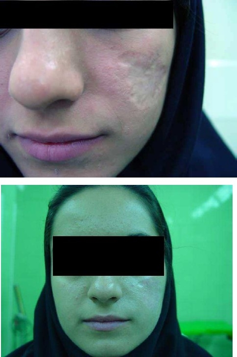
Cutaneous leishmaniasis is an endemic disease of Iran. Unfortunately, there is no definite treatment for this disease.1 We report a 21-year-old woman who was infected with cutaneous leishmaniasis about 16 years ago with disfiguring scar (Figure 1). Different methods of resurfacing were taken without significant results. To repair the scar, at first, a biopsy was performed from retroauricular area and was sent for Rooyan where culture of fibroblasts was performed. A mixture of 20 millions fibroblasts in 1 cc of serum was injected beneath the scar area with about 100% correction. After 2 months, fibroblast suspension was injected again. The scar area was then dermabraded until blood oozing was occurred. The dermabraded area was covered with a thin layer of Fibrin Glue, fibroblast and keratinocyte suspension. The dressing was removed 2 weeks later. The appearance of scar was significantly improved at 3-month follow-up. According to two blinded investigators and the patient herself, there was at least 80% and 90% improvement, respectively, in the cosmetic appearance of the scar (Figure 1).
Figure 1.

The appearance of scar before and after treatment
Leishmaniasis scars are usually depressed and atrophic. Patients affected by these scars usually have psychosocial and cosmetic complains.2 The result at 3 months follow up was very interesting and the cosmetic appearance of leishmaniasis scar was at least 90% improved according to the patient and investigators. More prolonged studies on further cases are recommended for better evaluation of this method in the treatment of atrophic scars.
Footnotes
Conflict of Interest
Authors have no conflict of interests.
References
- 1.Nilforoushzadeh MA, Jaffary F, Moradi S, Derakhshan R, Haftbaradaran E. Effect of topical honey application along with intralesional injection of glucantime in the treatment of cutaneous leishmaniasis. BMC Complement Altern Med. 2007;7:13. doi: 10.1186/1472-6882-7-13. [DOI] [PMC free article] [PubMed] [Google Scholar]
- 2.Bayat A, McGrouther A, Ferguson J. Skin scarring. BMJ. 2003;326(7380):88–92. doi: 10.1136/bmj.326.7380.88. [DOI] [PMC free article] [PubMed] [Google Scholar]


