Abstract
RNase Z is an endonuclease responsible for the removal of 3′ extensions from tRNA precursors, an essential step in tRNA biogenesis. Human cells contain a long form (RNase ZL) encoded by ELAC2, and a short form (RNase ZS; ELAC1). We studied their subcellular localization by expression of proteins fused to green fluorescent protein. RNase ZS was found in the cytosol, whereas RNase ZL localized to the nucleus and mitochondria. We show that alternative translation initiation is responsible for the dual targeting of RNase ZL. Due to the unfavorable context of the first AUG of ELAC2, translation apparently also starts from the second AUG, whereby the mitochondrial targeting sequence is lost and the protein is instead routed to the nucleus. Our data suggest that RNase ZL is the enzyme involved in both, nuclear and mitochondrial tRNA 3′ end maturation.
Introduction
tRNAs are generally synthesized as immature precursors with extensions on both ends [1]. Removal of these extra sequences is essential for tRNA function. Extraneous nucleotides at the 5′ end are removed by a single endonucleolytic cleavage catalyzed by an enzyme known as RNase P [2], [3]. 3′ end processing is either accomplished by precise endonucleolytic cleavage too, or by the concerted action of multiple exo- and endonucleases [1], [2], [4]. The enzyme responsible for the first, exclusively endonucleolytic pathway is called RNase Z (or tRNase Z; EC 3.1.26.11) [2], [5]–[8].
RNase Z enzymes belong to the metallo-β-lactamase superfamily. In addition to the diagnostic α–β/β–α β-lactamase-fold, a protruding flexible arm involved in binding tRNA precursors is characteristic for RNase Z structures. There are two forms of RNase Z, one of 280 to 360 amino acids (RNase ZS) found in archaea and many, but not all bacteria and eukarya, and one, more than twice this length (750–930 amino acids; RNase ZL), exclusively found in eukarya. The primary structure of RNase ZL suggests that it has evolved from RNase ZS by duplication. While all eukarya appear to have at least one RNase ZL gene, some also have RNase ZS, or more than one gene encoding a long and/or short form of RNase Z.
In the human genome RNase ZS is encoded by ELAC1 and RNase ZL by ELAC2 (the gene names refer to their homology to the Escherichia coli RNase Z gene elaC). Recombinant proteins derived from both genes have RNase Z activity, i.e., cleave tRNA precursors at their 3′ end in vitro [9]. The predicted mitochondrial targeting sequence of ELAC2 suggests that RNase ZL localizes to mitochondria, whereas RNase ZS might accordingly be assumed to be responsible for the processing of nuclear encoded tRNAs. However, due to a lack of systematic approaches, the available experimental evidence is largely inconclusive. RNase ZS was found mainly in the cytosol upon biochemical fractionation [10] and subcellular localization of RNase ZL (ELAC2) was either reported to be mostly cytosolic [11], exclusively mitochondrial [12], or ubiquitous (cytosol, nucleus, and mitochondria) [13].
To clarify the subcellular distribution of the two human RNase Z isoforms we studied the localization of enhanced green fluorescent protein (EGFP)-tagged variants in human cell lines. By bioinformatics and experimental manipulation of the translation initiation site of RNase ZL (ELAC2) we moreover revealed the basic mechanism underlying the observed dual nuclear/mitochondrial targeting of RNase ZL.
Results
Subcellular localization of EGFP-tagged RNase ZL and RNase ZS
Different bioinformatic tools invariably predicted human RNase ZL to be targeted to mitochondria by its N-terminal amino acid sequence. Moreover, amino acids 28 to 35 are predicted to comprise a nuclear localization signal. In contrast, RNase ZS appears to neither contain a mitochondrial targeting sequence nor a nuclear localization signal.
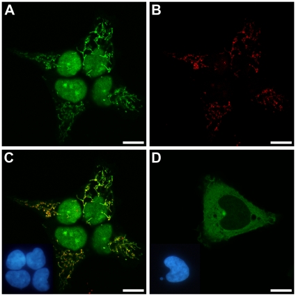
To address the localization of both proteins experimentally we expressed their complete coding sequences tagged by EGFP in human 293 cells. To avoid interfering with putative N-terminal sorting signals we fused the EGFP-tag to the C-termini of the proteins. RNase ZL localized to the nucleus and the mitochondria of transfected cells, but showed no cytosolic fluorescence (Figure 1A–C). RNase ZS on the other hand, was found primarily in the cytosol (Figure 1D). Still, RNase ZS-EGFP expressing cells also displayed some weak nuclear fluorescence. In contrast, EGFP alone distributed evenly throughout the cytosol and the nucleus (data not shown).
Figure 1. Subcellular localization of EGFP-tagged RNase ZL and RNase ZS in 293 cells.
(A) RNase ZL-EGFP; (B) immunofluorescence localization of subunit I of cytochrome c oxidase (mitochondrial staining) in the cells shown in (A); (C) overlay of (A) and (B) with nuclei shown as inset; (D) RNase ZS-EGFP with nuclei shown as inset. Confocal laser scanning microscopy (scale bar, 10 µm); insets showing nuclei by epifluorescent microscopy.
Dual nuclear/mitochondrial targeting of RNase ZL by alternative translation initiation
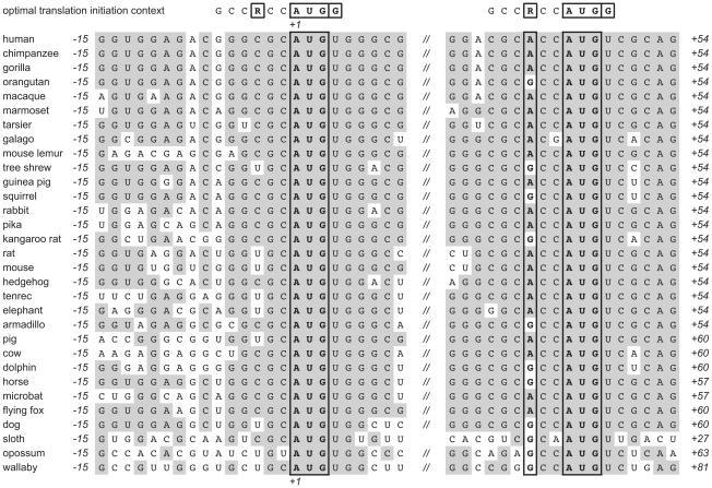
How does the nuclear fraction of RNase ZL escape the mitochondrial import pathway? The assigned start codon of human RNase ZL (ELAC2) appears to be in a suboptimal configuration for efficient initiation of translation (Figure 2). Initiation sites in mammalian mRNAs usually conform at least in part to the sequence GCCRCCAUGG [14]. The strongest determinants for translation initiation besides the AUG itself are a purine (R), preferably an A, at −3 and, especially in the absence of A−3, a G in position +4. The first AUG of human RNase ZL mRNA lacks both, A−3 and G+4. The next AUG, 45 nucleotides downstream, is preceded by ACC and thus in a much more favorable context for translation initiation. However, proteins translated from this second putative initiation site would lack 15 N-terminal amino acids and thereby most of the predicted mitochondrial targeting sequence. In fact, unlike full length RNase ZL (826 amino acids), the slightly shortened RNase ZL (811 amino acids) is no longer predicted to localize to mitochondria by any of the employed bioinformatic tools, whereas the predicted nuclear localization signal is not affected. The peculiar configuration of the two putative start codons is conserved among mammalian ELAC2 homologs (Figure 2). While position −3 is never occupied by a purine in the case of the first AUG, the second AUG is always preceded by a purine at −3. Also other, less important determinants for efficient translation initiation (C−4 and C−2) conform more closely to the consensus sequence in the case of the second putative initiation site. The second AUG thus invariably appears to be in a better initiation context than the first.
Figure 2. Alignment of the two putative initiation sites of mammalian RNase ZL mRNAs (ELAC2 homologs).
Optimal (consensus) translation initiation context of mammalian mRNAs [14] shown on top. R−3, AUGStart, and G+4 outlined; shading indicates identity to more than 50% of the sequences.
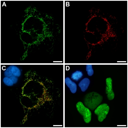
To test whether the observed dual nuclear/mitochondrial localization of human RNase ZL is indeed due to alternative translation initiation we prepared two variants of the ELAC2 cDNA and analyzed their subcellular localization. On the one hand, we changed the sequence preceding the first AUG to the more favorable initiation context GTCACC, on the other hand, we deleted the first 15 amino acids to allow translation from the second AUG only. Modification of the nucleotides preceding the first AUG to conform to the consensus gave rise to an RNase ZL exclusively localized to mitochondria (Figure 3A–C). Others also recently reported a mitochondria-restricted localization of RNase ZL; notably, their epitope-tagged RNase ZL was expressed from an (unintentionally) initiation context-optimized cDNA too [12]. As further predicted by our model, a cDNA devoid of the first few codons resulted in an RNase ZL variant exclusively found in the nucleus (Figure 3D; with the expected exception of cells undergoing mitosis).
Figure 3. Subcellular localization of EGFP-tagged RNase ZL variants with modified translation initiation sites in 293 cells.
(A) RNase ZL-EGFP variant with an optimized translation initiation context of the first AUG; (B) immunofluorescence localization of subunit I of cytochrome c oxidase (mitochondrial staining) in the cells shown in (A); (C) overlay of (A) and (B) with nuclei shown as inset; (D) RNase ZL-EGFP variant without the first 15 amino acids, nuclei shown as inset. Confocal laser scanning microscopy (scale bar, 10 µm); insets showing nuclei by epifluorescent microscopy.
In contrast to previous reports [11], [13] we did not observe any appreciable cytosolic RNase ZL in any of our experiments with the exception of cells currently undergoing mitosis. Apparently, nuclear RNase ZL was also completely re-imported after re-formation of the nuclear membrane. The localization pattern of RNase ZS, RNase ZL, and the two RNase ZL translation initiation variants was identical in 143B osteosarcoma cells (Figure S1).
Discussion
Determining an enzyme's subcellular localization is crucial to fully comprehend its biology. Eukaryal cells generally transcribe and process tRNA precursors in the nucleus and mitochondria (plants moreover in chloroplasts). All eukarya also have one RNase ZL gene, and it appears evolutionary optional to have further RNase Z genes encoding either short or additional long forms. Consistently, RNase ZL is targeted to the nuclear and the mitochondrial compartment in organisms where it is the only form of RNase Z, like Saccharomyces cerevisiae or Drosophila melanogaster [15], [16]. Yet, despite the additional presence of an RNase ZS, we show that human RNase ZL localizes to nuclei and mitochondria too, while RNase ZS is found mainly in the cytosol. Only if there is more than one RNase ZL gene, the general dual nuclear/mitochondrial localization of RNase ZL appears dispensable. In Arabidopsis thaliana one of the two RNases ZL is still dual targeted while the other one is found in mitochondria only [17]. In the yeast Schizosaccharomyces pombe finally, the two RNases ZL are confined to the nucleus and the mitochondria, respectively [18]. Interestingly, also in plants, where there are up to four RNases Z, only the long forms localize to nuclei and/or mitochondria, whereas the two RNases ZS are found in the cytosol and in chloroplasts, respectively [17].
The cytosolic localization of human RNase ZS indicates that it is not involved in the processing of tRNA primary transcripts. This conclusion is consistent with its low tRNA processing efficiency in vitro [19] and with the fact that its gene (ELAC1) is part of a homozygous deletion in a lung cancer cell line, and thus apparently dispensable for cell survival and growth [20]. Unfortunately, there are currently no more clues to the biological role of human RNase ZS.
In contrast, the subcellular localization of human RNase ZL suggests a role in the 3′ end maturation of nuclear and mitochondrial encoded tRNAs. Such a dual role has in fact been demonstrated for D. melanogaster RNase ZL [16]. It is unclear however, whether RNase ZL is generally the only nuclease responsible for tRNA 3′ end formation. While there is evidence for alternative, exonucleolytic pathways for nuclear tRNA maturation in different eukarya [1], the polycistronic nature of mitochondrial tRNA precursors appears to strictly require an endonuclease [21]. Notably, only mitochondrial and chloroplast RNases Z are essential in A. thaliana [17]. However, because both RNases ZL are targeted to mitochondria, only the double mutant is lethal in this case [17].
The basic mechanism underlying the dual nuclear/mitochondrial localization of human RNase ZL appears to be the alternative use of two translation initiation sites. The first AUG of mammalian RNase ZL is in a poor context for initiation and thus apparently passed by most scanning ribosomes, while the second AUG more closely conforms to a consensus initiation site and appears to be used preferentially. This mechanism of alternative translation initiation has been described as context-dependent leaky scanning [14]. In the case of mammalian RNase ZL downstream initiation results in the loss of most of the mitochondrial targeting signal, whereby the protein translated from the second AUG is no longer imported into the mitochondrial matrix, but routed to the nucleus via its nuclear localization signal. Accordingly, experimental optimization of the context of the first AUG resulted in an RNase ZL exclusively targeted to mitochondria, whereas its elimination produced nuclear RNase ZL only. This moreover implies that the mitochondrial targeting signal is either dominant over the nuclear localization signal, or the latter masked by the presence of the former [22]. A similar localization mechanism has been proposed for two other human nuclear/mitochondrial proteins, DNA ligase III and DNA topoisomerase IIIα [23], [24].
Context-dependent leaky scanning does not exclude that there are other, currently unknown mechanisms involved in regulating the balance between nuclear and mitochondrial RNase ZL. We previously reported a reduction in mitochondrial RNase Z activity in human cells lacking mitochondrial DNA (ρ0 cells) [25]. More recently, it was reported that the RNase ZL coding ELAC2 mRNA is in fact down-regulated in ρ0 cells [12]. It is not known whether this down-regulation is accompanied by the preferential targeting of the remaining RNase ZL to the nucleus or affects the cellular RNase ZL pool as a whole and thereby possibly contributes to the slower growth of ρ0 cells.
We did not observe any RNase ZL in the cytosol. Previously, others had reported a predominantly cytosolic localization of ELAC2 upon biochemical fractionation [11] and a ubiquitous cellular localization of RNase ZL by immunofluorescence [13]. Experimental design and methodological approach appear to account for these discrepancies. A protein tagged at its N-terminus by two FLAG epitopes was expressed from an optimized initiation site in the first study [11]. Thereby the N-terminal mitochondrial targeting signal was displaced and initiation from the downstream AUG, responsible for translation of nuclear RNase ZL, prevented. The N-terminal tag might have additionally masked the nuclear localization signal [22]. Some nuclear proteins moreover have a strong tendency to leak to the cytosol upon biochemical fractionation. Concerning the latter study [13], the uniform immunofluorescence throughout the cell casts doubts on the specificity of the antibody used. In the end, it is unlikely that the EGFP attached to the C-terminus of RNase ZL in our study prevented its cytosolic localization, as this was neither the case with EGFP alone nor with EGFP tagged RNase ZS.
Concluding remarks
Animal cells use two radically different enzymes for the processing of tRNA 5′ ends in the nucleus and mitochondria [26]–[28]. In contrast, key enzymatic components appear to be shared for the formation of tRNA 3′ ends. RNase ZL is localized to both compartments of tRNA biogenesis, and considering that only a single gene (TRNT1) for ATP(CTP):tRNA nucleotidyltransferase, the CCA-adding enzyme has been identified in the human genome, it appears reasonable to assume it is shared as well [29]. An analysis of TRNT1's N-terminus and putative translation initiation sites at least suggests that it employs the same basic mechanism for dual localization as RNase ZL (unpublished analysis).
Materials and Methods
Bioinformatics
For the prediction of subcellular localization we used the following tools: LOCtree [30], TargetP [31], MitoProt II [32], Predotar [33], and PredictNLS [34].
Mammalian RNase ZL gene sequences were retrieved from Ensembl [35]. Sequence alignments were generated with MacVector 6.5 (Oxford Molecular).
cDNA expression plasmids
The coding sequences of ELAC1 (NM_018696) and ELAC2 (NM_018127) including 15 nucleotides of 5′ untranslated region preceding the start codon were PCR amplified from HeLa cell (ATCC# CCL-2) cDNA using primers containing BamHI and in-frame XbaI restriction sites. PCR products were cloned into a pcDNA4/TO (Invitrogen) that was modified by insertion of the EGFP coding sequence from pEGFP-N3 (Invitrogen) into its ApaI site. Proteins expressed from these plasmids are composed of their complete native amino acid sequence fused at their C-terminus via a short linker peptide (SerArgGlyProAlaThr) to EGFP.
The native ELAC2 expression plasmid was modified by site-directed mutagenesis using the QuikChange (Stratagene) protocol: GTCACC replaced (i) the natural 5′ untranslated region, or (ii) the 5′ untranslated region plus the first 45 nucleotides of coding sequence.
Cell lines, staining, and microscopy
Stable T-REx-293 (Invitrogen) cell lines were generated as described recently [28]. For each expression cassette we isolated and analyzed several clones.
Cells were grown on cover slips and expression induced with tetracycline at 0.2 µg/ml. Paraformaldehyde fixed cells were stained with bisbenzimid H33342 and embedded in Kaiser's glycerol gelatin. For mitochondrial staining cells were fixed, permeabilized with 0.1% Triton X-100, and incubated with a monoclonal antibody specific for subunit I of cytochrome c oxidase (Molecular Probes A6403) at 5 ng/µl. For detection biotinylated anti mouse IgG and Texas Red conjugated streptavidin were used. A conventional epifluorescent and a dual-channel confocal laser scanning microscope (Olympus FluoView) were used. Pictures were processed with the FluoView application software (Olympus).
A 143B (ATCC# CRL-8303) derived cell line stably expressing DsRed2 (Clontech) targeted to mitochondria by the signal peptide of cytochrome c oxidase subunit VIII was selected. This cell line was transfected with the ELAC expression plasmids by a magnet assisted transfection procedure according to the instructions of the supplier (IBA BioTAGnology).
Supporting Information
Subcellular localization of EGFP-tagged RNase ZS, RNase ZL, and RNase ZL variants with modified translation initiation sites in 143B cells. (A) RNase ZS-EGFP; (B) RNase ZL-EGFP with native translation initiation context; (C) RNase ZL-EGFP variant with an optimized translation initiation context of the first AUG; (D) RNase ZL-EGFP variant without the first 15 amino acids; (E–H) DsRed2 labeled mitochondria of cells shown in the same row; (I–L) nuclear staining of cells shown in the same row.
(TIF)
Acknowledgments
I gratefully acknowledge the excellent technical assistance of Esther Löffler and Clemens Hufnagl.
Footnotes
Competing Interests: The author has declared that no competing interests exist.
Funding: The project was funded in part by the Austrian Science Fund (FWF): P17453. The funder had no role in study design, data collection and analysis, decision to publish, or preparation of the manuscript.
References
- 1.Phizicky EM, Hopper AK. tRNA biology charges to the front. Genes Dev. 2010;24:1832–1860. doi: 10.1101/gad.1956510. [DOI] [PMC free article] [PubMed] [Google Scholar]
- 2.Hartmann RK, Gößringer M, Späth B, Fischer S, Marchfelder A. The Making of tRNAs and More - RNase P and tRNase Z. Prog Nucleic Acid Res Mol Biol. 2009;85:319–368. doi: 10.1016/S0079-6603(08)00808-8. [DOI] [PubMed] [Google Scholar]
- 3.Lai LB, Vioque A, Kirsebom LA, Gopalan V. Unexpected diversity of RNase P, an ancient tRNA processing enzyme: challenges and prospects. FEBS Lett. 2010;584:287–296. doi: 10.1016/j.febslet.2009.11.048. [DOI] [PMC free article] [PubMed] [Google Scholar]
- 4.Deutscher MP. tRNA Processing Nucleases. In: Söll D, RajBhandary UL, editors. tRNA: Structure, Biosynthesis, and Function. Washington, D.C.: ASM Press; 1995. pp. 51–65. [Google Scholar]
- 5.Ceballos M, Vioque A. tRNase Z. Protein Pept Lett. 2007;14:137–145. doi: 10.2174/092986607779816050. [DOI] [PubMed] [Google Scholar]
- 6.Vogel A, Schilling O, Späth B, Marchfelder A. The tRNase Z family of proteins: physiological functions, substrate specificity and structural properties. Biol Chem. 2005;386:1253–1264. doi: 10.1515/BC.2005.142. [DOI] [PubMed] [Google Scholar]
- 7.Späth B, Canino G, Marchfelder A. tRNase Z: the end is not in sight. Cell Mol Life Sci. 2007;64:2404–2412. doi: 10.1007/s00018-007-7160-5. [DOI] [PMC free article] [PubMed] [Google Scholar]
- 8.Redko Y, Li de la Sierra-Gallay I, Condon C. When all's zed and done: the structure and function of RNase Z in prokaryotes. Nat Rev Microbiol. 2007;5:278–286. doi: 10.1038/nrmicro1622. [DOI] [PubMed] [Google Scholar]
- 9.Takaku H, Minagawa A, Takagi M, Nashimoto M. A candidate prostate cancer susceptibility gene encodes tRNA 3′ processing endoribonuclease. Nucleic Acids Res. 2003;31:2272–2278. doi: 10.1093/nar/gkg337. [DOI] [PMC free article] [PubMed] [Google Scholar]
- 10.Takahashi M, Takaku H, Nashimoto M. Regulation of the human tRNase ZS gene expression. FEBS Lett. 2008;582:2532–2536. doi: 10.1016/j.febslet.2008.06.020. [DOI] [PubMed] [Google Scholar]
- 11.Korver W, Guevara C, Chen Y, Neuteboom S, Bookstein R, et al. The product of the candidate prostate cancer susceptibility gene ELAC2 interacts with the γ-tubulin complex. Int J Cancer. 2003;104:283–288. doi: 10.1002/ijc.10945. [DOI] [PubMed] [Google Scholar]
- 12.Mineri R, Pavelka N, Fernandez-Vizarra E, Ricciardi-Castagnoli P, Zeviani M, et al. How do human cells react to the absence of mitochondrial DNA? PLoS ONE. 2009;4:e5713. doi: 10.1371/journal.pone.0005713. [DOI] [PMC free article] [PubMed] [Google Scholar]
- 13.Elbarbary RA, Takaku H, Uchiumi N, Tamiya H, Abe M, et al. Modulation of gene expression by human cytosolic tRNase ZL through 5′-half-tRNA. PLoS ONE. 2009;4:e5908. doi: 10.1371/journal.pone.0005908. [DOI] [PMC free article] [PubMed] [Google Scholar]
- 14.Kozak M. Pushing the limits of the scanning mechanism for initiation of translation. Gene. 2002;299:1–34. doi: 10.1016/S0378-1119(02)01056-9. [DOI] [PMC free article] [PubMed] [Google Scholar]
- 15.Hazbun TR, Malmström L, Anderson S, Graczyk BJ, Fox B, et al. Assigning function to yeast proteins by integration of technologies. Mol Cell. 2003;12:1353–1365. doi: 10.1016/s1097-2765(03)00476-3. [DOI] [PubMed] [Google Scholar]
- 16.Dubrovsky EB, Dubrovskaya VA, Levinger L, Schiffer S, Marchfelder A. Drosophila RNase Z processes mitochondrial and nuclear pre-tRNA 3′ ends in vivo. Nucleic Acids Res. 2004;32:255–262. doi: 10.1093/nar/gkh182. [DOI] [PMC free article] [PubMed] [Google Scholar]
- 17.Canino G, Bocian E, Barbezier N, Echeverria M, Forner J, et al. Arabidopsis Encodes Four tRNase Z Enzymes. Plant Physiol. 2009;150:1494–1502. doi: 10.1104/pp.109.137950. [DOI] [PMC free article] [PubMed] [Google Scholar]
- 18.Gan X, Yang J, Li J, Yu H, Dai H, et al. The fission yeast Schizosaccharomyces pombe has two distinct tRNase ZLs encoded by two different genes and differentially targeted to the nucleus and mitochondria. Biochem J. 2011;435:103–111. doi: 10.1042/BJ20101619. [DOI] [PubMed] [Google Scholar]
- 19.Yan H, Zareen N, Levinger L. Naturally Occurring Mutations in Human Mitochondrial Pre-tRNASer(UCN) Can Affect the Transfer Ribonuclease Z Cleavage Site, Processing Kinetics, and Substrate Secondary Structure. J Biol Chem. 2006;281:3926–3935. doi: 10.1074/jbc.M509822200. [DOI] [PubMed] [Google Scholar]
- 20.Yanaihara N, Kohno T, Takakura S, Takei K, Otsuka A, et al. Physical and transcriptional map of a 311-kb segment of chromosome 18q21, a candidate lung tumor suppressor locus. Genomics. 2001;72:169–179. doi: 10.1006/geno.2000.6454. [DOI] [PubMed] [Google Scholar]
- 21.Rossmanith W, Holzmann J. Processing mitochondrial (t)RNAs: new enzyme, old job. Cell Cycle. 2009;8:1650–1653. doi: 10.4161/cc.8.11.8502. [DOI] [PubMed] [Google Scholar]
- 22.Roberts BL, Richardson WD, Smith AE. The effect of protein context on nuclear location signal function. Cell. 1987;50:465–475. doi: 10.1016/0092-8674(87)90500-9. [DOI] [PubMed] [Google Scholar]
- 23.Lakshmipathy U, Campbell C. The human DNA ligase III gene encodes nuclear and mitochondrial proteins. Mol Cell Biol. 1999;19:3869–3876. doi: 10.1128/mcb.19.5.3869. [DOI] [PMC free article] [PubMed] [Google Scholar]
- 24.Wang Y, Lyu YL, Wang JC. Dual localization of human DNA topoisomerase IIIα to mitochondria and nucleus. Proc Natl Acad Sci USA. 2002;99:12114–12119. doi: 10.1073/pnas.192449499. [DOI] [PMC free article] [PubMed] [Google Scholar]
- 25.Rossmanith W, Karwan RM. Characterization of human mitochondrial RNase P: novel aspects in tRNA processing. Biochem Biophys Res Commun. 1998;247:234–241. doi: 10.1006/bbrc.1998.8766. [DOI] [PubMed] [Google Scholar]
- 26.Rossmanith W, Tullo A, Potuschak T, Karwan R, Sbisà E. Human mitochondrial tRNA processing. J Biol Chem. 1995;270:12885–12891. doi: 10.1074/jbc.270.21.12885. [DOI] [PubMed] [Google Scholar]
- 27.Rossmanith W, Potuschak T. Difference between mitochondrial RNase P and nuclear RNase P. Mol Cell Biol. 2001;21:8236–8237. doi: 10.1128/MCB.21.23.8236-8237.2001. [DOI] [PMC free article] [PubMed] [Google Scholar]
- 28.Holzmann J, Frank P, Löffler E, Bennett KL, Gerner C, et al. RNase P without RNA: identification and functional reconstitution of the human mitochondrial tRNA processing enzyme. Cell. 2008;135:462–474. doi: 10.1016/j.cell.2008.09.013. [DOI] [PubMed] [Google Scholar]
- 29.Reichert AS, Thurlow DL, Mörl M. A eubacterial origin for the human tRNA nucleotidyltransferase? Biol Chem. 2001;382:1431–1438. doi: 10.1515/BC.2001.176. [DOI] [PubMed] [Google Scholar]
- 30.Nair R, Rost B. Mimicking cellular sorting improves prediction of subcellular localization. J Mol Biol. 2005;348:85–100. doi: 10.1016/j.jmb.2005.02.025. [DOI] [PubMed] [Google Scholar]
- 31.Emanuelsson O, Brunak S, von Heijne G, Nielsen H. Locating proteins in the cell using TargetP, SignalP and related tools. Nat Protoc. 2007;2:953–971. doi: 10.1038/nprot.2007.131. [DOI] [PubMed] [Google Scholar]
- 32.Claros MG, Vincens P. Computational method to predict mitochondrially imported proteins and their targeting sequences. Eur J Biochem. 1996;241:779–786. doi: 10.1111/j.1432-1033.1996.00779.x. [DOI] [PubMed] [Google Scholar]
- 33.Small I, Peeters N, Legeai F, Lurin C. Predotar: A tool for rapidly screening proteomes for N-terminal targeting sequences. Proteomics. 2004;4:1581–1590. doi: 10.1002/pmic.200300776. [DOI] [PubMed] [Google Scholar]
- 34.Cokol M, Nair R, Rost B. Finding nuclear localization signals. EMBO Rep. 2000;1:411–415. doi: 10.1093/embo-reports/kvd092. [DOI] [PMC free article] [PubMed] [Google Scholar]
- 35.Flicek P, Aken BL, Ballester B, Beal K, Bragin E, et al. Ensembl's 10th year. Nucleic Acids Res. 2010;38:D557–562. doi: 10.1093/nar/gkp972. [DOI] [PMC free article] [PubMed] [Google Scholar]
Associated Data
This section collects any data citations, data availability statements, or supplementary materials included in this article.
Supplementary Materials
Subcellular localization of EGFP-tagged RNase ZS, RNase ZL, and RNase ZL variants with modified translation initiation sites in 143B cells. (A) RNase ZS-EGFP; (B) RNase ZL-EGFP with native translation initiation context; (C) RNase ZL-EGFP variant with an optimized translation initiation context of the first AUG; (D) RNase ZL-EGFP variant without the first 15 amino acids; (E–H) DsRed2 labeled mitochondria of cells shown in the same row; (I–L) nuclear staining of cells shown in the same row.
(TIF)


