Abstract
Background
Persons with post-stroke hemiparesis usually walk slowly and asymmetrically. Stroke severity and functional walking status are commonly predicted by post-stroke walking speed. The mechanisms that limit walking speed, and by extension functional walking status, need to be understood to improve post-stroke rehabilitation methods.
Methods
Three-dimensional forward dynamics walking simulations of hemiparetic subjects (and speed-matched controls) with different levels of functional walking status were developed to investigate the relationships between muscle contributions to walking subtasks and functional walking status. Muscle contributions to forward propulsion, swing initiation and power generation were analyzed during the pre-swing phase of the gait cycle and compared between groups.
Findings
Contributions from the paretic leg muscles (i.e., soleus, gastrocnemius and gluteus medius) to forward propulsion increased with improved functional walking status, with the non-paretic leg muscles (i.e., rectus femoris and vastii) compensating for reduced paretic leg propulsion in the limited community walker. Contributions to swing initiation from both paretic (i.e., gastrocnemius, iliacus and psoas) and non-paretic leg muscles (i.e., hamstrings) also increased as functional walking status improved. Power generation was also an important indicator of functional walking status, with reduced paretic leg power generation limiting the paretic leg contribution to forward propulsion and leg swing initiation.
Interpretation
These results suggest that deficits in muscle contributions to the walking subtasks of forward propulsion, swing initiation and power generation are directly related to functional walking status and that improving output in these muscle groups may be an effective rehabilitation strategy for improving post-stroke hemiparetic walking.
Keywords: forward dynamics simulation, modeling, biomechanics, gait, muscle function
1. Introduction
Post-stroke hemiparesis is seen in 50% of persons six months following stroke (Kelly-Hayes et al., 2003) and is often characterized by slow walking speed and asymmetry between the paretic and non-paretic legs (e.g., Olney and Richards, 1996). Walking speed is commonly used to predict stroke severity and assess functional walking status (i.e., household, limited community and community walking status) post-stroke (Bowden et al., 2008; Perry et al., 1995). Given that improving walking ability is a primary goal post-stroke (Bohannon et al., 1988), rehabilitation strategies focused on improving walking speed, and therefore functional walking status, are important. In order to develop more effective rehabilitation interventions, the underlying mechanisms that lead to different levels of walking status need to be fully understood. In particular, investigating the walking subtasks that are crucial to improving walking speed, including forward propulsion (i.e., pelvis forward acceleration), swing initiation (i.e., power delivery to the leg) and power generation (i.e., musculotendon power output), would provide insight into the impairments that limit functional walking status in hemiparetic subjects.
Previous studies have shown that the ankle plantarflexors, soleus (SOL) and gastrocnemius (GAS), are essential to the walking subtasks of forward propulsion, swing initiation and power generation in healthy walking and that pre-swing (i.e., double support phase preceding toe-off) is a critical region of the gait cycle for SOL and GAS to accomplish these subtasks (Liu et al., 2008; McGowan et al., 2008; Neptune et al., 2001; Neptune et al., 2008). SOL and GAS have unique contributions to the walking subtasks, with SOL contributing primarily to trunk forward propulsion while GAS contributes primarily to leg swing initiation (Neptune et al., 2001; Neptune et al., 2008). Plantarflexor weakness is commonly observed in hemiparetic walking and the output from the paretic plantarflexors has been correlated with walking speed (Nadeau et al., 1999; Olney et al., 1991; Parvataneni et al., 2007). We have recently suggested in a simulation study of two hemiparetic subjects that impaired performance in forward propulsion, swing initiation and power generation is at least partially due to decreased paretic plantarflexor contributions, specifically with reduced SOL contributions impairing forward propulsion, reduced GAS contributions impairing swing initiation and reduced SOL and GAS contributions impairing power generation (Peterson et al., 2010). However, the relationships between plantarflexor contributions to specific walking subtasks and functional walking status in hemiparetic subjects are not known.
Studies of healthy adults have shown the uniarticular hip flexors, iliacus and psoas (IL), provide swing initiation together with GAS (Neptune et al., 2004; Neptune et al., 2008) and that IL can compensate for overall plantarflexor weakness in some hemiparetic subjects (Nadeau et al., 1999; Olney and Richards, 1996). However, we have shown through simulation analyses that contributions of paretic IL to swing initiation and power generation are reduced in hemiparetic subjects relative to controls (Peterson et al., 2010). Thus, the relationship between paretic IL contributions to swing initiation and power generation and functional walking status in hemiparetic walking is also unclear.
The goal of this study was to build upon our previous work by identifying differences in pre-swing forward propulsion, swing initiation and power generation among hemiparetic subjects (and speed-matched controls) with different levels of functional walking status (i.e., limited community and community walkers) using muscle-actuated forward dynamics simulations. The simulations were used to identify changes in muscle coordination (i.e., muscle force production and timing) that reduce pre-swing deficits, increase walking speed and ultimately improve functional walking status in hemiparetic walking. Forward dynamics simulations provide an ideal framework to achieve this goal as they have been used to successfully quantify individual muscle contributions to the walking subtasks in healthy walking (e.g., Liu et al., 2008; Neptune et al., 2008) and in hemiparetic subjects (Higginson et al., 2006; Peterson et al., 2010). We expect that decreased muscle contributions to the walking subtasks during the pre-swing phase limits the functional walking status of hemiparetic subjects, with subjects who perform better in the three subtasks attaining higher walking speeds and higher functional walking status than subjects who have more impaired performance in one or more of the subtasks. We expect that the contributions of the paretic leg ankle plantarflexors (SOL and GAS) and the uniarticular hip flexors (IL) to forward propulsion, swing initiation and power generation will be reduced in hemiparetic subjects relative to controls, with the difference increasing (representing more impairment) as functional walking status decreases. By identifying the walking subtasks and specific muscle groups that limit functional walking status, this study will provide insight into the underlying mechanisms that contribute to reduced performance in the hemiparetic subjects and provide rationale for developing specific post-stroke rehabilitation interventions.
2. Methods
2.1. Musculoskeletal Model
A previously described 3D musculoskeletal model (Peterson et al., 2010) with 23 degrees-of-freedom was used to generate forward dynamics walking simulations of control and hemiparetic subjects. The model was developed using SIMM (MusculoGraphics, Inc., Santa Rosa, CA, USA) and included rigid segments representing the trunk, pelvis and two legs (thigh, shank, talus, calcaneus and toes). The pelvis had 6 degrees-of-freedom (3 translations and 3 rotations) with trunk and hip joints modeled by spherical joints. The knee, ankle, subtalar and metatarsophalangeal joints were modeled as single degree-of-freedom revolute joints. The foot-ground contact was modeled using 31 visco-elastic elements with coulomb friction attached to each foot (Neptune et al., 2000). Passive torques were applied at each joint to represent the forces applied by the ligaments, passive tissues and joint structures (Anderson, 1999; Davy and Audu, 1987). The dynamical equations of motion were generated using SD/FAST (PTC, Needham, MA, USA) and the forward dynamics simulations were produced using the framework provided by Dynamics Pipeline (MusculoGraphics, Inc., Santa Rosa, CA, USA).
The musculoskeletal model was actuated with 43 individual Hill-type musculotendon actuators per leg (Table 1). The muscle excitation patterns were defined using a bimodal Henning pattern as:
| (1) |
where e(t) is the excitation magnitude at time t and ai, onseti and offseti are the amplitude, onset and offset, respectively, of each mode, i. Musculotendon lengths and moment arms for each muscle were calculated using polynomial functions described by Menegaldo et al. (2004). The muscle contraction dynamics were governed by Hill-type muscle properties (Zajac, 1989) and the activation dynamics were modeled by a first-order differential equation (Raasch et al., 1997), with activation and deactivation time constants derived from Winters and Stark (1988). Nominal activation and deactivation time constants of 12 and 48 ms, respectively, were used for muscles not available in Winters and Stark (1988).
Table 1.
The 43 musculotendon actuators per leg were combined into 18 groups after analysis according to anatomical classification and how they contributed to the three walking subtasks.
| Muscle name | Analysis Group |
|---|---|
| Iliacus | IL |
| Psoas | IL |
| Adductor Longus | AL |
| Adductor Brevis | AL |
| Pectineus | AL |
| Quadratus Femoris | QF |
| Superior Adductor Magnus | AM |
| Middle Adductor Magnus | AM |
| Inferior Adductor Magnus | AM |
| Sartorius | SAR |
| Rectus Femoris | RF |
| Vastus Medialis | VAS |
| Vastus Lateralis | VAS |
| Vastus Intermedius | VAS |
| Anterior Gluteus Medius | GMED |
| Middle Gluteus Medius | GMED |
| Posterior Gluteus Medius | GMED |
| Piriformis | PIRI |
| Gemellus | GEM |
| Anterior Gluteus Minimus | GMIN |
| Middle Gluteus Minimus | GMIN |
| Posterior Gluteus Minimus | GMIN |
| Tensor Fascia Lata | TFL |
| Anterior Gluteus Maximus | GMAX |
| Middle Gluteus Maximus | GMAX |
| Posterior Gluteus Maximus | GMAX |
| Semitendinosus | HAM |
| Semimembranosus | HAM |
| Gracilis | HAM |
| Biceps Femoris Long Head | HAM |
| Biceps Femoris Short Head | BFSH |
| Medial Gastrocnemius | GAS |
| Lateral Gastrocnemius | GAS |
| Soleus | SOL |
| Tibialis Posterior | SOL |
| Peroneus Brevis | SOL |
| Peroneus Longus | SOL |
| Flexor Digitorum Longus | SOL |
| Flexor Hallucis Longus | SOL |
| Tibialis Anterior | TA |
| Extensor Digitorum Longus | TA |
| Peroneus Tertius | TA |
| Extensor Hallucis Longus | TA |
2.2 Dynamic Optimization
Forward dynamics simulations (from midstance to toe-off) were generated to emulate group-averaged experimentally measured kinematics and ground reaction forces (GRFs) of limited community and community hemiparetic subjects walking at their self-selected treadmill speed (limited community walkers: mean = 0.55 m/s (SD = 0.15 m/s); community walkers: mean = 0.92 m/s (SD = 0.05 m/s)) and similarly aged speed-matched control subjects walking at 0.6 and 0.9 m/s. The muscle excitation patterns (amplitude and timing) and the initial joint angular velocities were optimized using a simulated annealing algorithm (Goffe et al., 1994) that minimized differences between the simulated and experimental data. Quantities included in the cost function were the 3D pelvis translations and rotations, 3D trunk rotations, hip, knee and ankle joint angles and 3D GRFs. Muscle stress was also minimized in the cost function to ensure an even distribution across muscle groups.
2.3 Experimental Data Collection
Experimental GRF, kinematic and electromyographic (EMG) data were collected from 57 hemiparetic subjects (30 left hemiparesis; 37 men; age: mean = 61.4 years (SD = 11.4 years); time since stroke: mean = 5.0 years (SD = 5.5 years) and 21 similarly aged control subjects (4 men; age: mean = 65.2 years (SD = 9.6 years)). Each subject walked on an ADAL split-belt instrumented treadmill (Tecmachine, Andrézieux Bouthéon, France) while GRF, kinematic and EMG data were simultaneously collected for 30 second trials. Hemiparetic and similarly aged control subjects completed three walking trials at their self-selected speed. The control subjects completed additional walking trials at prescribed speeds of 0.3, 0.6 and 0.9 m/s. All subjects provided written informed consent approved by the Institutional Review Boards of the University of Florida and the University of Texas at Austin. 3D GRF data were measured at 2000 Hz and low pass filtered at 20 Hz. Reflective marker trajectories were recorded at 100 Hz with a 12-camera motion capture system (Vicon Motion Systems, Oxford, UK) and low pass filtered at 6 Hz. Surface EMG data were recorded bilaterally at 2000 Hz from the tibialis anterior, medial gastrocnemius, soleus, rectus femoris, biceps femoris long head, vastus medialis, semimembranosus and gluteus medius using a 16-channel EMG system (Konigsburg Instruments, Pasadena, CA, USA). The raw EMG data were high-pass filtered at 40 Hz, demeaned, rectified and low-pass filtered at 4 Hz. All data were processed using Visual3D (C-motion, Inc., Germantown, MD, USA). All data were synchronized and time normalized to the paretic (right) leg gait cycle for the hemiparetic (control) subjects and averaged across gait cycles within each subject for each condition. The average data for each subject were averaged across hemiparetic subjects within each functional group (limited community (n = 21); community (n = 5)) and across control subjects walking at prescribed speeds (0.6 m/s (n = 20); 0.9 m/s (n = 17)) to generate the tracking data for each optimization. The EMG data were used to constrain the timing (onset and offset) for each muscle excitation pattern in the optimizations to ensure muscles were producing force at the appropriate point in the gait cycle.
2.4 Analysis of Muscle Function
Performance of the walking subtasks was quantified by individual muscle contributions to forward propulsion, swing initiation and power generation. A muscle-induced acceleration and segment power analysis (Fregly and Zajac, 1996) was performed to quantify individual muscle contributions to the walking subtasks (e.g., Neptune et al., 2001; Neptune et al., 2004; Neptune et al., 2008). Each muscle’s contribution to forward propulsion was determined by quantifying its average contribution to the horizontal pelvis acceleration during pre-swing. Muscle-induced mechanical power generated, absorbed or transferred to or from each segment within the leg was summed and averaged over the pre-swing phase to determine each muscle’s contribution to swing initiation. Power generation for each muscle was calculated as the average musculotendon power during pre-swing with positive contributions representing concentric shortening and negative contributions representing eccentric lengthening. Contributions from individual muscles were grouped after analysis according to anatomical classification and how they contributed to the walking subtasks (Table 1). The contributions of the muscle groups to each of the waking subtasks were compared across the hemiparetic and control groups to identify the relationships between performance of the subtasks and functional walking status.
3. Results
The kinematic and GRF data from the control and hemiparetic simulations agreed well with the experimental data, with an average absolute difference of 5.0 degrees (SD = 0.71 degrees) and 3.8% BW (SD = 0.77% BW) for all simulations (Table 2). The differences between simulated and experimental data were within the average of two standard deviations of the experimental data for all subjects (angle: mean = 13.6 degrees (SD = 0.86 degrees); GRF: mean = 9.8% BW (SD = 1.2% BW)). The timing of the optimized excitation patterns generally agreed with the experimentally collected EMG data as in our previous work (e.g., Neptune et al., 2001; Neptune et al., 2004).
Table 2.
The average difference between the experimental and simulated kinematic angles and ground reaction forces (GRFs) compared to the average standard deviation (SD) of the experimental data. For each quantity: the average difference (2 SD) is reported.
| Controls at 0.6 m/s |
Controls at 0.9 m/s |
Limited Community |
Community | |||
|---|---|---|---|---|---|---|
| Kinematic Angles (degrees) | Pelvis | Obliquity | 2.7 (3.9) | 1.5 (3.8) | 1.5 (7.7) | 2.4 (4.3) |
| Rotation | 2.0 (7.5) | 3.5 (7.6) | 2.3 (13.5) | 2.9 (9.9) | ||
| Tilt | 6.3 (13.5) | 9.6 (12.7) | 3.1 (13.2) | 5.6 (12.8) | ||
| Trunk | Obliquity | 3.0 (5.5) | 3.4 (4.9) | 2.2 (8.5) | 2.4 (8.6) | |
| Rotation | 6.3 (11.8) | 7.9 (11.2) | 4.8 (9.7) | 5.0 (12.6) | ||
| Tilt | 0.5 (15.4) | 3.2 (16.3) | 2.3 (13.8) | 6.7 (16.7) | ||
|
Ipsilateral/ Paretic Leg |
Hip Adduction | 3.3 (5.2) | 2.1 (5.9) | 2.8 (9.5) | 7.3 (8.9) | |
| Hip Rotation | 8.4 (27.2) | 5.2 (28.2) | 8.9 (22.7) | 16.9 (13.1) | ||
| Hip Flexion | 6.1 (18.5) | 7.4 (17.7) | 13.5 (22.0) | 12.7 (18.2) | ||
| Knee Flexion | 4.5 (14.6) | 4.3 (13.2) | 14.2 (24.9) | 5.2 (17.9) | ||
| Ankle Dorsiflexion | 3.2 (11.4) | 6.6 (12.1) | 3.9 (11.4) | 9.8 (9.7) | ||
|
Contralateral/ Non-Paretic Leg |
Hip Adduction | 2.1 (9.0) | 1.2 (7.9) | 10.2 (12.7) | 2.4 (12.0) | |
| Hip Rotation | 11.4 (29.2) | 4.8 (26.0) | 1.6 (19.5) | 2.5 (18.9) | ||
| Hip Flexion | 5.0 (15.3) | 5.8 (15.4) | 2.9 (21.6) | 6.2 (22.5) | ||
| Knee Flexion | 8.4 (14.8) | 2.5 (12.9) | 4.5 (17.2) | 2.9 (19.7) | ||
| Ankle Dorsiflexion | 2.3 (10.6) | 2.4 (11.3) | 1.7 (10.3) | 6.1 (7.3) | ||
| Forces (%BW) |
Ipsilateral/ Paretic Leg |
A/P GRF | 2.4 (5.1) | 3.2 (5.5) | 1.1 (3.7) | 1.9 (4.9) |
| Vertical GRF | 7.0 (18.2) | 12.7 (19.4) | 6.1 (23.8) | 10.9 (27.5) | ||
| M/L GRF | 1.8 (2.7) | 3.1 (2.8) | 2.1 (2.6) | 0.7 (1.7) | ||
|
Contralateral/ Non-Paretic Leg |
A/P GRF | 3.1 (4.5) | 1.1 (4.8) | 0.7 (4.5) | 0.5 (4.6) | |
| Vertical GRF | 7.6 (17.0) | 7.7 (18.5) | 8.7 (26.7) | 4.4 (24.3) | ||
| M/L GRF | 1.4 (3.2) | 1.9 (3.3) | 1.4 (3.0) | 1.1 (3.3) | ||
| Average Angle Error (degrees) | 4.1 (13.4) | 4.5 (12.9) | 5.0 (14.9) | 6.1 (13.3) | ||
| Average GRF Error (%BW) | 3.9 (8.5) | 4.9 (9.1) | 3.3 (10.7) | 3.3 (11.0) | ||
3.1 Forward Propulsion
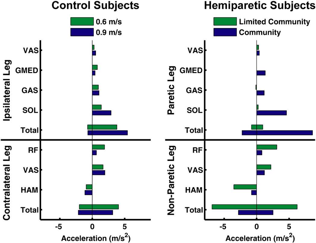
In the control subjects, contributions to forward propulsion from the ipsilateral and contralateral leg muscles increased and decreased, respectively, with increased walking speed (Fig. 1: Total). Contributions from the ipsilateral and contralateral leg muscles to pelvis deceleration (negative values) were similar as walking speed increased (Fig. 1: Total).
Figure 1.
Average muscle contributions to forward propulsion (i.e., horizontal pelvis acceleration) by the control subjects during the ipsilateral pre-swing phase and the hemiparetic subjects during the paretic pre-swing phase, where Total is the sum of the positive and negative contributions from all muscles for the respective leg. Contributions from the ipsilateral leg muscles to forward propulsion increased as walking speed increased from 0.6 to 0.9 m/s in the control subjects. Similarly, contributions from the paretic leg muscles (i.e., SOL, GAS and GMED) to forward propulsion increased with improved functional walking status. The non-paretic leg muscles (i.e., RF and VAS) contributed to forward propulsion in the limited community walkers to compensate for reduced paretic leg muscle contributions.
In the hemiparetic subjects, contributions from the paretic and non-paretic leg muscles to forward propulsion increased and decreased, respectively, with improved functional walking status (Fig. 1: Total). Similarly, contributions from the paretic and non-paretic leg muscles to pelvis deceleration increased and decreased, respectively, as functional walking status improved (Fig. 1: Total).
3.2 Swing Initiation
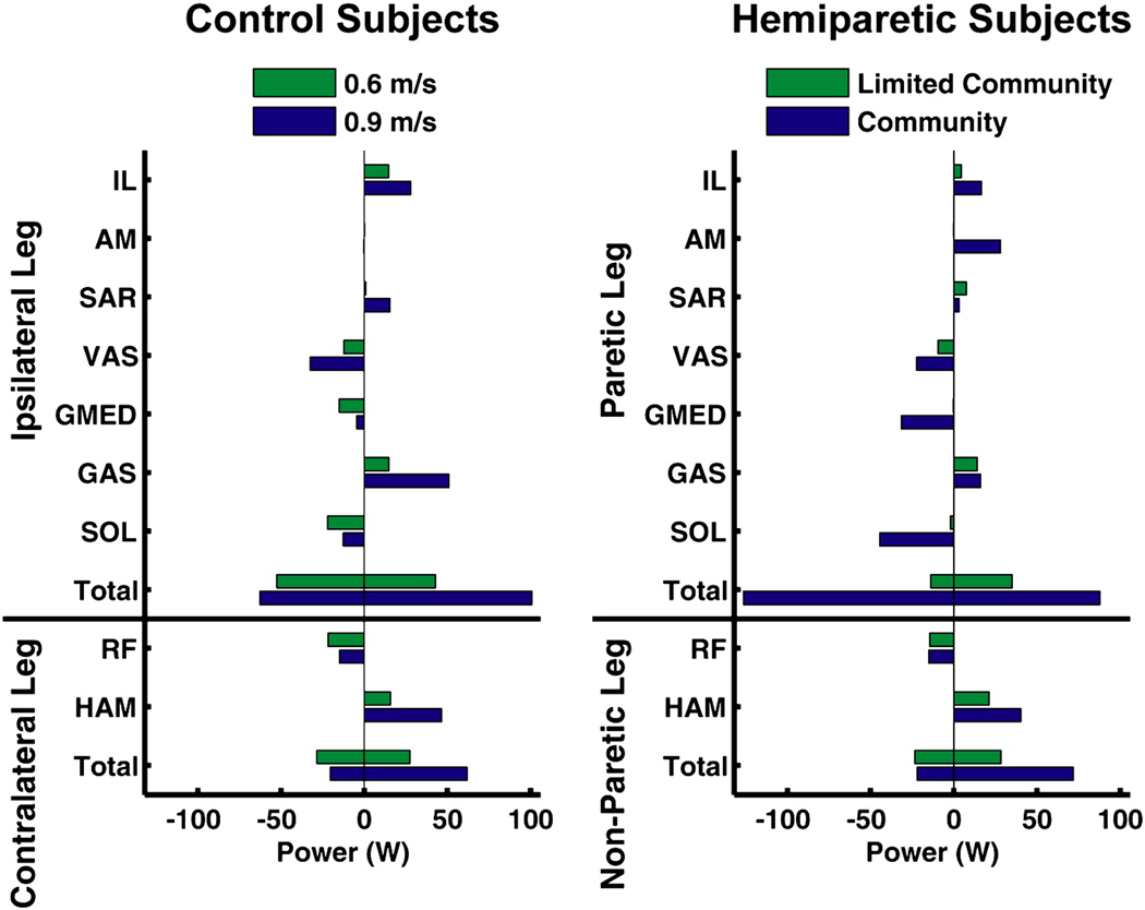
In the control subjects, positive contributions to swing initiation from the ipsilateral and contralateral leg muscles increased with increased walking speed (Fig. 2: Total). The negative contributions to swing initiation from the ipsilateral and contralateral leg muscles increased and decreased, respectively, as walking speed increased from 0.6 to 0.9 m/s (Fig. 2: Total).
Figure 2.
Average muscle contributions to swing initiation (i.e., net power transferred to/from the ipsilateral/paretic leg) by the control subjects during the ipsilateral pre-swing phase and the hemiparetic subjects during the paretic pre-swing phase, where Total is the sum of the positive and negative contributions from all muscles for the respective leg. In the control subjects, both the ipsilateral leg muscles (i.e., GAS, IL and SAR) and contralateral leg muscles (i.e., HAM) increased their contributions to swing initiation with increased walking speed. In a similar manner, both the paretic leg muscles (i.e., GAS and IL) and non-paretic leg muscles (i.e., HAM) increased their contributions to swing initiation with improved functional walking status. In the community walkers, paretic AM and GMED contributed positively and negatively, respectively to swing initiation, while these muscles did not contribute to swing initiation in the limited community walkers.
In the hemiparetic subjects, positive contributions to swing initiation from the paretic and non-paretic leg muscles increased as functional walking status improved (Fig. 2: Total) while the negative contributions to swing initiation from the paretic and non-paretic leg muscles increased and decreased, respectively (Fig. 2: Total).
3.3 Power Generation
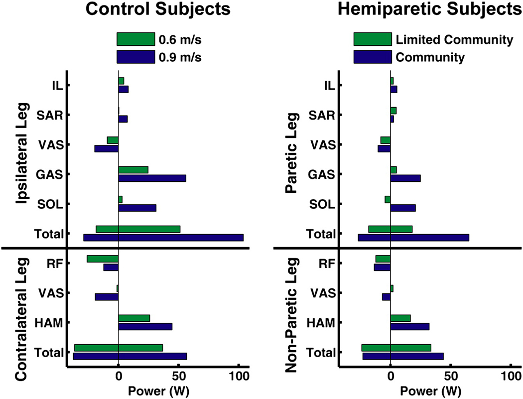
In the control subjects, ipsilateral and contralateral leg muscles increased power generation as speed increased from 0.6 m/s to 0.9 m/s (Fig. 3: Total). Power absorption increased in the ipsilateral leg and did not change in the contralateral leg, with increased walking speed (Fig. 3: Total).
Figure 3.
Average power generation (i.e., net musculotendon power) by the control subjects during the ipsilateral pre-swing phase and the hemiparetic subjects during the paretic pre-swing phase, where Total is the sum of the positive and negative contributions from all muscles for the respective leg. Power generation by the ipsilateral leg muscles (i.e., SOL, GAS, IL and SAR) and the contralateral leg muscles (i.e., HAM) increased in the control subjects as speed increased from 0.6 to 0.9 m/s. In the hemiparetic subjects, as functional walking status improved, the paretic leg muscles (i.e., SOL and GAS) and the non-paretic leg muscles (i.e., HAM) generated more power.
Paretic and non-paretic leg muscles generated more power as functional walking status improved (Fig. 3: Total). Power absorption was increased and did not change with improved functional walking status in the paretic and non-paretic legs, respectively (Fig. 3: Total).
4. Discussion
We previously showed using simulation analyses that output from the paretic leg ankle plantarflexor and hip flexor muscles is reduced in hemiparetic subjects relative to controls, which leads to impaired performance in the walking subtasks of forward propulsion, swing initiation and power generation (Peterson et al., 2010). The purpose of this study was to build upon this work by analyzing hemiparetic subjects with different levels of functional walking status to identify the relationships between individual muscle contributions to these subtasks and walking status.
The results showed that decreased paretic leg muscle contributions to these subtasks were indeed associated with functional walking status. In both the control and hemiparetic subjects, ipsilateral (paretic) SOL was an important contributor to forward propulsion during pre-swing and was critical to increasing walking speed and corresponded with higher functional walking status (Fig. 1). This result is consistent with previous studies showing SOL to be a primary contributor to forward propulsion (e.g., McGowan et al., 2008; Neptune et al., 2001) and a mechanism for attaining higher walking speeds in healthy walking (Liu et al., 2008; Neptune et al., 2008) and in the hemiparetic population (Nadeau et al., 1999; Olney et al., 1991; Parvataneni et al., 2007). The contralateral (non-paretic) VAS also had a large contribution to forward propulsion during the ipsilateral (paretic) leg pre-swing phase in the control (hemiparetic) subjects (Fig. 1). As a result, both ipsilateral (paretic) SOL and contralateral (non-paretic) VAS were important determinants of walking speed in our subjects.
In the hemiparetic subjects, a strong relationship was found between the contribution of the paretic leg muscles to forward propulsion and functional walking status. In the community walkers, the paretic leg muscles generated the majority of the forward propulsion similar to the control subjects (Fig. 1). However, the limited community walkers’ muscle contributions were altered compared to the community walkers and the control subjects, with the paretic leg muscles (SOL, GAS and GMED) contributing little to forward propulsion and the non-paretic leg muscles (RF and VAS) compensating for the reduced paretic leg output (Fig. 1). This result is consistent with Bowden et al (2006) who showed that the non-paretic leg’s contribution to the A/P GRF increased as hemiparetic severity increased. In addition, the non-paretic leg muscles (primarily HAM) increased their contributions to pelvis deceleration in the limited community walkers compared to the community walkers, resulting in a net (sum of all paretic and non-paretic leg muscles) deceleration of the pelvis during pre-swing.
Leg swing initiation was provided primarily by ipsilateral GAS, IL and contralateral HAM in the control subjects and increased contribution from these muscles was required to increase walking speed from 0.6 to 0.9 m/s (Fig. 2), which is consistent with previous simulations of healthy walking at self-selected speed (e.g., Neptune et al., 2001; Neptune et al., 2004) and increased walking speeds (Neptune et al., 2008). In the hemiparetic subjects, these muscles have a clear impact on functional walking status. The community walkers’ muscle contributions to swing initiation were similar to the muscle contributions seen in the control subjects with paretic GAS, IL and contralateral HAM contributing strongly to paretic leg swing initiation. In addition to these muscles, AM and GMED contributed positively and negatively, respectively, to swing initiation. The contributions from AM and GMED were similar in magnitude and resulted in a small net negative contribution (−3.5 W) to swing initiation. Clear deficits existed in the paretic and non-paretic leg muscle contributions to swing initiation in the limited community walkers (Fig. 2), which is consistent with previous studies showing reduced paretic leg kinetic energy at toe-off, suggesting impaired swing initiation in the paretic leg (e.g., Chen and Patten, 2008). Paretic GAS, IL and non-paretic HAM contributions were reduced in the limited community walkers compared to the community walkers (Fig. 2). The negative contributions from the paretic leg muscles (VAS, GMED, SOL) were also greatly reduced (Fig. 2) in the limited community walkers, allowing the leg to accelerate into swing.
Power generation by both ipsilateral and contralateral leg muscles during pre-swing in the control subjects generally increased with walking speed (Fig. 3) and showed consistent trends to previous studies (e.g., Neptune et al., 2004; Neptune et al., 2008). Power generation was also an important indicator of functional walking status in the hemiparetic subjects. Power generation by muscles in the community walkers closely resembled those of the control subjects. However, in the limited community walkers, the paretic leg muscles, specifically GAS and IL, generated less power, consistent with their reduced contributions to forward propulsion (GAS) and swing initiation (GAS and IL). In addition, paretic SOL absorbed power in the limited community walkers in contrast with the community walkers and control subjects, where it generated power in pre-swing. The power absorbed by SOL in the limited community walkers limited its ability to contribute to forward propulsion.
The limitations of forward dynamics simulations and analyses have been previously discussed (e.g., Neptune et al., 2001; Neptune et al., 2004), including necessary modeling assumptions and constraints. Specific to this study, a potential limitation is that model parameters for all the simulations were based on measurements obtained from healthy control subjects. It is likely that these properties are altered post-stroke, as has been shown in a recent study (Gao et al., 2009). However, the optimization was able to compensate for altered model properties by modulating the magnitude of the muscle excitation to produce the necessary muscle force output to replicate the subject’s walking mechanics. Muscle redundancy is also present in the neuromuscular system such that multiple muscle coordination patterns may exist that produce the same kinematic and kinetic patterns. In this study, the timing of the muscle excitation patterns were constrained to closely match the experimental EMG patterns and muscle stress was minimized in the cost function to reduce unnecessary co-contraction. We expect that hemiparetic subjects who generate similar muscle forces to those in this study would have similar muscle contributions to the walking subtasks. In addition, the results of this study are specific to hemiparetic subjects who walk with similar kinematic and kinetic patterns to the groups simulated in this study. Because of the heterogeneity of the post-stroke hemiparetic population, the extent to which these results can be generalized to the post-stroke population as a whole is not clear. Future work should focus on developing simulations of a large number of hemiparetic subjects to gain further insight into the changes in muscle function in this population.
In summary, the analyses showed that deficits in the walking subtasks of forward propulsion, swing initiation and power generation are related to functional walking status in the hemiparetic walkers. Increased contributions from the paretic leg muscles (i.e., ankle plantarflexors and hip flexors) and reduced contributions from the non-paretic leg (i.e., knee and hip extensors) to the walking subtasks were critical in achieving higher functional walking status. These results provide rationale for developing locomotor therapies that focus on these muscle groups in order to improve the functional walking status of persons with post-stroke hemiparesis.
Acknowledgements
The authors would like to thank Erin Carr, Dr. Mark Bowden, Dr. Bhavana Raja, Dr. Cameron Nott, Dr. Chitra Balasubramanian, Kelly Rooney and Ryan Knight for help with the data collection and processing and the members of the Neuromuscular Biomechanics Lab at The University of Texas at Austin for their insightful comments on the manuscript. This work was funded by NIH grant RO1 HD46820 and the Rehabilitation Research & Development Service of the VA. The contents are solely the responsibility of the authors and do not necessarily represent the official views of the NIH, NICHD or VA.
Footnotes
Publisher's Disclaimer: This is a PDF file of an unedited manuscript that has been accepted for publication. As a service to our customers we are providing this early version of the manuscript. The manuscript will undergo copyediting, typesetting, and review of the resulting proof before it is published in its final citable form. Please note that during the production process errors may be discovered which could affect the content, and all legal disclaimers that apply to the journal pertain.
References
- Anderson FC. Doctoral Dissertation. Austin, TX: The University of Texas at Austin; 1999. A dynamic optimization solution for a complete cycle of normal gait. [Google Scholar]
- Bohannon RW, Andrews AW, Smith MB. Rehabilitation goals of patients with hemiplegia. Int. J. Rehabil. Res. 1998;11(2):181–184. [Google Scholar]
- Bowden MG, Balasubramanian CK, Neptune RR, Kautz SA. Anterior-posterior ground reaction forces as a measure of paretic leg contribution in hemiparetic walking. Stroke. 2006;37(3):872–876. doi: 10.1161/01.STR.0000204063.75779.8d. [DOI] [PubMed] [Google Scholar]
- Bowden MG, Balasubramanian CK, Behrman AL, Kautz SA. Validation of a speed-based classification system using quantitative measures of walking performance poststroke. Neurorehabil. Neural Repair. 2008;22(6):672–675. doi: 10.1177/1545968308318837. [DOI] [PMC free article] [PubMed] [Google Scholar]
- Chen G, Patten C. Joint moment work during the stance-to-swing transition in hemiparetic subjects. J. Biomech. 2008;41(4):877–883. doi: 10.1016/j.jbiomech.2007.10.017. [DOI] [PubMed] [Google Scholar]
- Davy DT, Audu ML. A dynamic optimization technique for predicting muscle forces in the swing phase of gait. J. Biomech. 1987;20(2):187–201. doi: 10.1016/0021-9290(87)90310-1. [DOI] [PubMed] [Google Scholar]
- Fregly BJ, Zajac FE. A state-space analysis of mechanical energy generation, absorption, and transfer during pedaling. J. Biomech. 1996;29(1):81–90. doi: 10.1016/0021-9290(95)00011-9. [DOI] [PubMed] [Google Scholar]
- Gao F, Grant TH, Roth EJ, Zhang LQ. Changes in passive mechanical properties of the gastrocnemius muscle at the muscle fascicle and joint levels in stroke survivors. Arch. Phys. Med. Rehabil. 2009;90(5):819–826. doi: 10.1016/j.apmr.2008.11.004. [DOI] [PubMed] [Google Scholar]
- Goffe WL, Ferrier GD, Rogers J. Global optimization of statistical functions with simulated annealing. J. Econometrics. 1994;60(1–2):65–99. [Google Scholar]
- Higginson JS, Zajac FE, Neptune RR, Kautz SA, Delp SL. Muscle contributions to support during gait in an individual with post-stroke hemiparesis. J. Biomech. 2006;39(10):1769–1777. doi: 10.1016/j.jbiomech.2005.05.032. [DOI] [PubMed] [Google Scholar]
- Kelly-Hayes M, Beiser A, Kase CS, Scaramucci A, D'Agostino RB, Wolf PA. The influence of gender and age on disability following ischemic stroke: The Framingham study. J. Stroke Cerebrovasc. Dis. 2003;12(3):119–126. doi: 10.1016/S1052-3057(03)00042-9. [DOI] [PubMed] [Google Scholar]
- Liu MQ, Anderson FC, Schwartz MH, Delp SL. Muscle contributions to support and progression over a range of walking speeds. J. Biomech. 2008;41(15):3243–3252. doi: 10.1016/j.jbiomech.2008.07.031. [DOI] [PMC free article] [PubMed] [Google Scholar]
- McGowan CP, Neptune RR, Kram R. Independent effects of weight and mass on plantar flexor activity during walking: Implications for their contributions to body support and forward propulsion. J. Appl. Physiol. 2008;105(2):486–494. doi: 10.1152/japplphysiol.90448.2008. [DOI] [PMC free article] [PubMed] [Google Scholar]
- Menegaldo LL, de Toledo Fleury A, Weber HI. Moment arms and musculotendon lengths estimation for a three-dimensional lower-limb model. J. Biomech. 2004;37(9):1447–1453. doi: 10.1016/j.jbiomech.2003.12.017. [DOI] [PubMed] [Google Scholar]
- Nadeau S, Gravel D, Arsenault AB, Bourbonnais D. Plantarflexor weakness as a limiting factor of gait speed in stroke subjects and the compensating role of hip flexors. Clin. Biomech. 1999;14(2):125–135. doi: 10.1016/s0268-0033(98)00062-x. [DOI] [PubMed] [Google Scholar]
- Neptune RR, Wright IC, Van Den Bogert AJ. A method for numerical simulation of single limb ground contact events: Application to heel-toe running. Comput. Methods Biomech. Biomed. Engin. 2000;3(4):321–334. doi: 10.1080/10255840008915275. [DOI] [PubMed] [Google Scholar]
- Neptune RR, Kautz SA, Zajac FE. Contributions of the individual ankle plantar flexors to support, forward progression and swing initiation during walking. J. Biomech. 2001;34(11):1387–1398. doi: 10.1016/s0021-9290(01)00105-1. [DOI] [PubMed] [Google Scholar]
- Neptune RR, Zajac FE, Kautz SA. Muscle force redistributes segmental power for body progression during walking. Gait Posture. 2004;19(2):194–205. doi: 10.1016/S0966-6362(03)00062-6. [DOI] [PubMed] [Google Scholar]
- Neptune RR, Sasaki K, Kautz SA. The effect of walking speed on muscle function and mechanical energetics. Gait Posture. 2008;28(1):135–143. doi: 10.1016/j.gaitpost.2007.11.004. [DOI] [PMC free article] [PubMed] [Google Scholar]
- Olney SJ, Griffin MP, Monga TN, McBride ID. Work and power in gait of stroke patients. Arch. Phys. Med. Rehabil. 1991;72(5):309–314. [PubMed] [Google Scholar]
- Olney SJ, Richards C. Hemiparetic gait following stroke. Part I: Characteristics. Gait Posture. 1996;4(2):136–148. [Google Scholar]
- Parvataneni K, Olney SJ, Brouwer B. Changes in muscle group work associated with changes in gait speed of persons with stroke. Clin. Biomech. 2007;22(7):813–820. doi: 10.1016/j.clinbiomech.2007.03.006. [DOI] [PubMed] [Google Scholar]
- Perry J, Garrett M, Gronley JK, Mulroy SJ. Classification of walking handicap in the stroke population. Stroke. 1995;26(6):982–989. doi: 10.1161/01.str.26.6.982. [DOI] [PubMed] [Google Scholar]
- Peterson CL, Hall AL, Kautz SA, Neptune RR. Pre-swing deficits in forward propulsion, swing initiation and power generation by individual muscles during hemiparetic walking. 2010;43(12):2348–2355. doi: 10.1016/j.jbiomech.2010.04.027. [DOI] [PMC free article] [PubMed] [Google Scholar]
- Raasch CC, Zajac FE, Ma B, Levine WS. Muscle coordination of maximum-speed pedaling. J. Biomech. 1997;30(6):595–602. doi: 10.1016/s0021-9290(96)00188-1. [DOI] [PubMed] [Google Scholar]
- Winters JM, Stark L. Estimated mechanical properties of synergistic muscles involved in movements of a variety of human joints. J. Biomech. 1988;21(12):1027–1041. doi: 10.1016/0021-9290(88)90249-7. [DOI] [PubMed] [Google Scholar]
- Zajac FE. Muscle and tendon: Properties, models, scaling, and application to biomechanics and motor control. Crit. Rev. Biomed. Eng. 1989;17(4):359–411. [PubMed] [Google Scholar]


