

Abstract
Cholecystectomy is the most commonly performed operation in surgery. Variations inanatomical disposition are not infrequent. However variations in number of cystic ductand gall bladder is quiet rare. This poses a diagnostic and management problem withcomplications during surgery and missed gall bladder being reported in world literature. We here by report a case of double gall bladder with double cystic duct that was managed by laparoscopic surgery.
Keywords: double gall bladder
Introduction
Variations in anatomy are a frequently encountered entity in medical science. They pose management dilemma both diagnostic and therapeutic. These variations are frequently the cause of complications which can increase morbidity and mortality.
Anatomical variation in number of cystic duct and gall bladder is a rare entity. Still when diseased they have a common presentation of signs and symptoms. As these entities are rare they are frequently overlooked during investigation, and as they are unexpected during surgery, they may complicate the course of surgery. Even more devastating is the fact that the diseased entity may be missed and end up with complications [1].
Case Report
A 42 year old female presented to us with complaints suggestive of acute cholecystitis. On ultrasonography calculus cholecystitis was diagnosed.
During surgery after dissection of calots triangle two tubular structures where identified joining gallbladder with common bile duct. Further dissection revealed there were two gallbladders with a long common mesentery. Each ducts and cystic artery were clipped separately and gallbladder dissected out. (Photos 1, 2 and 3)
Photo 1.
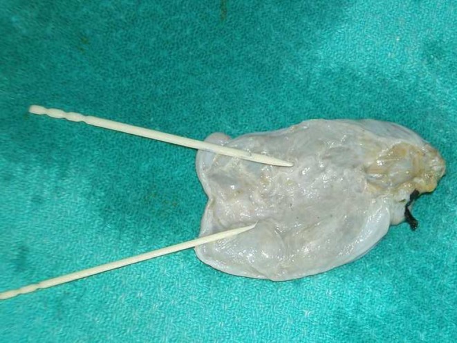
Double gall bladder with common mesentery
Photo 2.
Gall bladder with mesentery separated
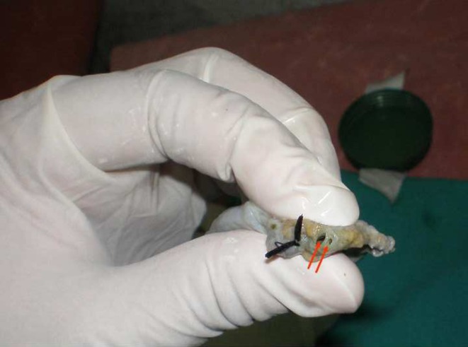
Photo 3.

Two openings of cystic duct artery tagged by black silk
The specimen was a double gallbladder with separate cystic duct opening in common bile duct.(H shaped)
Patient was discharged on second postoperative day and had uneventful recovery.
Histopathology revealed both gall bladders had stones and were inflamed.
Discussion
Vesica fellea duplex or a true double gall bladder as it is called is a rare entity which is usually not suspected and can complicate the diagnosis and course of surgery. The patient presents with symptoms and signs attributable to cholecystitis and are put up for surgery.
During surgery this may lead to various complications as confusion in anatomy or injury to adjoining structures. Even one entity may be left over which if pathological can lead to various complications as perforation or recurrent attacks of inflammation etc [2].
The two main types of duplications are vesica fellea divisa or bilobed gallbladder and vesica fellea duplex or true duplication, with two different cystic duct. The true duplication is sub classified into Y shaped type (two cystic duct unites before entering into the common bile duct, usually the two gallbladder and adherent and occupy the same fossa) and the H shaped type or ductular type (two separate gallbladder and cystic ducts entering separately into the common bile duct).
The true duplication occurs due to bifurcation of gallbladder primodium during the 5th and early 6th week of embryonic life [1, 3–6].
There are many classifications of double gallbladder. Boyden classified double gallbladder in bilobed gallbladder and in true duplicated gallbladder, with two types according to the connection of the cystic ducts [3].
Any or both of the entity can be pathological and hence must be removed when diagnosed.
Ultrasonography can suspect double gallbladder. When suspected diagnosis can be made by endoscopic retrograde cholangiopancreatography (ERCP) which is gold standard examination for confirming the diagnosis. The disadvantages of this approach include invasiveness and a high rate of false-negatives [7]. In centers where MR cholangiography is the initial investigation of choice for biliary pathologies the detection rate is very high.
When diagnosed, whether preoperatively or preoperatively surgery is the treatment of choice.
References
- 1.Ozgen A, Akata D, Arat A, Demirkazik FB, Ozmen MN, Akhan O. Gallbladder duplication: imaging findings and differential considerations. Abdom Imaging. 1999;24(3):285–288. doi: 10.1007/s002619900496. [DOI] [PubMed] [Google Scholar]
- 2.Borghi F, Giraudo G, Geretto P, Ghezzo L. Perforation of missed double gallbladder after primary laparoscopic cholecystectomy: endoscopic and laparoscopic management. J Laparoendosc Adv Surg Tech. 2008;18(3):429–431. doi: 10.1089/lap.2007.0088. [DOI] [PubMed] [Google Scholar]
- 3.Boyden EA. The accessory gallbladder-an embryological and comparative study of aberrant biliary vesicles occurring in man and domestic mammals. Am J Anast. 1926;38:177–231. doi: 10.1002/aja.1000380202. [DOI] [Google Scholar]
- 4.Mazziotti S, Minutoli F, Blandino A, Vinci S, Salamone I, Gaeta M. Gallbladder duplication: MR cholangiography demonstration. Abdom Imaging. 2001;26:287–289. doi: 10.1007/s002610000157. [DOI] [PubMed] [Google Scholar]
- 5.Gray SW, Skandalakis JE. Embryology for surgeons. Philadelphia: Saunders; 1972. [Google Scholar]
- 6.Shivhare R, Sikora S. Double cystic duct: a rare biliary anomaly encountered at laparoscopic cholecystectomy. J Laparoendosc Adv Surg Tech A. 2002;12:391–392. doi: 10.1089/109264202320884162. [DOI] [PubMed] [Google Scholar]
- 7.Vitellas KM, El-Dieb A, Vaswani KK, et al. Using contrast-enhanced MR cholangiography with IV mangafodipir trisodium (Teslascan) to evaluate bile duct leaks after cholecystectomy: a prospective study of 11 patients. AJR. 2002;179:409–416. doi: 10.2214/ajr.179.2.1790409. [DOI] [PubMed] [Google Scholar]