Abstract
Retroperitoneal lipomas are known for their rarity and varied presentations. We are reporting a case of giant retroperitoneal lipoma which presented as inguinal hernia.
Keywords: Retoperitoneal lipoma, Orchidectomy, Diffuse lipomatosis, Panniculitis
Introduction
Lipomas are benign variant of liposarcoma located in the peritoneal cavity and especially in the retro peritoneum. They have been reported very rarely in medical literature including journals [1, 2]. Retroperitoneal lipomas must be carefully differentiated from liposarcomas of low grade malignancy in order to provide the correct treatment and post operative follow up [3].
Case Report
A 65 years old male patient farmer by occupation presented to us, with complaints of painless swelling in left inguinal region since 3 years. There were complaints of occasional upper abdominal pain with vomiting. There was no history of anorexia and weight loss. Bladder and bowel habits were normal. On examination, there was a swelling of 15 × 6 cms in the left inguinoscrotal region. Its consistency varied from firm to hard. It was irreducible and cough impulse was absent. Abdominal examination did not revealed any abnormality except gross central obesity. Left hemiscrotum was enlarged burrowing the penis. Both testes were normal. A provisional diagnosis of left irreducible sliding inguinal hernia was established.
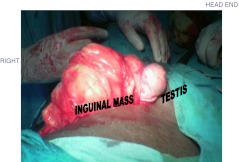
However, ultrasonography of the abdomen & pelvis did not reveal any abnormality. Patient was posted for left hernioplasty. On opening the inguinal canal definite hernial sac could not be identified. Cord contents were pushed anteriorly. Posterior wall of the inguinal canal was completely occupied by the diffuse lipomatous mass. Contents of the cord could not be separated from this mass. Since the mass was extending superiorly, we suspected retroperitoneal lipoma. Midline laparotomy was carried out. A giant retroperitoneal diffuse lipomatous mass measuring 25 × 12 cms in size was found (Fig. 1).
Fig. 1.
Lipoma mass in the inguinal region
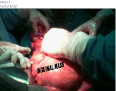
The mass was located below the transverse mesocolon on either sides of the midline, extending inferiorly into the pelvis and both inguinal canals, pushing and engulfing the cord on either sides. The small bowel mesentery was pushed entirely to the right. The small bowel was found in collapsed state in right paravertebral & subhepatic region. The mass was extending retroperitoneally towards inguinal region, occupying both inguinal canals. The mass was noncapsulated & soft, but could be separated from surrounding structures. Debulking of the mass was done (Fig. 2). Left orchidectomy and closure of inguinal canal with non absorbable sutures was done. Abdominal incision was closed in layers. Post operative course was uneventful.
Fig. 2.
Lipomatous mass
Histopathological examination showed diffuse lipomatosis with foci of panniculitis and fat necrosis. Absence of variation in size and shape of fat cells, absence of multivacuolated lipoblasts and hyperchromatic nuclei excluded possibility of liposarcoma. Patient was followed up for eighteen months postoperatively and has been asymptomatic.
Discussion
Primary retroperitoneal tumors are rare and are of varied histological variety. They may originate from retroperitoneal adipose tissue [4], muscle, connective, lymphatic and nerve tissue and from the urogenital tract [5, 6]. Commonest amongst them is adipose tissue.
When lipomas affect retroperitoneum, they attain considerable dimensions [7]. Surgery is the only treatment for giant lipoma. Attempt should be made for complete excision of the giant lipoma. If it is not possible debulking of lipoma should be done as far as possible. Recurrence has to be again managed surgically.
Ultrasonography examination of retroperitoneal lipoma is not possible in the presence of dilated air filled bowel loops overlying the retroperitoneal structures. Pelvis also cannot be assessed in empty bladder and if pelvis is occupied by dilated ileal loops.
However, a search of literature (Table 1) has not revealed any case of retroperitoneal lipoma presenting as a inguinal hernia, perhaps this is a first case of its kind.
Table 1.
Reported cases
| Carlos Augusto Real Martinez et al. Arquivas de Gastroenterlogia [8]. | It was a case a giant retroperitoneal lipoma in 32 year old female, presented with abdominal pain and mass. Laprotomy showed a large encapsulated tumour measuring 20 × 13 cms and weighing 3,400 gms. |
| Naoyuki Yokoyama et al. Department of Surgery and department of pathology, Okayama University Medical School, Japan [9]. | They have reported a case in which a retroperitoneal liposarcoma presented as indirect inguinal hernia. |
Histopathological examination did not reveal any malignancy in this lipomatosis.
Lipomas are benign tumors. Following excision they may recur locally. The recurrence rate is less than 5% of all tumors. But deep lipomas like retroperitoneal lipomas have great tendency to recur, presumably because of the difficulty of complete removal. Malignant changes in a lipoma are exceedingly rare, in fact, authors have never encountered such a case and only a few examples have been reported in the literature [10].
Lipomas are occasionally altered by admixture of other mesenchymal elements that form intrinsic part of the tumor. The most common of these is fibrous connective tissue to form fibrolipoma. Other altered types are myxolipomas in which portions of the tumor are replaced by mucoid substances, myolipoma, a lipoma with a distinct smooth muscle component, and chondroid lipoma, a tumor displaying features of both chondrolipoma and hibernoma [10].
Secondary changes in lipoma occur as the result of impaired blood supply or traumatic injury. Prolonged ischemia may lead to infarction, hemorrhage and calcification and may terminate in cyst like changes. Similarly, infection or trauma may cause fat necrosis and local liquefaction of fat [10].
References
- 1.Marshall MT, Rosem P, Berlin R, Gresson N. Appendicitis Masquerading as tumour. J Emerge Med. 2001;2:397–99. doi: 10.1016/S0736-4679(01)00422-X. [DOI] [PubMed] [Google Scholar]
- 2.Shouzhu Z, Xinhau Y, Xumin L, Shulian L, Xianzhi W. Giant retroperitoneal pleomorphic lipoma. Am J Surg Pathol. 1987;11:557–62. doi: 10.1097/00000478-198707000-00008. [DOI] [PubMed] [Google Scholar]
- 3.Susini T, Taddei G, Massi D, Massi G. Giant pelvic retroperitoneal liposarcoma. Obstet Gynaecol. 2000;95:1002–4. doi: 10.1016/S0029-7844(00)00818-8. [DOI] [PubMed] [Google Scholar]
- 4.KawashimaO KM, Ishikawa S, Morshita Y. Retroperitoneal lipoma through Foramen of Bochdalek detected as a mass on chest roento genogram. Report of a case. 1999;52:1141–3. [PubMed] [Google Scholar]
- 5.Montbrun E, Pereio R, Barbeito J, Nastasi A, Montbrun M, Gomez J, Gonsalvis I, Baez L. Tumours malignos retroperitoneales primarios. Arch Hosp Vargas. 1990;32:101–6. [Google Scholar]
- 6.Montesions MR, Falco JE, Sinaagra DL, Mazzadri NA, Curutchet P. Sarcomas retroperitoneales. Rev Argent Cir. 2000;78:1–5. [Google Scholar]
- 7.Zhang SZ, Yue XH, Liu XM, Lo SL, Wang SZ. Giant retroperitoneal pleomorphic lipoma. Am J Surg Pathol. 1987;11:557–62. doi: 10.1097/00000478-198707000-00008. [DOI] [PubMed] [Google Scholar]
- 8.Real Martinez CA, Palma RT, Sau Paulo JW (2003) Giant retroperitoneal lipoma: a case report. Arq Gastroenterol 40(4) [DOI] [PubMed]
- 9.Yokoyama N, Shirai Y, Yamazaki H, Hatake K. An inguinal hernia sac tumor of extra of extra hepatic cholangiocarcinoma origin. World J Oncol. 2006;4:13. doi: 10.1186/1477-7819-4-13. [DOI] [PMC free article] [PubMed] [Google Scholar]
- 10.Enzinger FM, Weiss SW. Soft tissue tumors. Benign lipomatous tumors. 3. St Louis: Mosby publications; 1995. pp. 386–389. [Google Scholar]


