Abstract
Background
Chronic leg ulcers are defined as those that show no tendency to heal after 3 months of appropriate treatment or are still not fully healed at 12 months. In this article, we present an approach to the challenging problem of chronic leg ulcers that is based on the principles of evidence-based medicine, i.e., the explicit use of the best available scientific evidence as a guide to treatment.
Methods
Selective review of the relevant literature, including current guidelines and meta-analyses, concerning diagnostic and therapeutic strategies for chronic leg ulcers.
Results
The main types of causally directed treatment are: vein surgery to eliminate pathological reflux, interventions to improve the circulation in arterial occlusive disease, and treatment of underlying diseases, such as diabetes mellitus.
Conclusion
Physicians providing modern evidence-based management of chronic leg ulcers should make use of their own clinical experience in combination with the best current scientific evidence. It seems clear that the many available treatment options should be evaluated critically in an interdisciplinary setting. In particular, causally directed treatment must be provided in addition to symptomatic, stage-based local wound treatment.
Chronic wounds are a significant issue not only in specialist facilities but also in daily practice for family physicians and specialists across a wide variety of disciplines. These chronic wounds are primarily chronic lower-limb ulcerations resulting from chronic venous insufficiency which may be complicated by concomitant arterial macro- or micro-angiopathy. In a cohort of 3072 volunteers (1, e1) the Bonn Vein Study showed a prevalence of 0.6% for healed venous ulcer and of 0.1% for florid ulcer. Moffat et al. revealed similar numbers in a survey of 252 000 London citizens in 2004 (prevalence 0.45/1000) (e2). The numbers of chronic wounds in diabetes mellitus or neuropathic ulcers should not be underestimated. Between 2% and 10% of all people with diabetes mellitus suffer from foot ulcers. The incidence rate is 2.2 to 5.9% annually. These data refer to cross-sectional studies in diabetics (2, e3).
The present study is based on current evidence-based guidelines. These include the S3 guideline for the diagnosis and treatment of venous stasis ulcers published by the German Society for Phlebology (DGP, Deutsche Gesellschaft für Phlebologie) (3), the evidence-based guideline for diabetic foot syndrome of the German Diabetes Association (DDG, Deutsche Diabetes Gesellschaft) (2), and the current German treatment guideline for the prevention and treatment of foot complications in type 2 diabetes mellitus (4). The guidelines are based on current systematic literature reviews meaning that another systematic literature review using different databases was not done. A selective literature search in medical databases (PubMed, Cochrane Library) and medical journals was carried out for individual topics.
The study aims to ensure that routine daily medical practice does not become mundane, particularly when treating chronic wounds. Information with regard to different aspects of evidence-based medicine can be helpful to critically scrutinize one’s own actions in a positive sense and to incorporate more causal treatment options into therapy planning.
Evidence-based management
Evidence-based management (EBM) is defined as the deliberate, explicit and judicious use of the best information available for making decisions when caring for an individual patient (5, e4). EBM implies the use of current clinical medicine based on clinical trials and medical publications that confirm or refute facts.
Because the whole body of medical knowledge is currently doubling in size every five years, however (6), even a skillful physician feels increasingly overwhelmed when trying to determine what is significant amongst the abundance of existing and emerging knowledge.
Information about the evidence supporting diagnostic approaches and the safety and efficacy of treatments is most important for the management of chronic wounds.
Hierarchical levels of evidence and different grades of recommendations are differentiated for different treatment modalities. Level of evidence and recommendation grades are indicated in the following in square brackets. An overview of evidence levels and recommendation grades is provided in Table 1.
Table 1. Causal treatment of venous stasis ulcers.
| Causal treatment options of chronic venous leg ulcers | Modalities of the causal treatment | level of evidedence / grade of recommendation* |
| Compression therapy | Phlebological compression bandages, medical compression stockings, intermittent pneumatic compression | Ia/A |
| Varicose surgery | Crossectomy and surgical stripping of the saphenous veins, selective varicose exeresis, mini-phlebectomy, CHIVA, perforator vein division, subfascial endoscopic perforator surgery (SEPS) | Ib/A |
| Endovascular therapies | Endovascular laser therapy (various laser systems), radiofrequency ablation, radiofrequency-induced thermotherapy (RFITT) | Ib/A |
| Sclerosing therapy | Injection sclerotherapy, ultrasound-guided foam sclerotherapy (direct puncture, catheter sclerosing) | Ib/A |
| Surgical ulcer treatment | Shaving surgery with graft coverage, ulcer excision with fasciectomy with graft coverage | III/B |
*Based on the evidence classification of the Oxford Centre for Evidence-Based Medicine
Evidence level:
Ia: Evidence from meta-analyses of several randomized controlled trials
Ib: Evidence based on at least one randomized controlled trial
IIa: Evidence based on at least one well-designed but not randomized and controlled trial
IIb: Evidence based on at least one well-designed quasi-experimental trial
III: Evidence based on well-designed, non-experimental descriptive trials such as comparative studies, correlation studies or case-control studies
IV: Evidence based on reports in expert panels or expert opinions or clinical experience of recognized authorities
Recommendation grade:
A: Key recommendation:there is at least one randomized controlled trial of overall good quality (evidence levels Ia and Ib)
B: Preferred recommendation: there are well-implemented clinical trials with direct reference to the recommendation (evidence levels II or III)
C: Optional recommendation: expert opinion and/or clinical
Chronic wounds
A wound that is present for more than three months is considered chronic (7). The clinically most significant chronic wounds in terms of epidemiology and health economics are venous stasis ulcers, wounds and wound healing disorders in diabetes mellitus, and pressure ulcers in immobile patients with reduced general condition (5).
In general, evidence-based management of chronic wounds includes the following:
Comprehensive diagnosis, medical history and documentation of findings [III; B]
Relevant data include pain, allergization and tetanus vaccine protection [I; A]
Known thrombophilia [I; A]
Decreased levels of zinc, iron, folate, albumin, vitamin C and selenium as a result of poor diet must be eliminated in cases of large (>100 cm2) ulcerations and corrected if necessary (3).
The significance of bacterial colonization of chronic wounds is unclear as is the use of antiseptics [IV; C]. Debridement, also using maggots (biosurgery) or vacuum-assisted closure (VAC) technique, is recommended in cases of wound infection [Ib; A]. Wound debridement is, however, recommended in general [III, B] as is maintaining a moist wound environment [Ia, A].
Refractory and/or morphologically abnormal ulcerations must be histologically evaluated [I, A].
Venous stasis ulcers
Venous ulcers are the result of ambulatory venous hypertension with chronic venous insufficiency (CVI). Venous ulcerations generally develop on skin that typically exhibits the stigmata of CVI such as hyperpigmentation, varicose eczema, lipodermatosclerosis or atrophie blanche. The skin around the ulcer therefore exhibits typical trophic changes. Problems are caused by refractory ulcers that do not show signs of healing after 3 months’ therapy or have not healed after 12 months of adequate treatment. The information about evidence in the following section refers to the S3 guideline for the diagnosis and treatment of venous stasis ulcers (3).
Diagnosis
As a matter of principle extensive vascular Doppler or duplex ultrasonographic imaging is required. In addition, phlebological diagnosis is used not only to precisely determine the clinical symptoms but also to plan causal treatment procedures [II; B].
Causal treatment
Reducing the pressure and volume overload in the venous system is the main causal treatment (8) [Ia; A]. This can be achieved using compression therapy (phlebological compression bandages, medical compression stockings) or using targeted correction of pathological reflux (Table 1).
Conservative causal treatment
Compression therapy is still considered the basic treatment for venous ulcers, and its ability to heal venous ulcers is clearly supported by a body of evidence from many studies (9). The healing rate increases and the recurrence rate decreases with increasing interface pressures of the compression bandages or stockings [Ia; A]. A meta-analysis showed that adequate compression therapy is not only important for the healing of venous stasis ulcers but that continuing the compression after the ulcer has healed is also critical for the recurrence-free interval.
Compression stockings have proven to be effective for venous ulcers (10). The benefits of compression stocking therapy with venous stasis ulcers are derived both from the constant pressure of the stocking and better compliance of patients (e6) [Ia; A].
Surgical causal treatment
The aim is to eliminate venous reflux because reflux is relevant to prevent wound healing and causes a considerable increase in the ulcer recurrence rate. The effects of varicose correction measures on venous hemodynamics can be determined using duplex ultrasonography, or plethysmographically, or using direct venous pressure measurement.
The efficacy of all types of reflux correction measures in terms of accelerated ulcer healing has been proven (11). The choice of method can therefore be tailored according to benefits and drawbacks in each individual patient. Classical surgical techniques, endovascular thermal procedures (e7, 12), and (foam) sclerotherapy (13) are all options [Ib; A]. There is less benefit gained from regenerating the superficial venous system in patients with an insufficient deep venous system than in patients with a sufficient deep venous system in terms of the healing of venous stasis ulcers and in terms of the prolongation of the recurrence-free interval [IIa; B].
The significance of insufficient perforating veins in hemodynamics and the relevance of isolated treatment of these veins has not been clarified [Ia; A]
Surgical ulcer treatment
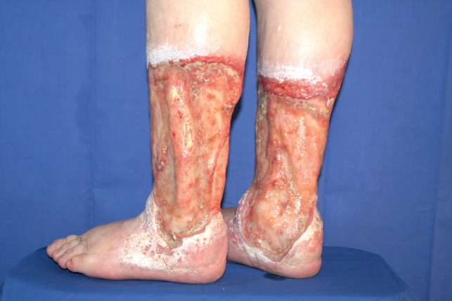
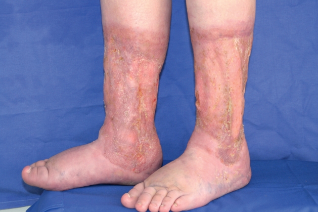
If a trophic disorder (dermatolipofasciosclerosis) is present around the ulcer, radical ablation of the entire trophically destroyed tissue with subsequent coverage of the defect is recommended [II; B]. The use of tangential excision of the affected tissue has become established as shave therapy (Figures 1 and 2). This technique accelerates ulcer healing. Simultaneous removal of the crural fascia is optional and is still controversial (14, 15) [III; B].
Figure 1.
Ulceration in the gaiter zone due to chronic venous insufficiency; no tendency to heal for years
Figure 2.
Postoperative result five years after shaving surgery
Coverage of the defect using autologous mesh grafts is preferable to a full-thickness skin transplant and transplantation of free vascularized musculofasciocutaneous flaps because it achieves comparable results with less effort [III; B].
Symptomatic treatment
The requirements for ideal wound dressings are listed in Box 1.
Box 1. Requirements for an ideal wound dressing.
Reduces pain and itching
Absorbs wound exudate without drying out the wound
Made from inert or at least hypoallergenic or non-irritating material
Changing of the dressing should be possible without irritating the wound
Does not leave traces of dressing components on the wound
Allows gaseous exchange from the wound (O2/CO2)
Protects against physical, chemical and bacterial exposure
Adapts to the prevalent wound healing phase in the wound
Easy to change
Biologically compatible and environmentally friendly
Suitable dressing materials include non-medicated paraffin gauze, foams, alginates, hydrogels, hydrocolloids, and hydroactive dressings. Wound dressings that provide a moist wound environment have been proven to have a general benefit. There is also consensus about the need for an appropriate balance in the moisture content of the dressing. There is evidence of pain reduction with the use of hydrocolloidal and foam wound dressings [Ia; A]. General superiority of one particular wound dressing compared to another has not yet been demonstrated (16) [Ia; A]. However, physicians must be mindful of the high sensitization rate to external dressings and their constituents in ulcer patients [I; A]. There is thus an increased risk of developing type IV allergies in the form of allergic contact eczema around the ulcer.
Chronic wounds in diabetes mellitus
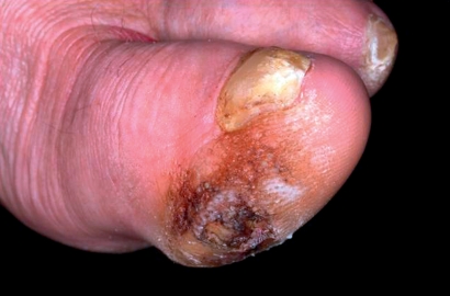
Poorly healing ulceration, predominantly around the feet, can develop as a complication of diabetes. Often amputation is the last resort (2, e3). The risk of reamputation in patients with diabetes is high. In a longitudinal study the cumulative risk of being reamputated was 27% after one year, 48% after 3 years, and 61% after 5 years (17). The risk factors for diabetic foot ulcer are listed in Table 2. Peripheral sensorimotor neuropathy is particularly important [III; B]. Polyneuropathy without concomitant vascular disease often gives rise to the development of diabetic foot ulcer. Repeated trauma, which is often not perceived, leads to the formation of excessive callusing (Figure 3). Subkeratotic hematomas develop below these calluses as a result of the persistent action of pressure and shear forces. Finally, ulceration develops at the exposed sites (e8). The most important cause of foot ulceration is unsuitable footwear [II; A]. Information about evidence for the following section in based on the German treatment guideline for type 2 diabetes and the guideline for diabetic foot syndrome from the German Diabetes Association (2, e3, 4).
Table 2. Factors that promote diabetic foot ulcer (guideline of the German Diabets Association, DDG).
| Risk factor | Risk assessment and clinical parameters |
| Poor glycemic control | HbA1c |
| Previous ulcer/ amputation | Medical history, physical examination |
| Neuropathy | Abnormal sensorimotor perception of vibration |
| Reduction in visual acuity | Ophthalmological examination |
| Trauma | Poorly fitting shoes, pressure, burns |
| Biomechanics | Limited joint mobility, bony prominences, foot deformity/osteoarthritis, callus |
| Peripheral arterial occlusive disease | Ancle-brachial index |
| Socioeconomic status | Poor access to medical facilities, lack of compliance/neglect, no or inadequate education |
Figure 3.
Hyperkeratosis with bleeding and incipient ulceration with diabetic neuropathy
Diagnostic clarification
As for venous ulcers, a vascular examination of the vessels supplying blood to the extremities is also required with diabetic foot. As a matter of priority, Doppler-derived arterial wedge pressures with calculation of the ankle/brachial index should be measured [II; B] while taking into account any potential medial sclerosis.
The diagnostic criteria for diabetic neuropathy that are of particular relevance for diabetic foot syndrome include, analogous to the Neuropathy Deficit Score (NDS), examination of the achilles tendon reflex, and vibration, pain, temperature and touch perception. Examination of the achilles tendon reflex using a reflex hammer is used to determine the depth sensitivity. Vibration perception is examined using biothesiometry which has a high predictive value for ulcer formation (e9) [III; A]. The Rydel-Seiffer tuning fork is a simple and practical alternative to biothesiometry [Ib; A]. It also makes sense to differentiate the neuropathic and arterial risk factors. In addition, the wound should be classified using the Wagner and Armstrong classification system. The conventional classification by Wagner describes diabetic foot ulcers based on the extent of existing tissue destruction. The University of Texas Wound Classification System (the Armstrong classification) is superior to the Wagner classification in terms of an estimate of the likely success of treatment because of the additional description of infection and ischemia (Table 3) (18).
Table 3. Classification of diabetic ulcers according to Wagner and Armstrong (e10).
| 0 | 1 | 2 | 3 | 4 | 5 | |
| A | Callus or scar | Superficial wound | Wound penetrating to tendon/capsule | Wound penetrating to bone/joint | Necrotic foot areas | Necrotic foot, entire |
| B | Infection | Infection | Infection | Infection | Infection | Infection |
| C | Ischemia | Ischemia | Ischemia | Ischemia | Ischemia | Ischemia |
| D | Infection + ischemia | Infection + ischemia | Infection + ischemia | Infection + ischemia | Infection + ischemia | Infection + ischemia |
Vascular causal treatment
Appropriate systemic therapy [Ia; A], interventional or surgical revascularization [III; A] should be implemented with verified hemodynamically relevant peripheral arterial occlusive disease (PAOD).
Biomechanical treatment options/pressure relief
Several studies have confirmed the importance of pressure relief for the healing of diabetic ulcers [III; B]. Patient education is also critical [II; A]. The patient must understand that even a few weight-bearing steps on an ulcerated foot can delay healing. The effectiveness of the biomechanical approach is therefore highly dependent on patient compliance.
An important study about pressure relief demonstrated using hidden activity sensors in a double-shelled total-contact cast (TCC) that the TCC was actually only worn on average for 28% of daily activities (e10). Very good data are available for the non-removable cast or walker. They revealed outstanding healing rates using two non-removable relieving aids with no difference between a TCC and TCC with an additional fixed ready-made mobility orthosis (19) [III; A].
Symptomatic treatment
The same guidelines apply to the local treatment of diabetic foot ulcer as in chronic venous ulcer. Amputation should be considered as a symptomatic treatment option if no improvement could be achieved despite consistent causal treatment or if there is a risk of a severe systemic infection arising from the wound [III; B].
Secondary prophylaxis
Secondary prophylaxis plays a critical role for patients with diabetes in light of the high recurrence rates and the high risk of developing an additional foot ulcer. Regular check-ups are recommended in accordance with the particular risk profile (Table 4). It is important to ensure the patient is aware of his/her disease and the importance of adequate pressure relief.
Table 4. Recommended frequency of check-ups according to risk profile (2).
| Risk profile | Examinations |
| No sensory neuropathy | 1×/year |
| Sensory neuropathy | Every 6 months |
| Sensory neuropathy and signs of peripheral arterial occlusive disease and/or foot deformities | Every 3 months |
| Previous ulcer | Every 1 to 3 months |
Pressure ulcers of the leg
A pressure ulcer is a site of localized damage to the skin and/or the underlying tissue, usually over a bony prominence as a result of pressure, shearing forces, and/or friction. Pressure ulcers are divided into four stages based on the definitions of the European Pressure Ulcer Advisory Panel (Box 2).
Box 2. Clinical stages of pressure ulcers*.
-
Stage 1
Non-blanchable erythema of intact skin. Skin discolorations, warmth, edema or induration can also be indicators of stage 1, particularly in dark-skinned individuals.
-
Stage 2
Partial-thickness loss of skin with damage to the epidermis and/or the dermis. The ulcer is superficial and presents clinically as an abrasion or blister.
-
Stage 3
Full-thickness loss of skin including damage to or necrosis of the subcutaneous tissue that may extend to, but not through, the fascia.
-
Stage 4
Extensive tissue necrosis possibly including muscle, bone or supporting structures
*Classification from the Pressure Ulcer Treatment Guidelines (EPUAP 1998)
A range of influential factors are associated with a pressure ulcer, the significance of which have not yet been definitively clarified (20). Although risk assessments are recommended, randomized controlled trials that would verify their benefit have not been published to date (20). The most important endogenous risk factor for the development of a leg ulcer is peripheral sensorimotor neuropathy such as that which occurs in diabetes mellitus, for example. The only facts verified with a high level of evidence are that pressure relief by repositioning protects against a pressure ulcer and that mattresses with higher specifications have a greater protective effect than standard foam mattresses (21) [III; B]. Even if the problems and their prevention and treatment are known and recognized in principle, there is nevertheless no solid evidence verified by appropriate studies for diagnostic and therapeutic approaches (21, 22). The following comments are based primarily on expert opinions, case-control studies, or randomized controlled trials with low power. The use of electrical stimulation, which has demonstrably enhanced wound healing in pressure ulcers, is an exception (e11).
Prevention of pressure damage to the skin and the underlying tissue is an essential part of treatment in at-risk patients. Adequate risk assessment and subsequent risk reduction is crucial (23).
Conservative causal treatment
The foundation of any pressure ulcer treatment is targeted local pressure relief by repositioning and the use of aids (24). In contrast, wound cleansing does not appear to play a critical role (e12).
Surgical causal treatment
Surgical strategies should be considered for pressure ulcers from stage II onwards that do not heal using conservative measures. If no tendency to heal becomes apparent using surgical debridement with the aid of the above named conservative measures, plastic surgery reconstruction is indicated where appropriate. Surgical measures must be planned and undertaken in the context of the general condition of the patient. Geriatric patients in particular often suffer from multiple comorbidities.
Symptomatic treatment / local treatment
For local treatment absolute pressure relief is critical for wound healing, otherwise the same guidelines apply as for chronic venous ulcers (24).
Summary
The treatment of chronic wounds of the lower extremities still presents a therapeutic challenge. There is clear evidence suggesting that causal treatment should have priority. A comprehensive diagnostic evaluation including vascular, metabolic and physical aspects is therefore essential at the start of treatment.
With ulcers that are predominantly venous in origin, reduction of venous hypertension is critical, with compression therapy occupying an important place in achieving this. Modern compression stocking systems supplied by various manufacturers are promising. Nevertheless, options such as plastic surgery and shave therapy for venous ulcers, which has “only” evidence level III, is an important component of the overall treatment plan.
Appropriate pressure relief has priority for neuropathic, diabetic or pressure ulcers.
Key Messages.
For refractory and/or morphologically abnormal ulcerations malignancies must be histologically excluded.
Information about the evidence supporting diagnostics, the safety, and the efficacy of treatments is most important for the management of chronic wounds.
A precise diagnosis is essential for the planning of individualized causal treatment.
For venous stasis ulcers treatment of venous hypertension using compression together with reflux correction has priority.
For foot ulcerations in diabetes mellitus pressure relief around the wound is important for healing.
Acknowledgments
Translated from the original German by language & letters.
Footnotes
Conflict of interest statement
PD Kahle receives sponsorship for conferences from the following companies:Bauerfeind AG Phlebologie, Biolitec AG; Covidien Deutschland GmbH, VNUS
Medical Technologies, medi GmbH & Co. KG, Villa sana GmbH & Co. medizinische Produkte KG, Leo Pharma GmbH, Stiefel GmbH, Sigvaris GmbH, Chemische Fabrik Kreussler GmbH, GE Ultraschall Deutschland GmbH&Co KG.
She receives financial support for participation or implementation of trials and observational studies from the following companies:
Bauerfeind AG Phlebologie, BSN-JOBST GmbH, Chemische Fabrik Kreussler GmbH, Karl Beese GmbH & Co KG, Söring GmbH Medizintechnik, Paul Hartmann AG, URGO GmbH, OM Pharma, medi GmbH & Co. KG, Villa sana GmbH & Co. medizinische Produkte KG, Chemische Fabrik Kreussler GmbH, Böhringer Ingelheim Pharma GmbH & Co. KG.
She also recieved honoraria for speaking from: Bauerfeind AG Phlebologie, Johnson and Johnson, Ethicon, BSN-JOBST GmbH, Medi GmbH & Co KG, and Chemische Fabrik Kreussler GmbH.
Dr. Hermanns and Dr. Gallenkemper declare that no conflict of interest exists.
References
- 1.Rabe E, Pannier-Fischer F, Bromen K, et al. Bonner Venenstudie der Deutschen Gesellschaft für Phlebologie. Epidemiologische -Untersuchung zur Frage der Häufigkeit und Ausprägung von chronischen Venenkrankheiten in der städtischen und ländlichen Wohnbevölkerung. Phlebologie. 2003;32:1–14. [Google Scholar]
- 2.Morbach S, Müller E, Reike H, Risse A, Spraul M. Scherbaum WA, Kiess W, Landgraf R, editors. Diagnostik, Therapie, Verlaufskontrolle und Prävention des Diabetischen Fußsyndroms. Evidenzbasierte Leitlinie der Deutschen Diabetes-Gesellschaft. Diabetes und Stoffwechsel. 2004;Band 13(Suppl 2) [Google Scholar]
- 3.Leitlinie der Deutschen Gesellschaft für Phlebologie. Diagnostik und Therapie des Ulcus cruris venosum AWMF online AWMF Register Nr 037/009. Phlebologie. 2004;33:166–185. und www.uni-duesseldorf.de/AWMF/ll/037-009.htm. [Google Scholar]
- 4.Nationale Versorgungsleitlinie Typ-2. www.versorgungsleitlinien.de. 2010. Februar Diabetes Präventions - und Behandlungsstrategien für Fußkomplikationen Version 2.8. [Google Scholar]
- 5.Sackett DL, Rosenberg WMC, Gray JAM, Haynes RB, Richardson WS. Evidence-based Medicine: What it is and what it isn’t. In: BMJ. 1996;312:71–72. doi: 10.1136/bmj.312.7023.71. [DOI] [PMC free article] [PubMed] [Google Scholar]
- 6.Dietzel GTW. Von eEurope 2002 zur elektronischen Gesundheitskarte: Chancen für das Gesundheitswesen. Dtsch Arztebl. 2002;99(21) [Google Scholar]
- 7.Dissemond J. Wann ist eine Wunde chronisch? Hautarzt. 2006;57 doi: 10.1007/s00105-005-1048-9. [DOI] [PubMed] [Google Scholar]
- 8.Nicolaides AN, Allegra C, Bergan JJ, et al. Management of chronic venous disorders of the lower limbs. Guidelines according to scientific evidence. Int Ang. 2008;27:1–59. [PubMed] [Google Scholar]
- 9.Cullum N, Nelson EA, Fletcher AW, Sheldon TA. In: The Cochrane Library. Issue 4. Oxford: Update Software; 2002. Compression for venous leg ulcers (Cochrane Review) [Google Scholar]
- 10.Jünger M, Wollina U, Kohnen R, Rabe E. Efficacy and tolerability of an ulcer compression stocking for therapy of chronic venous ulcer compared with a below-knee compression bandage: results from a prospective, randomized, multicentre trial. Current Medical Research & Opinion. 2004;20:1613–1623. doi: 10.1185/030079904X4086. [DOI] [PubMed] [Google Scholar]
- 11.Barwell JR, Davies CE, Deacon J, et al. Comparison of surgery and compression alone in chronic venous ulceration (ESCHAR study): randomised controlled trial. Lancet. 2004;363:1854–1859. doi: 10.1016/S0140-6736(04)16353-8. [DOI] [PubMed] [Google Scholar]
- 12.Pannier F, Rabe E. Endovenöse Lasertherapie mit dem 980-nm-Diodenlaser bei Ulcus cruris venosum. Phlebologie. 2007;36:179–185. [Google Scholar]
- 13.Stücker M, Reich S, Hermes N, Altmeyer P. Sicherheit und Effektivität der periulzerösen Schaumsklerosierung bei Patienten mit postthrombotischem Syndrom und/oder oraler Antikoagulation mit Phenprocoumon. JDDG. 2006;4:734–738. [Google Scholar]
- 14.Hermanns HJ, Gallenkemper G, Waldhausen P, Hermann V. Die Behandlung des therapieresistenten Ulcus cruris durch Shave-Therapie - „mid-term results“. ZfW. 2002;3 [Google Scholar]
- 15.Hermanns HJ, Gallenkemper G, Kanya S, Waldhausen P. Die Shave-Therapie im Konzept der operativen Behandlung des therapieresistenten Ulcus cruris venosum. Aktuelle Langzeitergebnisse. Phlebologie. 2005;34:209–215. [Google Scholar]
- 16.Nelson EA, Bradley MD. Dressings and topical agents for arterial leg ulcers. Cochrane Database of Systematic Reviews. 2007;(Issue 1) doi: 10.1002/14651858.CD001836.pub2. Art. No.: CD001836. DOI: 10.1002/14651858.CD001836.pub2. [DOI] [PubMed] [Google Scholar]
- 17.Izumi Y, Satterfield K, Lee S, Harkless LB. Risk of reamputation in diabetic patients stratified by limb and level of amputation: A 10-year observation. Diabetes Care. 2006;29:566–570. doi: 10.2337/diacare.29.03.06.dc05-1992. [DOI] [PubMed] [Google Scholar]
- 18.Oyibo SO, Jude EB, Tarawneh I, Nguyen HC, Harkless LB, Boulton AJ. A comparison of two diabetic foot ulcer classification systems: the Wagner and the University of Texas wound classification systems. Diabetes Care. 2001;24:84–88. doi: 10.2337/diacare.24.1.84. [DOI] [PubMed] [Google Scholar]
- 19.Katz IA, Harlan A, Miranda-Palma B, et al. A randomized trial of two irremovable off-loading devices in the management of plantar neuropathic diabetic foot ulcers. Diabetes Care. 2005;28:555–559. doi: 10.2337/diacare.28.3.555. [DOI] [PubMed] [Google Scholar]
- 20.Moore ZEH, Cowman S. Risk assessment tools for the prevention of pressure ulcers. Cochrane Database of Systematic Reviews. 2008;(Issue 3) doi: 10.1002/14651858.CD006471.pub2. Art. No.: CD006471. DOI: 10.1002/14651858.CD006471.pub2. [DOI] [PubMed] [Google Scholar]
- 21.European Pressure Ulcer Advisory Panel and National Pressure Ulcer Advisory Panel. Washington DC: National Pressure Ulcer Advisory Panel; 2009. Treatment of pressure ulcers: Quick Reference Guide. [Google Scholar]
- 22.McInnes E, Cullum NA, Bell-Syer SEM, Dumville JC. Support surfaces for pressure ulcer prevention. Cochrane Database of Systematic Reviews. 2008;(Issue 4) doi: 10.1002/14651858.CD001735.pub3. Art. No.: D001735. DOI: 10.1002/14651858.CD001735.pub3. [DOI] [PubMed] [Google Scholar]
- 23.Anders J, Heinemann A, Leffmann C, et al. Decubitus ulcers: pathophysiology and primary prevention. Dtsch Arztebl Int. 2010;107(21):371–382. doi: 10.3238/arztebl.2010.0371. [DOI] [PMC free article] [PubMed] [Google Scholar]
- 24.European Pressure Ulcer Advisory Panel and National Pressure Ulcer Advisory Panel. Washington DC: National Pressure Ulcer Advisory Panel; 2009. Treatment of pressure ulcers: Quick Reference Guide. [Google Scholar]
- e1. www.weyergans.ch/upload/publikationen/BonnerVenenstudie.pdf.
- e2.Moffat CJ, Franks PJ, Doherty DC, et al. Prevalence of leg ulceration in a London Population. Q J Med. 2004;97:431–437. doi: 10.1093/qjmed/hch075. [DOI] [PubMed] [Google Scholar]
- e3.Morbach S, Müller E, Reike H, Risse A, Spraul M. Diabetisches Fußsyndrom. Diabetologie. 2008;3(Suppl 2):175–180. [Google Scholar]
- e4.Guyatt GH, Oxman AD, Kunz R, et al. GRADE Working Group. Going from evidence to recommendations. BMJ. 2008;336:1049–1051. doi: 10.1136/bmj.39493.646875.AE. [DOI] [PMC free article] [PubMed] [Google Scholar]
- e5.Brocatti LK. Versorgungsqualität bei chronischen Wunden: Gesundheitsökonomische Bewertung der Behandlung des Ulcus cruris in Hamburg. Dissertationsschrift der medizinschen Fakultät Hamburg. 2008 [Google Scholar]
- e6.Jünger M, Häfner HM. Interface pressure under a ready made compression stocking developed for the treatment of venous ulcers over a period of six weeks. Vasa. 2003;32:87–90. doi: 10.1024/0301-1526.32.2.87. [DOI] [PubMed] [Google Scholar]
- e7.Barwell JR, Davies CE, Deacon J, et al. Comparison of surgery and compression alone in chronic venous ulceration (ESCHAR study): randomised controlled trial. Lancet. 2004;363:1854–1859. doi: 10.1016/S0140-6736(04)16353-8. [DOI] [PubMed] [Google Scholar]
- e8.Murray HJ, Young MJ, Hollis S, Boulton AJM. The association between callus formation,high pressure and neuropathy in diabetic foot ulceration. DiabetMed. 1996;13:979–982. doi: 10.1002/(SICI)1096-9136(199611)13:11<979::AID-DIA267>3.0.CO;2-A. [DOI] [PubMed] [Google Scholar]
- e9.Young MJ, Breddy JL, Veves A, Boulton AJM. The prediction of neuropathic foot ulceration using vibration perception thresholds. Diabetes Care. 1994;17:557–561. doi: 10.2337/diacare.17.6.557. [DOI] [PubMed] [Google Scholar]
- e10.Armstrong DG, Lavery LA, Kimbriel HR, Nixon BP, Boulton AJM. Activity patterns of patients with diabetic foot ulceration. Diabetes Care. 2003;26:2595–2597. doi: 10.2337/diacare.26.9.2595. [DOI] [PubMed] [Google Scholar]
- e11.Olyaee Manesh A, Flemming K, Cullum NA, Ravaghi H. Electromagnetic therapy for treating pressure ulcer. Cochrane Database of Systematic Reviews. 2006;(Issue 2) doi: 10.1002/14651858.CD002930.pub3. Art. No.: CD002930. DOI: 10.1002/14651858.CD002930.pub3. [DOI] [PubMed] [Google Scholar]
- e12.Moore ZEH, Cowman S. Wound cleansing for pressure ulcers. Cochrane Database of Systematic Reviews. 2005;(Issue 4) doi: 10.1002/14651858.CD004983.pub2. Art. No.: CD004983. DOI: 10.1002/14651858.CD004983.pub2. [DOI] [PubMed] [Google Scholar]


