Abstract
Objective
To compare, landing mechanics and electromyographic activity of the lower extremities during side hopping and crossover hopping maneuvers, in noninjured women and women with anterior cruciate ligament (ACL) reconstruction.
Design
A case-control study.
Setting
A 3-dimensional motion analysis laboratory.
Participants
Twenty-eight young women (range, 21–35 years) (15 control subjects and 13 subjects with ACL reconstruction).
Patients and Methods
All participants performed a side-to-side hopping task that consisted of hopping single-legged 10 times consecutively from side to side across 2 lines marked 30 cm apart on 2 individual force plates. The task was designated as a side hopping when the hop was to the opposite side of the stance leg and as crossover hopping when the hop was toward the side of the stance leg.
Main Outcome Measurements
Peak hip-/knee-joint angles; peak knee extension/abduction joint moments; electromyographic studies of the gluteus maximus, gluteus medius, rectus femoris, and hamstring muscles; and quadriceps/hamstring co-contraction ratio were compared between the groups by means of 2 × 2 multivariate analysis of variance tests (group × maneuver).
Results
Noninjured women and women with ACL reconstruction exhibited similar hip-and knee-joint angles during both types of hopping. Hip-joint angles were greater during the crossover hopping in both groups, and knee-joint angles did not differ between the groups or hops. Knee-joint moments demonstrated a significant group × maneuver interaction. Greater knee extension and valgus moments were noted in the control group during crossover hopping, and greater knee abduction moments were noted in the ACL group during side hopping. Electromyographic data revealed no statistically significantly differences between the groups.
Conclusions
Women with ACL reconstruction exhibited the restoration of functional biomechanical movements such as hip-/knee-joint angles and lower extremity neuromuscular activation during side-to-side athletic tasks. However, not all biomechanical strategies are restored years after surgery, and women who have undergone a procedure such as ACL reconstruction may continue to exhibit knee-joint abduction moments that increase the risk of additional knee injury.
INTRODUCTION
Side-to-side maneuvers are some of the most injurious drills encountered in sports [1,2]. These maneuvers create an excessive load on the knee joint and increase the predisposition to ligamentous injuries such as anterior cruciate ligament (ACL) rupture [1–5]. These maneuvers are performed most often in a high-speed manner during which an opponent is being avoided during sports or play [3,6,7]. Side-to-side maneuvers increase the likelihood of uncontrolled movements, thereby presenting an opportunity for injury [1–3,7].
Women participating in sports are more likely than men to experience an ACL injury during such maneuvers [4–7]. Typically, women tend to exhibit lesser hip and knee flexion joint angles, greater valgus moments, and decreased neuromuscular recruitment of the lower extremity muscle groups during those maneuvers when compared with their male counterparts [4–7]. Although the effects of gender differences during sports have been well studied, the biomechanical differences between women who have undergone ACL surgery and noninjured women have not been well assessed during those maneuvers. Women who have undergone ACL reconstruction have exhibited mechanics similar to noninjured women during landing activities, but they also exhibited joint moments and lower extremity neuromuscular recruitment strategies that continue to predispose them to reinjury [8]. However, knee-joint moments and neuromuscular recruitment strategies have not been studied extensively during side-to-side maneuvers in women with ACL reconstruction.
The purpose of this investigation was to compare hip- and knee-joint angles, knee extension and abduction joint moments, quadriceps and hamstrings electromyographic (EMG) amplitudes, and quadriceps/hamstring co-contraction ratios in women with ACL reconstruction and noninjured women during side-hopping and crossover- hopping maneuvers. We hypothesized that the women with ACL reconstruction would exhibit relatively smaller hip and knee flexion joint angles, greater hip adduction and knee abduction joint angles, greater knee extension and abduction moments, lower quadriceps and hamstring EMG amplitudes, and a lower quadriceps/hamstrings co-contraction ratio of the reconstructed knee during both maneuvers. After reviewing reports from Lloyd and Buchanan [2,9], we hypothesized that measures taken during side hopping and crossover hopping would differ between our study groups.
PATIENTS AND METHODS
Study Participants
Fourteen physically active young women with ACL reconstruction (age, 25.4 ± 3.1 years; height, 167.5 ± 5.9 cm; body mass, 63.2 ± 6.7 kg) and 15 physically active, healthy, noninjured young women (age, 24.6 ± 2.6 years; height: 164.7 ± 6.5 cm; body mass: 58.4 ± 8.9 kg) were recruited for this study. Physically active was operationally defined as participating in recreational fitness activities such as jogging, running, and weightlifting. None of the participants participated in any intercollegiate, varsity, or competitive sport teams. The noninjured subjects were young women recruited from the collegiate community. Women who had ACL surgery were recruited by word of mouth from local outpatient physical therapy sports medicine clinics in surrounding areas.
We were not able to control for surgery type, physician, or postsurgical rehabilitation protocol because of the nature of the recruitment process. Nevertheless, the participants reported similarities in their respective rehabilitation protocols. The protocol shared the following characteristics: initial bracing in full extension, modalities for inflammation and pain management during the acute and subacute phase, neuromuscular re-education and closed and open kinetic chain strengthening exercises for the hip, knee, and ankle after the subacute phase, and functional training that included plyometrics and sport drills to enable return to physical activity. The mean time after surgery for the women with ACL reconstruction was 7.2 ± 4.2 years (range, 1–16 years after reconstruction). Among the 14 women with ACL reconstruction, 9 had undergone patellar tendon graft reconstructions, 2 had undergone Achilles tendon allograft reconstructions, and 3 had undergone gracilis-semitendinosus graft reconstructions.
The inclusion criteria for the ACL group were as follows: women who ranged in age from 18 to 35 years and who had an ACL reconstruction that had been performed at least 1 year before the initiation of the study. Women with ACL reconstruction were excluded if they: (1) exhibited a leg-to-leg difference of more than 3 mm of anterior tibial displacement, indicating that they had an unstable graft as measured by a knee arthrometer (KT-1000; Med-Metric Corp, San Diego, CA) according to manufacturer’s testing procedures; (2) had undergone multiple surgeries on the same knee; or (3) were unable to perform the 2 screening jumps on the operated leg. Patients with concomitant meniscal injuries that had been surgically addressed simultaneously at time of the ACL reconstruction were allowed to participate given the 91% incidence of concomitant ACL and meniscal injuries in persons with ACL injuries [10,11]. The inclusion criteria for the noninjured group were female gender and age between 18 and 35 years. Women were excluded if they: (1) reported a low-back or lower extremity surgery; (2) reported other injuries or medical problems that would affect the lower extremities or trunk; or (3) were unable to perform 2 single-legged screening single hops for distance and crossover hops.
The participants were asked to wear loose spandex clothing, a sports bra, and athletic shoes. Before participating in the study and after having been informed of all possible risks and discomforts associated with participation, each participant was asked to read and sign a written informed consent form that had been approved by the university institutional review board. One of the recruited women among those with ACL reconstruction was not able to perform the screening and practice trials for both tasks and was excluded from the study. Therefore a total of 13 participants with ACL reconstruction were included in the study.
Procedures
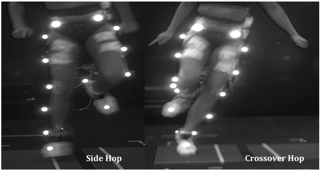
After informed consent was provided, we measured weight, height, and distance between anterior superior iliac spines for each participant. Leg dominance was determined for all healthy participants by ascertaining which leg the participant preferred to use to perform a single hop for distance [12,13]. The dominant leg served as a control against the reconstructed limb of the participants who had undergone surgery. A warm-up protocol was performed before recording the side-to-side hopping task. The warm-up consisted of 5 minutes of cycling at 40 to 60 rpm on a cycle ergometer, 10 half squats, 5 continuous vertical jumps (countermovement jumps), and 2 practice trials of the side-to-side hopping task. The side-to-side hopping task was performed as standardized by Itoh et al [14] (Figure 1). To perform that task, each participant stood facing 2 force plates that had been placed on the floor. Each individual force plate had a line marked with tape that was positioned 30 cm apart from the line on the other force plate. When the participant was ready to start the task, she stood on the force plate of her preference and started jumping single-legged from one force plate to another for 10 consecutive times across the marked lines.
Figure 1.
Side-to-side hop test: During this task participants hopped 10 times side to side across 2 lines marked 30 cm apart in 2 individual force plates. One hop was equivalent to hopping from and onto the same force plate; a successful trial was considered when 10 jumps were completed.
One jump was defined as jumping away and back to the same force plate. As a result, 20 single hops were included in the task. The side-to-side task was subdivided into side and crossover hopping components similar to the description of Lloyd [2]. A side-hopping maneuver was defined as the direction of movement to the opposite side of the weight-bearing leg (Figure 1). A crossover hop was defined as the direction of movement toward the same side of the weight-bearing leg (Figure 1). For example, if the participant jumped with her right leg toward her left leg, the maneuver was considered to be a side hop. If the subject jumped toward her right side, the maneuver was considered to be a crossover hop.
Participants were allowed to use their arms freely during task performance for balance purposes. Each participant was allowed to rest no less than 1 minute (or as long as she wanted) to prevent fatigue between trials. Each participant performed 2 testing trials that were timed from beginning to end of the 10 hops; the fastest trial was selected for analysis. Noninjured women performed both trials with their predetermined dominant leg, and women with ACL reconstruction performed the task with their reconstructed leg. Only the reconstructed leg of the women with ACL reconstruction was used for comparison, because in other studies [15–17] neuromuscular impairments were identified in the nonreconstructed leg; thus the contralateral leg is not a reliable control.
Instrumentation
The participants wore 21 retro-reflective markers attached over both anterior superior iliac spines, the second sacral vertebra, greater trochanters, lateral femoral epicondyles, mid distance between greater trochanters and lateral femoral epicondyles, medial femoral epicondyles, lateral malleoli, mid distance between lateral femoral epicondyles and lateral malleoli, medial malleoli, calcaneal tuberosities, and the area of the second metatarsophalangeal joints over the shoe. The motion capture system consisted of 4 digital cameras (60-Hz sampling rate) time synchronized to 2 force plates (AMTI, Watertown, MA) at a sampling rate of 1 kHz. The video and force plate data were captured with VSOL MutiDV Capture software (VSOL Inc, Seoul, Korea) and KwonGRF 2.1 software (VSOL Inc), respectively. Before data collection, the volume of the recording space was calibrated according to manufacturer’s recommendation with a 12-point, 81.5-cm3 cube using an 11-parameter direct linear transformation method. Also captured was a static trial of the participant standing still with her arms across her chest to align the joint coordinates to the laboratory recording instruments and to estimate joint centers on the basis of each subject’s anthropometric characteristics. After the static trial, the medial epicondyle and medial malleolus markers were removed to prevent interference between the medial markers and the lower extremities during side-to-side hopping.
A surface EMG study was recorded with 8 bipolar, self-adhesive, Ag/AgCl preamplified surface electrodes (M-00-S; Ambu, Ølstykke, Denmark, with overall gain = 2000 mV; total electrode contact area, 4.1 × 3.4 cm; and 1.32-cm2 sensor areas). The electrodes were placed, according to the recommendations of Cram et al [18], on the skin over the gluteus maximus, quadriceps, and lateral and medial hamstrings after the skin had been cleansed with an alcohol-soaked gauze. A self-adhesive reference electrode was placed over the anterior tibial crest. All electrodes were secured with hypoallergenic adhesive tape to reduce movement artifact. EMG data were collected with a telemetry system consisting of a transmitter and receiver units (Noraxon Inc, Scottsdale, AZ). Raw muscle activity recordings were transmitted via an FM signal from the transmitter each participant wore on a belt. The signal was filtered at a bandwidth of 10 to 500 Hz with 130 dB common-mode rejection in the transmitter. In the receiver, the signal was converted from analog to digital and transmitted through a USB A/D converter to the computer monitor, on which raw muscle signals were displayed.
Data Reduction
As we previously mentioned, the side-to-side hopping task was divided into side maneuvers and crossover-hopping maneuvers as defined by Lloyd [2]. During a side-hopping maneuver, the direction of movement was toward the opposite side of the weight bearing leg (Figure 1). In a crossover hop, the direction of movement was toward the same side of the weight-bearing leg (Figure 1). Joint angles, ground reaction forces, and knee-joint moment data were synchronized and analyzed with Kwon3D 3.1 software (VSOL Inc). The joint angles were derived from the 3-dimensional trajectory of retro-reflective markers filtered through a second-order, low-pass Butterworth filter (6 Hz). The hip- and knee-joint angles were defined in the sagittal, frontal, and transverse planes as the first, second, and third rotations, respectively. Joint moments were derived by an inverse dynamics method instrumented in the software. The kinematic and kinetic data of interest in the side-to-side hopping task were the peak values during the ground contact phase, from initial contact to push-off as identified by each force plate [8]. The first 2 and last 2 jumps of the total 10 were excluded from the analysis to account for acceleration and deceleration variability at the beginning and end of the task; thus the peak kinematic and kinetic values of the middle 6 jumps were averaged for analyses. As a result, a total of 6 side hops and 6 crossover hops were used for analyses.
All EMG data were time-synchronized to the force plates. EMG raw data were amplified (×1000) and full-wave rectified with use of Myoresearch software (Noraxon Inc, Scottsdale, AZ). Normalization for the EMG data was accomplished by using a dynamic normalization procedure in which the average signal for each muscle group in the window of interest was divided by the maximum signal generated during the specific trial analyzed. This method has been widely used to analyze EMG activity during dynamic tasks [9,19–23] and has been shown to reduce participant variability to a greater extent than do maximal isometric voluntary contractions [23,24]. In addition, this procedure controls for the variability among trials that is caused by fatigue during dynamic tasks [22].
Because the hamstring muscle group was separated into medial and lateral compartments, the normalized results were summed and averaged to represent the hamstring group in its entirety [9,23]. The hamstring values were averaged and were used to calculate the knee muscle co-contraction ratio [9,23]. That co-contraction ratio was calculated by first obtaining the normalized values for both the quadriceps and hamstring muscle groups during the targeted window of time [9,23]. The hamstring value was used as the divisor if it was greater than the quadriceps value; however, the quadriceps value was used if it was greater than the hamstrings value [9,23]. Therefore the co-contraction ratio value was always less than or equal to 1 [9,23]. A co-contraction ratio closer to 1 indicated excellent co-contraction, and values closer to 0 represented poor co-contraction between the quadriceps and hamstring muscle groups [9,23]. That ratio represents a component of joint stability that allows for the relative activation of the knee flexor and extensor muscle groups that cross the joint [9,23].
Data Analyses
The dependent variables of this investigation were as follows: hip flexion, hip adduction, knee flexion, and knee valgus angles; knee extension and abduction joint moments; gluteus, quadriceps, and hamstrings normalized-rectified EMG study results; and the quadriceps/hamstrings co-contraction ratio. All variables were screened for normality assumptions and outliers through the use of box plots and scatter plots. Three split-plot multivariate analyses of variance with the maneuver as the within-group factor were used to compare the hip- and knee-joint angles, knee internal moments, and EMG variables between the groups. Follow-up analyses were performed when indicated.
RESULTS
Hip-Joint Angles
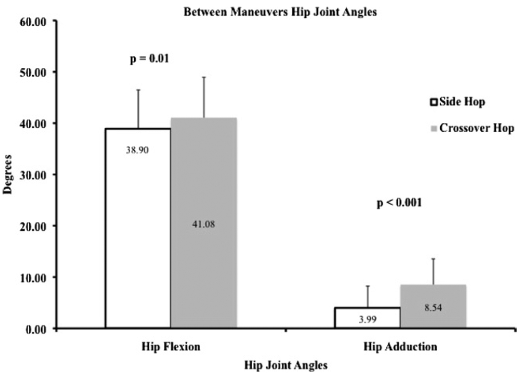
The group × maneuver interaction (F2,24 = 1.51; P = .241; effect size: 0.11; power: 0.29) and group main effect (F2,24 = 0.283; P = .756; effect size: 0.023; power: 0.090) were not statistically significant. However, there was a statistically significant main effect for maneuver (F2,24 = 29.32; P < .001; effect size: 0.71; power: 1.0) with greater hip adduction (P < .001) and flexion (P = .01) joint angles during the crossover-hopping maneuver than during the side-hopping maneuver (Figure 2).
Figure 2.
Hip flexion and hip adduction joint angles between maneuvers for control and ACL groups combined. The crossover maneuver elicited greater hip flexion joint angles and greater hip adduction joint angles for both groups.
Knee-Joint Angles
In neither study group was group × maneuver interaction (F2,24 = 1.03, P = .21; effect size: 0.08; power: 0.21), group main effect (F2,24 = 0.583; P = .57; effect size: 0.046; power: 0.14), or maneuver main effect (F2,24 = 0.528; P = .60; effect size: 0.042; power: 0.13) statistically significant for knee-joint angles.
Knee-Joint Moments
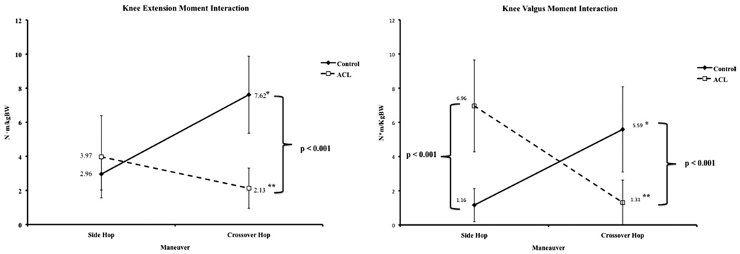
With respect to knee-joint moments, group × maneuver interaction was statistically significant (F2,24 = 58.33; P < .001; effect size: 0.83; power: 1.0). Figure 3 shows the group × maneuver interaction. Follow-up analyses of simple main effects of group showed significantly greater knee extension (P < .001) and abduction (P < .001) moments in the control group compared with the ACL group during the crossover-hopping task but significantly greater knee abduction moments (P < .001) in the ACL group than in the control group during the side-hopping task (Figure 3). Follow-up analyses of the simple main effects of the maneuver in individual groups revealed significantly greater moments during the crossover-hopping task (P < .001) than during the side-hopping task in the control group and greater moments during the side-hopping task than in the crossover-hopping task in the ACL group (P < .001; Figure 3).
Figure 3.
Group × maneuver interactions. *Knee extension and valgus moments were greater during the crossover hop for the control group. **Knee extension and valgus moments were greater during the side-hopping maneuver for the ACL group.
EMG Variables
In neither group did group × maneuver interaction (F4,22 = 1.05; P = .402; effect size: 0.16; power: 0.28), group main effect (F4,22 = 2.05; P = .12; effect size: 0.27; power: 0.52), or maneuver main effect (F4,22 = 2.20; P = .10; effect size: 0.29; power: 0.55) in the gluteus, rectus femoris, and hamstrings muscles or the co-contraction ratios reach statistical significance.
DISCUSSION
The purpose of this investigation was to compare landing mechanics between noninjured women and women with ACL reconstruction during side-hopping and crossover-hopping maneuvers. Differences between side-hopping and crossover-hopping maneuvers for both groups also were examined. The main findings of this investigation were the differences in the study subjects between maneuvers for hip joint angles (Figure 2) and maneuvers involving the variables of knee extension and abduction joint moments (Figure 3).
Hip- and Knee-Joint Angles
The hypothesis stating that women with ACL reconstruction are at greater risk than their healthy counterparts for injury-predisposing hip and knee joint angles was not supported by the results of this investigation. The authors of a previous study reported that landing mechanics from a drop jump and up-down hopping task were similar in noninjured women and women with ACL reconstruction [8]. Therefore it appears that in side-hopping and crossover-hopping maneuvers, lower extremity biomechanics are similar in women with or without ACL reconstruction. Conversely, Ristanis et al [25] reported greater knee rotational joint angles in male subjects 1 year after ACL reconstruction than in noninjured men during a landing/pivoting maneuver. Those authors concluded that ACL reconstruction may not restore rotational stability during high-level activities regardless of anteroposterior stability [25].
We did not assess knee external/internal rotation joint angles in our investigation; we addressed only sagittal and coronal planes. We found differences among maneuvers for hip flexion and adduction angles across both study groups. Side-to-side maneuvers have been shown to be one of the most dangerous athletic tasks in sports [5]. Greater knee valgus during side-to-side maneuvers has been shown to create excessive knee loads capable of causing injury [2]. The combination of small hip joint flexion and large adduction angles has been found to be one of the main causes of valgus at the knee leading to ligamentous injury [26,27]. For those reasons, women athletes seem to be at greater risk than their male counterparts for lower extremity injuries during side-to-side maneuvers.
The results we observed in both of our study groups contradicted our hypothesis because of the greater hip flexion and hip adduction angles that we observed during crossover hopping. Our investigation revealed a combination of both protective and high-risk movement patterns during crossover hopping: greater hip flexion and hip adduction, respectively. The crossover hop may inherently create large anteroposterior (hip flexion) and mediolateral (hip adduction) angles related to trunk movements while the stance leg is in a closed kinetic chain [28–30]. Therefore further analyses studying trunk movements during cutting/pivoting maneuvers are needed to clarify the ways in which trunk movements influence lower extremity biomechanics during side-hopping and crossover-hopping maneuvers.
Knee-Joint Moments
Our knee extension and abduction joint moment data supported in part our study hypothesis. The observed finding in which the control group exhibited greater knee extension and abduction joint moments than did the ACL group during the crossover-hopping maneuver (indicating greater anterior translatory knee load during that maneuver) contradicted our study hypothesis. Conversely, the ACL group exhibited greater knee abduction moments during the side-hop maneuver, supporting our study hypothesis.
Knee abduction moments have been reported to be the most dangerous biomechanical deviation leading to ACL injury [2]. Therefore women with ACL reconstruction continue to be at risk for knee injury during side-hopping maneuvers. This finding concurs with those of previous studies in which the authors evaluated joint moments during landing activities (studies indicating a long-term biomechanical deficiency in women with ACL reconstruction) that were predisposing to reinjury [8].
Knee extension and valgus moments during the crossover maneuver were greater in the control group than in the women with ACL reconstruction, a finding that is not readily explained. During the crossover maneuver, there may be more sources of variation, such as proximal movements of the trunk and pelvis that affect lower extremity biomechanics. A possible explanation for the discrepancies between our study and the previous literature are the differences in rotational demands between the tasks used. Our study incorporated solely a side-to-side maneuver instead of a sidestepping or crossover pivot maneuver after a running approach, making these comparisons unequal.
EMG Variables
The hypothesis that noninjured women would exhibit greater EMG values than do women with ACL reconstruction during both tasks studied was not supported by our investigation. Our findings suggest that with respect to factors such as high quadriceps activation, low hamstring muscle activation, and a low quadriceps/hamstring cocontraction ratio, all of which increase the risk of knee injury, women with ACL reconstruction recover their neuromuscular recruitment strategies and exhibit results similar to those in noninjured women. Therefore it appears that women with ACL reconstruction are capable of reaching compensatory neuromuscular stability of the knee joint during functional tasks [8,31,32]. These neuromuscular stabilization strategies allowed women with ACL reconstruction to perform without any long-term giving-way episodes during functional tasks.
The results for the co-contraction ratios did not differ between the groups, indicating good dynamic anteroposterior knee stability in the ACL group. Most studies in which the authors assessed neuromuscular activation strategies by using EMG have performed between-gender comparisons [5,7,33,34]. Hence comparisons between injury and reconstruction status specifically in women have not been adequately addressed in the literature. In studies in which the authors compared EMG amplitudes of the quadriceps and hamstring muscle groups between men and women, greater quadriceps activation and lower hamstring activation have been linked to other factors that increase the risk of biomechanical injury in women [7,34]. However, other researchers have reported greater neuromuscular stability of both those muscle groups in women compared with men [5]. These conflicting results are evidence of the complexity of the neuromuscular system in creating dynamic knee stability during athletic tasks.
The results of this investigation need to be evaluated within the scope of its limitations. First, the sample size recruited for this study probably underpowered some of the results. Second, the 60-Hz sampling rate may have introduced variability into the data on joint angles. However, the high-frequency components for the hopping tasks (especially during impact with the force plate) were filtered through the 6-Hz low-pass filter. Therefore the 60-Hz sampling rate with a 6-Hz Butterworth filter seems reasonable given the data of interest were peak hip and knee joint moments during the ground contact phase.
Typically, men are used as control subjects, representing correct biomechanics and neuromuscular control during the performance of athletic tasks. In our investigation we studied only women, but even our subjects without ACL reconstruction could have presented injury-predisposing factors during side-hopping and crossover-hopping maneuvers if they had been compared with a healthy noninjured group of men. If that had been the case, on the basis of previous research, it can be hypothesized that women with ACL reconstruction continue to be at risk of reinjury during side-to-side maneuvers. The women with ACL reconstruction who volunteered to participate in our investigation or who passed the screening procedures might have been at a greater level of performance than average women after ACL surgery. Thus women with poor postsurgical biomechanical compensations might not have been represented in our study. We suggest that future investigations include women at both ends of the spectrum—those that have been able to return to their previous level of physical activity and those not able to perform dynamic activities—to assess the biomechanical factors that are not recovered after surgery.
The physical activity of the women who participated in our investigation was at a recreational level; thus our results should not be extrapolated to women participating at elite or professional levels of physical activity. The variation in years after ACL reconstruction shows that the sample of women with ACL reconstruction was heterogeneous. Because of the sample size of our study, no stratification for the surgical procedure used could be performed; different surgical procedures might create different biomechanical and neuromuscular adaptations that warrant further analyses. In addition, transverse plane movements were not assessed in our investigation. Although Ristanis et al [25] found that ACL reconstruction did not restore rotational stability of the knee, their investigation assessed only men. Findings in female soccer players have shown that transverse plane movements of the hip and knee do not involve biomechanical factors that increase the risk for knee injury in female athletes [35]. These contradictory findings suggest that transverse hip and knee joint angles should be further assessed to confirm their potential contribution to knee injury.
CONCLUSION
This investigation revealed that women with ACL reconstruction exhibited landing joint angles, quadriceps and hamstrings EMG amplitudes, and a quadriceps/hamstrings co-contraction ratio similar to those in noninjured women. These findings reveal the restoration of biomechanical strategies during side-to-side athletic tasks. However, during side-hopping maneuvers, our subjects with ACL reconstruction continue to exhibit valgus joint moments that increase the likelihood of injury during such maneuvers. Side-hopping and crossover-hopping maneuvers were shown to exert different stresses on the lower extremity in both study groups; this finding adds to the challenge of identifying factors that predispose women to biomechanical injury.
ACKNOWLEDGMENTS
We acknowledge research assistants Luis Rivas, Glorimar Garcia, and Carmen Capo for their help in digitization and data reduction processes.
This project was supported in part by NIH grants G12RR03051, 1P20 RR11126, and R25RR17589 and the NSCA Foundation.
Footnotes
Disclosure: nothing to disclose
Contributor Information
Alexis Ortiz, Physical Therapy Program, School of Health Professions and Department of Anatomy & Neurobiology, School of Medicine, University of Puerto Rico-Medical Sciences Campus, PO Box 365067, San Juan, PR 00936; and University of Puerto Rico-Rio Piedras Campus, San Juan, PR..
Sharon Olson, School of Physical Therapy, Texas Woman’s University, Houston, TX.
Elaine Trudelle-Jackson, School of Physical Therapy, Texas Woman’s University, Dallas, TX.
Martin Rosario, Department of Anatomy & Neurobiology, School of Medicine, University of Puerto Rico-Medical Sciences Campus, San Juan, PR.
Heidi L. Venegas, Department of Biostatistics & Epidemiology, School of Public Health, University of Puerto Rico-Medical Sciences Campus, San Juan, PR.
REFERENCES
- 1.Besier TF, Lloyd DG, Cochrane JL, Ackland TR. External loading of the knee joint during running and cutting maneuvers. Med Sci Sports Exerc. 2001;33:1168–1175. doi: 10.1097/00005768-200107000-00014. [DOI] [PubMed] [Google Scholar]
- 2.Lloyd DG. Rationale for training programs to reduce anterior cruciate ligamet injuries in Australian football. J Orthop Sports Phys Ther. 2001;31:645–654. doi: 10.2519/jospt.2001.31.11.645. [DOI] [PubMed] [Google Scholar]
- 3.Besier TF, Lloyd DG, Ackland TR, Cochrane JL. Anticipatory effects on knee joint loading during running and cutting maneuvers. Med Sci Sports Exerc. 2001;33:1176–1181. doi: 10.1097/00005768-200107000-00015. [DOI] [PubMed] [Google Scholar]
- 4.Pollard CD, Sigward SM, Powers CM. Gender differences in hip joint kinematics and kinetics during side-step cutting maneuver. Clin J Sport Med. 2007;17:38–42. doi: 10.1097/JSM.0b013e3180305de8. [DOI] [PubMed] [Google Scholar]
- 5.Sell TC, Ferris CM, Abt JP, et al. The effect of direction and reaction on the neuromuscular and biomechanical characteristics of the knee during tasks that simulate the noncontact anterior cruciate ligament injury mechanism. Am J Sports Med. 2006;34:43–54. doi: 10.1177/0363546505278696. [DOI] [PubMed] [Google Scholar]
- 6.Ford KR, Myer GD, Toms HE, Hewett TE. Gender differences in the kinematics of unanticipated cutting in young athletes. Med Sci Sports Exerc. 2005;37:124–129. [PubMed] [Google Scholar]
- 7.Landry SC, McKean KA, Hubley-Kozey CL, Stanish WD, Deluzio KJ. Neuromuscular and lower limb biomechanical differences exist between male and female elite adolescent soccer players during an unanticipated run and crosscut maneuver. Am J Sports Med. 2007;35:1901–1911. doi: 10.1177/0363546507307400. [DOI] [PubMed] [Google Scholar]
- 8.Ortiz A, Olson SL, Libby CL, et al. Landing mechanics between noninjured women and women with ACL reconstruction during two jump tasks. Am J Sports Med. 2008;36:149–157. doi: 10.1177/0363546507307758. [DOI] [PMC free article] [PubMed] [Google Scholar]
- 9.Lloyd DG, Buchanan TS. Strategies of muscular support of varus and valgus isometric loads at the human knee. J Biomech. 2001;34:1257–1267. doi: 10.1016/s0021-9290(01)00095-1. [DOI] [PubMed] [Google Scholar]
- 10.Fithian DC, Paxton LW, Goltz DH. Fate of the anterior cruciate ligament-injured knee. Orthop Clin North Am. 2002;33:621–636. doi: 10.1016/s0030-5898(02)00015-9. [DOI] [PubMed] [Google Scholar]
- 11.Ross MD, Irrgang JJ, Denegar CR, McCloy CM, Unangst ET. The relationship between participation restrictions and selected clinical measures following anterior cruciate ligament reconstruction. Knee Surg Sports Traumatol Arthrosc. 2002;10:10–19. doi: 10.1007/s001670100238. [DOI] [PubMed] [Google Scholar]
- 12.Chappell JD, Yu B, Kirkendall DT, Garrett WE. A comparison of knee kinetics between male and female recreational athletes in stop-jump tasks. Am J Sports Med. 2002;30:261–267. doi: 10.1177/03635465020300021901. [DOI] [PubMed] [Google Scholar]
- 13.Wikstrom EA, Powers ME, Tillman MD. Dynamic stabilization time after isokinetic and functional fatigue. J Athl Train. 2004;39:247–253. [PMC free article] [PubMed] [Google Scholar]
- 14.Itoh H, Kurosaka M, Yoshiya S, Ichihashi N, Mizuno K. Evaluation of functional deficits determined by four different hop tests in patients with anterior cruciate ligament deficiency. Knee Surg Sports Traumatol Arthrosc. 1998;6:241–245. doi: 10.1007/s001670050106. [DOI] [PubMed] [Google Scholar]
- 15.Ageberg E. Consequences of a ligament injury on neuromuscular function and relevance to rehabilitation—using the anterior cruciate ligament-injured knee as model. J Electromyogr Kinesiol. 2002;12:205–212. doi: 10.1016/s1050-6411(02)00022-6. [DOI] [PubMed] [Google Scholar]
- 16.Friden T, Roberts D, Ageberg E, Walden M, Zatterstrom R. Review of knee proprioception and the relation to extremity function after an anterior cruciate ligament rupture. J Orthop Sports Phys Ther. 2001;31:567–576. doi: 10.2519/jospt.2001.31.10.567. [DOI] [PubMed] [Google Scholar]
- 17.Roberts D, Friden T, Stomberg A, Lindstrand A, Moritz U. Bilateral proprioceptive defects in patients with a unilateral anterior cruciate ligament reconstruction: A comparison between patients and healthy individuals. J Orthop Res. 2000;18:565–571. doi: 10.1002/jor.1100180408. [DOI] [PubMed] [Google Scholar]
- 18.Cram JR, Kasman GS, Holtz J. Introduction to Surface Electromyography. Gaithersburg, MD: Aspen Publishers; 1998. [Google Scholar]
- 19.Manolopoulos E, Papadopoulos C, Kellis E. Effects of combined strength and kick coordination training on soccer kick biomechanics in amateur players. Scand J Med Sci Sports. 2005;16:102–110. doi: 10.1111/j.1600-0838.2005.00447.x. [DOI] [PubMed] [Google Scholar]
- 20.Rodacki AL, Fowler NE, Bennett SJ. Multi-segment coordination: Fatigue effects. Med Sci Sports Exerc. 2001;33:1157–1167. doi: 10.1097/00005768-200107000-00013. [DOI] [PubMed] [Google Scholar]
- 21.Rodacki AL, Fowler NE, Bennett SJ. Vertical jump coordination: Fatigue effects. Med Sci Sports Exerc. 2002;34:105–116. doi: 10.1097/00005768-200201000-00017. [DOI] [PubMed] [Google Scholar]
- 22.Croce RV, Russell PJ, Decoster LC. Knee muscular response strategies differ by developmental level but not gender during jump landing. Electromyogr Clin Neurophysiol. 2004;44:339–348. [PubMed] [Google Scholar]
- 23.Besier TF, Lloyd DG, Ackland TR. Muscle activation strategies at the knee during running and cutting maneuvers. Med Sci Sports Exerc. 2003;35:119–127. doi: 10.1097/00005768-200301000-00019. [DOI] [PubMed] [Google Scholar]
- 24.Soderberg GL, Knutson LM. A guide for use and interpretation of kinesiologic electromyographic data. Phys Ther. 2000;80:485–498. [PubMed] [Google Scholar]
- 25.Ristanis S, Stergiou N, Patras K, Vasiliadis HS, Giakas G, Georgoulis AD. Excessive tibial rotation during high-demand activities is not restored by anterior cruciate ligament reconstruction. Arthroscopy. 2005;21:1323–1329. doi: 10.1016/j.arthro.2005.08.032. [DOI] [PubMed] [Google Scholar]
- 26.Powers CM. The influence of altered lower-extremity kinematics on patellofemoral joint dysfunction: A theoretical perspective. J Orthop Sports Phys Ther. 2003;33:639–646. doi: 10.2519/jospt.2003.33.11.639. [DOI] [PubMed] [Google Scholar]
- 27.Powers CM, Ward SR, Fredericson M, Guillet M, Shellock FG. Patellofemoral kinematics during weight-bearing and non-weight-bearing knee extension in persons with lateral subluxation of the patella: A preliminary study. J Orthop Sports Phys Ther. 2003;33:677–685. doi: 10.2519/jospt.2003.33.11.677. [DOI] [PubMed] [Google Scholar]
- 28.Hewett TE, Torg JS, Boden BP. Video analysis of trunk and knee motion during non-contact anterior cruciate ligament injury in female athletes: Lateral trunk and knee abduction motion are combined components of the injury mechanism. Br J Sports Med. 2009;43:417–422. doi: 10.1136/bjsm.2009.059162. [DOI] [PMC free article] [PubMed] [Google Scholar]
- 29.Blackburn JT, Padua DA. Sagittal-plane trunk position, landing forces, and quadriceps electromyographic activity. J Athl Train. 2009;44:174–179. doi: 10.4085/1062-6050-44.2.174. [DOI] [PMC free article] [PubMed] [Google Scholar]
- 30.Blackburn JT, Padua DA. Influence of trunk flexion on hip and knee joint kinematics during a controlled drop landing. Clin Biomech. 2008;23:313–319. doi: 10.1016/j.clinbiomech.2007.10.003. [DOI] [PubMed] [Google Scholar]
- 31.Rozzi SL, Lephart SM, Gear WS, Fu FH. Knee joint laxity and neuromuscular characteristics of male and female soccer and basketball players. Am J Sports Med. 1999;27:312–319. doi: 10.1177/03635465990270030801. [DOI] [PubMed] [Google Scholar]
- 32.Rudolph KS, Axe MJ, Buchanan TS, Scholz JP, Snyder-Mackler L. Dynamic stability in the anterior cruciate ligament deficient knee. Knee Surg Sports Traumatol Arthrosc. 2001;9:62–71. doi: 10.1007/s001670000166. [DOI] [PubMed] [Google Scholar]
- 33.Hanson AM, Padua DA, Troy Blackburn J, Prentice WE, Hirth CJ. Muscle activation during side-step cutting maneuvers in male and female soccer athletes. J Athl Train. 2008;43:133–143. doi: 10.4085/1062-6050-43.2.133. [DOI] [PMC free article] [PubMed] [Google Scholar]
- 34.Malinzak RA, Colby SM, Kirkendall DT, Yu B, Garrett WE. A comparison of knee joint motion patterns between men and women in selected athletic tasks. Clin Biomech. 2001;16:438–445. doi: 10.1016/s0268-0033(01)00019-5. [DOI] [PubMed] [Google Scholar]
- 35.Imwalle LE, Myer GD, Ford KR, Hewett TE. Relationship between hip and knee kinematics in athletic women during cutting maneuvers: A possible link to noncontact anterior cruciate ligament injury and prevention. J Strength Cond Res. 2009;23:2223–2230. doi: 10.1519/JSC.0b013e3181bc1a02. [DOI] [PMC free article] [PubMed] [Google Scholar]


