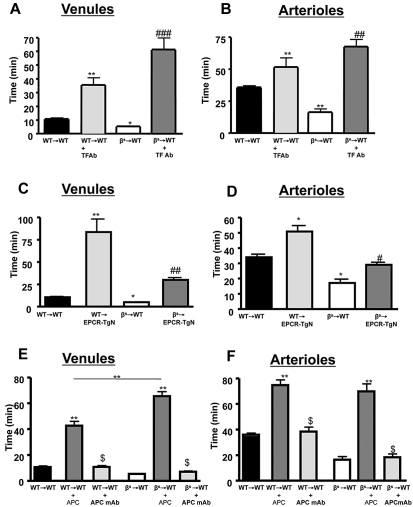
Figure 3.
Effects of TF immunoneutralization and role of EPCR and APC on light/dye–induced thrombus formation in cerebral vessels. Administration of TF-blocking antibody to WT→WT and βs→WT chimeras extended blood flow-cessation time in cerebral venules (A) and arterioles (B). The accelerated thrombus formation observed in βs→WT chimeras was significantly reversed in βs→EPCR-TgN mice in both cerebral venules (C) and arterioles (D). WT (WT→WT) or sickle cell–transgenic (βs→WT) bone marrow chimeras were treated with either APC (40 μg/mouse) or an APC-neutralizing mAb (10 μg/mouse) 5 minutes before inducing thrombosis in both venules (E) and arterioles (F). Data are shown as the means ± SEM. *P < .05 compared with the corresponding control group (WT→WT or βs→WT); **P < .01 compared with the corresponding control group (WT→WT or βs→WT); #P < .05 compared with βs→WT or WT-EPCR-TgN; ##P < .01 compared with βs→WT or WT-EPCR-TgN; ###P < .001 compared with βs→WT or WT-EPCR-TgN; $P < .001 compared with WT→WT + APC or βs→WT + APC. n = 5-6 mice/group.

