Abstract
Background
This randomized phase III study was to evaluate the efficacy and safety of cytoreductive surgery (CRS) plus hyperthermic intraperitoneal chemotherapy (HIPEC) for the treatment of peritoneal carcinomatosis (PC) from gastric cancer.
Methods
Sixty-eight gastric PC patients were randomized into CRS alone (n = 34) or CRS + HIPEC (n = 34) receiving cisplatin 120 mg and mitomycin C 30 mg each in 6000 ml of normal saline at 43 ± 0.5°C for 60–90 min. The primary end point was overall survival, and the secondary end points were safety profiles.
Results
Major clinicopathological characteristics were balanced between the 2 groups. The PC index was 2–36 (median 15) in the CRS + HIPEC and 3–23 (median 15) in the CRS groups (P = 0.489). The completeness of CRS score (CC 0–1) was 58.8% (20 of 34) in the CRS and 58.8% (20 of 34) in the CRS + HIPEC groups (P = 1.000). At a median follow-up of 32 months (7.5–83.5 months), death occurred in 33 of 34 (97.1%) cases in the CRS group and 29 of 34 (85.3%) cases of the CRS + HIPEC group. The median survival was 6.5 months (95% confidence interval 4.8–8.2 months) in CRS and 11.0 months (95% confidence interval 10.0–11.9 months) in the CRS + HIPEC groups (P = 0.046). Four patients (11.7%) in the CRS group and 5 (14.7%) patients in the CRS + HIPEC group developed serious adverse events (P = 0.839). Multivariate analysis found CRS + HIPEC, synchronous PC, CC 0–1, systemic chemotherapy ≥ 6 cycles, and no serious adverse events were independent predictors for better survival.
Conclusions
For synchronous gastric PC, CRS + HIPEC with mitomycin C 30 mg and cisplatin 120 mg may improve survival with acceptable morbidity.
Gastric cancer (GC) is a pathophysiologically heterogeneous disease, associated with predominantly lymphatic spread, hematogenous metastasis or intra-abdominal spread. For the lymphatic and hematogenous metastases, reasonably extended lymphadenectomy, regional radiotherapy, and adjuvant antitumor chemotherapy have been proved effective, as demonstrated by several large scale international studies such as INT-0116 trial, the MAGIC trial, and the ACTS-GC trial.1–3 These trials all show the same pattern of cancer recurrence, that is either the patients were treated by surgery alone or surgery plus peri- or postoperative chemoradiotherapy, regional recurrence (typically abdominal carcinomatosis) is the most common pattern of first recurrence.
These results reflect the fact that GC is a disease with easy intra-abdominal spread, largely because free cancer cells in peritoneal washings could be detected in up to 24% of stage IB GC, and up to 40% of those with stage II or III diseases.4 In diffuse type GC, peritoneal seeding (recurrence) is a characteristic feature of cancer spread.5 More than 50% of potentially curable advanced GC patients died of peritoneal recurrence.6 In addition, established peritoneal carcinomatosis (PC) is detected in more than 30% of patients with advanced GC. Accordingly, almost 60% of all causes of GC death is from PC.7
PC from GC is characterized by the presence of tumor nodules of various size, number and distribution on parietal or/and visceral peritoneal surfaces, with very poor prognosis and a median survival of less than 6 months.8 As PC is currently regarded as a variant of systemic spread of disease, treatments for such patients are palliative systemic chemotherapy and best support care, with limited efficacy. These nihilistic treatment approaches are ill conceived, as data supporting systemic therapy for secondary PC is derived from clinical trials reporting the results of treatment of distinctively different tumor biology-visceral metastasis of hematogenous origin, not PC.9 To tackle this problem, a more aggressive treatment strategy called cytoreductive surgery (CRS) plus hyperthermic intraperitoneal chemotherapy (HIPEC) has been developed over the past 3 decades, taking advantages of surgery to reduce visible tumor burden, and regional hyperthermic chemotherapy to eradicate micro-metastases, expanding cancer surgery from resection of primary tumor to surgical management of metastatic diseases.10
Cohort studies suggested CRS plus HIPEC could improve outcome of patients with PC from GC.11–13 Nonrandomized comparative studies suggested the superior efficacy of CRS + HIPEC over CRS alone for the treatment of gastric PC.14,15 So far, however, only a few prospective randomized clinical trials have been conducted to support such treatment strategy, which is a major reason for skepticism and criticism among the oncology community.16,17 This phase III prospective randomized clinical trial was to evaluate the efficacy and safety of CRS plus HIPEC for PC from GC.
Patients and Methods
Patients
Sixty-eight gastric PC patients, including 35 men and 33 women, aged 24–75 years (median 50 years) were recruited onto this study. Before treatment, all patients had signed informed consent, and the study protocol was approved by the ethics committee of Zhongnan Hospital of Wuhan University. These patients were randomized into the CRS group and CRS + HIPEC group according to a computer-generated randomize number. Detailed study information is available at ClinicalTrials.gov (http://www.clinicaltrials.gov/ct2/show/NCT00454519). Routine preoperative studies included thorough physical examination, blood test, serum biochemistry and electrolytes, liver and renal function evaluation and coagulation studies. Other examinations included chest x-ray, contrast-enhanced three-dimensional abdominal-pelvic computed tomography, and cardiac function assessments. Patient inclusion criteria were: (1) age 20–75 years old; (2) Karnofsky performance status of >50; (3) life expectancy of >8 weeks; (4) normal peripheral blood white blood cells count ≥3500/mm3 and platelet count ≥80,000/mm3; (5) acceptable liver function, with bilirubin no greater than 2 times the upper limit of normal (ULN), and aspartic aminotransferase and alanine aminotransferase no greater than 2 ULN; (6) acceptable renal function, with serum creatinine no greater than 1.5 mg/dl; and (7) cardiovascular pulmonary and other major organ functions can stand major operation. Major exclusion criteria were: (1) age <20 years or >75 years; (2) any lung metastasis, liver metastasis, or prominent retroperitoneal lymph node metastasis during preoperative assessment; (3) serum bilirubin level >3 ULN; (4) liver enzymes >3 ULN; and (5) serum creatinine level >1.5 mg/dl.
CRS and HIPEC
All CRS and HIPEC procedures were performed by a designated team of surgical oncologists, anesthesiologist and operating room staff, with the principal investigator (Y.L.) as the chief surgeon. The abdominal exploration was performed under general anesthesia and hemodynamic monitoring, through a midline xiphoid-pubic incision. Once the abdominal wall was open, detailed evaluation of peritoneal carcinomatosis index (PCI) was conducted, taking into consideration the size and distribution, according to the principle of Sugarbaker.18 We defined PCI < 20 as low PCI and PCI ≥ 20 as high PCI. The characteristics of ascites were also recorded. After evaluation, maximal CRS was performed, including the resection of the primary tumor with acceptable margins, any involved adjacent structures, lymphadenectomy, peritoneotomies where peritoneal surfaces were involved by tumor, according to the peritonectomy procedure developed by Sugarbaker.18
After surgery, HIPEC was performed before closure of abdominal cavity, as this open technique is believed to provide optimal thermal homogeneity and spatial diffusion, with 120 mg of cisplatin and 30 mg of mitomycin C each dissolved 6 l of heated saline (drug concentration cisplatin 20 μg/ml, mitomycin C 5 μg/ml).19,20 An outflow tube for perfusion was placed in Douglas’ pouch just before HIPEC. The heated perfusion solution was infused into the peritoneal cavity at a rate of 500 ml/min through the inflow tube introduced from an automatic hyperthermia chemotherapy perfusion device (ES-6001, Wuhan E-sea Digital Engineering, Wuhan, China). The skin of the abdomen is attached to a retractor ring and a plastic sheet covered the open wound to keep the temperature stable. The perfusion in the peritoneal cavity was stirred manually with care not to infuse directly on the bowel surface. The temperature of the perfusion solution in peritoneal space was kept at 43.0 ± 0.5°C and monitored with a thermometer on real time. The total HIPEC time was 60–90 min, after which the perfusion solution in the abdominal cavity was removed through the suction tube, and drainage tubes were placed at appropriate sites depending on the type of primary operation. The wound was closed with relaxing suture, and patient was delivered to the intensive care unit for recovery. When the condition stabilized, usually 24–48 h later, the patients were transferred to the surgical oncology ward.
The extent of CRS was determined by Sugarbaker’s criteria on the completeness of cytoreduction (CC).18,21 A score of CC-0 indicates no residual peritoneal disease after CRS; CC-1, less than 2.5 mm of residual disease; CC-2, residual tumor between 2.5 mm and 2.5 cm; and CC-3, more than 2.5 cm of residual tumor or the presence of a sheet of unresectable tumor nodules.
The primary end point was disease specific overall survival (OS), defined as time interval from randomization to death due to disease. The secondary end points were serious adverse events (SAE), defined as severe local and/or systemic infection, abdominal leakage, or death related to the procedure.
Statistical Analysis
All patients were regularly followed up for detailed monitoring of disease status. Data were obtained from a database of clinical records, surgical reports, medical imaging reports, laboratory and pathology reports, and follow-up records. Patients alive at the time of analysis were censored at the last follow-up. OS was estimated by the Kaplan–Meier method, stratified by PCI and CC, and tested with the log-rank test.
This trial was designed to detect a 30% absolute difference in OS. With a statistical power of 90% to detect such difference and 5% significance level, at least 60 patients had to be entered. Categorized variables in the two groups were compared by chi square test or Fisher’s exact test. The numerical data were directly recorded, and the category data were recorded into different categories. The Kaplan–Meier method was used to compare the survival, with log rank test. Data were analyzed by the Statistical Package for Social Sciences (SPSS, Chicago, IL), version 13.0, with a 2-sided P value of <0.05 considered statistically significant.
Results
Baseline Data and Surgical Intervention
A total of 68 patients were randomized into CRS alone (n = 34) and CRS + HIPEC (n = 34) groups. These patients were well balanced and comparable regarding major baseline clinicopathological characteristics and surgical procedures (Table 1).
Table 1.
Clinicopathological characteristics of 68 patients with peritoneal carcinomatosis
| Variable | With HIPEC (N = 34) | CRS alone (N = 34) | P value |
|---|---|---|---|
| Age (years) | |||
| Median (range) | 50 (24–74) | 51 (28–75) | NS |
| Gender (n) | |||
| Male/female | 16/18 | 19/15 | NS |
| PCI | |||
| Range | 2–36 | 3–23 | NS |
| Median | 15 | 15 | |
| PCI > 20 | 14 (41.2%) | 9 (26.5%) | 0.206 (NS) |
| Histological diagnosis | |||
| Well/intermediately differentiated adenocarcinoma | 10 (29.4%) | 6 (17.8%) | NS |
| Poorly/undifferentiated adenocarcinoma | 19 (55.9%) | 24 (70.4%) | |
| Signet ring cell carcinoma | 4 (11.8%) | 0 (0.0%) | |
| Mucous carcinoma | 0 (0.0%) | 2 (5.9%) | |
| Squamous-cell carcinoma | 1 (2.9%) | 2 (5.9%) | |
| Organ resections | |||
| Total gastrectomy | 8 | 3 | NS |
| Subtotal gastrectomy | 25 | 31 | |
| Splenectomy | 2 | 1 | |
| Ovariectomy | 12 | 5 | |
| Colectomy | 0 | 2 | |
| Partial hepatectomy | 1 | 0 | |
| Peritonectomy locations | |||
| Right diaphragmatic copula | 10 | 7 | NS |
| Left diaphragmatic copula | 15 | 16 | |
| Greater omentum | 34 | 34 | |
| Lesser omentum | 34 | 34 | |
| Omental bursa | 34 | 34 | |
| Right colon gutter | 15 | 16 | |
| Left colon gutter | 20 | 21 | |
| Douglas pouch | 20 | 14 | |
| Anterior wall peritoneum | 15 | 10 | |
| Mesenteric fulguration | 23 | 9 | |
| CC 0–1 | 20/34 (58.8%) | 20/34 (58.8%) | |
| Median blood loss during operation, ml (range) | 800 (500–3000) | 600 (400–1200) | NS |
| Median operating time (excluding HIPEC), h (range) | 5.0 (4.0–7.5) | 4.0 (3.0–5.5) | NS |
CC Completeness of cytoreduction, NS not significant
Survival Analysis
At the time of this writing, the median follow-up was 32 months (7.5–83.5 months). Disease specific death had occurred in 33 of 34 (97.1%) cases in the CRS group and 29 of 34 (85.3%) cases in the CRS + HIPEC group. Follow-up, therefore, is long enough to demonstrate any impact of this new therapy on survival, and the data are mature for final analysis.
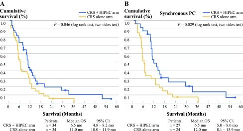
The median OS was 6.5 months (95% confidence interval [CI] 4.8–8.2 months) in CRS group and 11.0 months (95% CI 10.0–11.9 months) in the CRS + HIPEC group (P = 0.046, log rank test) (Fig. 1a). The 1-, 2-, and 3-year survival rates were 29.4, 5.9 and 0% for CRS group, and 41.2, 14.7 and 5.9% for CRS + HIPEC group.
Fig. 1.
CRS + HIPEC provides far better survival advantage than the CRS alone group in patients with gastric PC (a), particularly in patients with synchronous gastric PC (b)
In patients with synchronous PC (n = 51), the median OS was 12.0 months (95% CI 8.1–15.9 months) in CRS + HIPEC group (n = 24) and 6.5 months (95% CI 5.0–8.0 months) in the CRS group (n = 27) (P = 0.029) (Fig. 1b).
There were 17 patients with metachronous PC, including 10 in the CRS + HIPEC group and 7 in the CRS alone group. The number was too small for any definite conclusion, although the median OS was shorter in the CRS + HIPEC group (5.5 months) than in the CRS alone group (11.0 months).
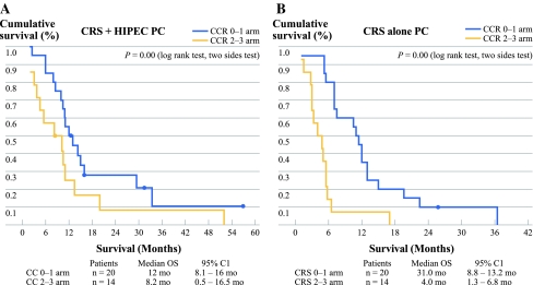
We further investigated the impact of CC on survival. In the CRS + HIPEC group, the median OS was 12.0 months (95% CI 8.1–16.0 months) in CC 0–1 subgroup (n = 20) and 8.2 months (95% CI 0.5–16.5 months) in CC 2–3 subgroup (n = 14) (P = 0.000) (Fig. 2a). In CRS group, the median OS was 11.0 months (95% CI 8.8–13.2 months) in CC 0–1 subgroup (n = 20) and 4.0 months (95% CI 1.3–6.8 months) in CC 2–3 subgroup (n = 14) (P = 0.000) (Fig. 2b). In patients with incomplete cytoreduction, HIPEC + CRS brought longer OS than CRS alone (median OS 8.2 vs. 4.0 months, P = 0.024).
Fig. 2.
In either CRS + HIPEC group (a) or CRS alone group (b), patients with CC 0–1 cytoreduction had better survival advantage
The impact of PCI on survival was also analyzed. In the high PCI group (n = 23), the median OS was 13.5 months (95% CI 8.7–18.3 months) in CRS + HIPEC subgroup (n = 14), and 3.0 months (95% CI 2.4–3.6 months) in CRS subgroup (n = 9) (P = 0.012, log-rank test). In the low PCI group (n = 45), the median OS was 10.2 months (95% CI 9.3–11.1 months) in CRS + HIPEC subgroup (n = 20), and 10.5 months (95% CI 4.0–17.0 months) in CRS subgroup (n = 25) (P = 0.464, log-rank test).
Multivariate analysis by Cox regression model identified CRS + HIPEC, synchronous PC, CC 0–1, systemic chemotherapy >6 cycles, and no SAE as major independent predictors for better survival, while age, sex and PCI were not independent survival factors (Table 2). Compared with CRS alone, CRS + HIPEC is about 2.6 times likely to improve survival (hazard ratio = 2.617; 95% CI 1.436–4.769).
Table 2.
Multivariate analysis on factors influencing survival
| Covariate | χ2 | P value | Hazard ratio | 95% CI |
|---|---|---|---|---|
| Sex (M vs. F) | 0.099 | 0.753 | 1.101 | 0.605–2.002 |
| Age (<60 years vs. ≥60 years) | 0.638 | 0.425 | 1.275 | 0.702–2.317 |
| PCI (low PCI vs. high PCI) | 0.292 | 0.589 | 1.222 | 0.590–2.529 |
| Treatment (CRS + HIPEC vs. CRS alone) | 9.871 | 0.002 | 2.617 | 1.436–4.769 |
| PC state (synchronous vs. metachronous) | 5.438 | 0.02 | 2.228 | 1.136–4.367 |
| CC (0–1 vs. 2–3) | 8.585 | 0.003 | 2.794 | 1.405–5.556 |
| Chemotherapy (≥6 vs. <6 cycles) | 15.649 | 0 | 3.344 | 1.838–6.061 |
| SAE (no vs. yes) | 13.765 | 0 | 4.295 | 1.989–9.274 |
PCI Peritoneal carcinomatosis index, CRS cytoreductive surgery, HIPEC hyperthermic intraperitoneal chemotherapy, PC peritoneal carcinomatosis, CC completeness of cytoreduction, SAE serious adverse events
Adverse Events
SAE had occurred in 9 patients, 4 in the CRS group (11.7%) and 5 in the CRS + HIPEC group (14.7%) (P = 0.839). These SAE included wound infection and sepsis, respiratory failure, gastrointestinal bleeding, severe bone marrow suppression, and intestinal obstruction (Table 3). SAE had a marked negative impact on survival. The median OS of patients who developed SAE was 5.0 months in CRS group and 3.0 months in the CRS + HIPEC group, although the number was too small for definite statistical analysis.
Table 3.
Distribution of SAE in the 2 treatment groups
| Characteristic | CRS group (n = 34) | CRS + HIPEC (n = 35) | P value |
|---|---|---|---|
| Total SAE | 4 | 5 | 0.839 |
| Respiratory system | |||
| Respiratory failure | 1 | 1 | |
| Respiratory fungi infection | 1 | 0 | |
| Wound infection and sepsis | 0 | 2 | |
| Massive gastrointestinal bleeding | 1 | 1 | |
| Anastomosis leakage | 1 | 0 | |
| Intestinal obstruction | 0 | 1 | |
SAE Serious adverse event, CRS cytoreductive surgery, HIPEC hyperthermic intraperitoneal chemotherapy
Patterns of Treatment Failure
Disease specific death had occurred in 33 of 34 (97.1%) cases in the CRS group and 29 of 34 (85.3%) cases of the CRS + HIPEC group. Among the 29 deaths in the CRS + HIPEC group, 1 was due to massive mediastinal lymph nodes and brain metastases leading to intracranial hemorrhage after radiotherapy, 1 was due to respiratory failure, and the remaining 27 were due to abdominal recurrence leading to progressive intestinal obstruction. Among the 33 deaths in the CRS group, 1 was due to widespread bone metastasis leading to bone marrow failure, 1 was due to massive systemic metastases involving the lungs, the liver, and the brain, 2 due to respiratory failure, and the remaining 27 were due to abdominal obstruction secondary to PC recurrence or progression.
Discussion
There is no standard treatment for PC from GC. CRS plus HIPEC represent a multidisciplinary approach to this problem. It was first reported in 1988 by Fujimoto et al on 15 patients with PC secondary to advanced GC, with a mean survival of 7.2 ± 4.6 months with acceptable morbidity.22 This new treatment modality gradually gains acceptance in many countries. Although the reported studies use different PCI scoring system to evaluate the extent of PC and different HIPEC approaches, they produce similar results that CRS plus HIPEC is an appropriate treatment option for a selected subgroup of GC patients with PC, and for advanced GC with high risk of developing PC. A systematic review and meta-analysis of 13 acceptable quality randomized controlled trials also have established that HIPEC is associated with marked improvement in survival in advanced GC, in comparison with the current standard treatments.17 As a result, a panel of international experts strongly recommend that CRS plus HIPEC be the current standard treatment for advanced GC.23 Nevertheless, controversy over this treatment modality remains, and more high quality clinical studies are required to clarify the value and the usefulness of this strategy.
In this prospective randomized study, CRS and HIPEC has been demonstrated to provide survival benefit for gastric PC. The median OS was 6.5 months for CRS group and 11.0 months for CRS + HIPEC group. The results were similar to those reported by Glehen et al. (OS 10.3 months) and Yonemura et al. (OS 11.5 months).13,24 Compared with CRS alone, CRS + HIPEC could extend the OS by nearly 70% (6.5 vs. 11.0 months). This is similar to the 76% improvement of OS (22.2 vs. 12.6 months) favoring CRS + HIPEC in the landmark phase III clinical trial in colorectal PC by the Netherlands Cancer Institute, which has provided level 1 evidence to support HIPEC with CRS for colorectal PC.16 Taking together, these results suggest that in either gastric or colorectal PC, CRS + HIPEC could provide similar survival advantage in selected cases.
Our results also demonstrated that patients with metachronous PC had worse survival than those with synchronous PC, in agreement with Glehen et al.13 The usefulness of CRS + HIPEC was evident for synchronous PC (median OS 12.0 vs. 6.5 months). For metachronous PC, the number was too small for any definite conclusion, although the median OS was shorter in the CRS + HIPEC group (5.5 months) than in the CRS alone group (11.0 months). More studies with greater sample size are required to clarify this issue.
This study demonstrated again the importance of complete cytoreduction for long term survival. Whether patients underwent CRS alone or CRS + HIPEC, CC 0–1 was independently associated with longer survival (Fig. 2). Therefore, efforts should be focused on complete CRS.
The synergistic effects of CRS to remove the macroscopic tumor and HIPEC to eradicate microscopic residual diseases are major advantages of this combined approach. However, such a procedure also brings greater risks for major morbidity and mortality. Major complications are directly related to the magnitude of the procedure, including the extent of resections and peritonectomy, the number of anastomoses, the duration of surgery, and the doses of cytotoxic chemotherapeutic drugs used in HIPEC.25 As a result of extensive resection, more blood loss, and more complex gastrointestinal reconstruction, complications become more frequent. To minimize potential complications, all patients in our group required blood, plasma and cryoprecipitation transfusion and large doses of antibiotics during and after operation. All patients were in intensive care for at least 24 h after the procedure. Even in intensified medical and surgical care, complications did occur. In terms of SAE, there were no statistically significant differences in the incidence of SAE between CRS group (11.7%) and CRS + HIPEC group (14.7%). But the CRS + HIPEC group had 3 cases with wound infection and sepsis, possibly as a result of long wound exposure during the lengthy operation. Our results are similar to the 19% grade IV complication rate reported by Sugarbaker et al.26 Our multivariate analysis demonstrated that SAE was an independent factor for worse survival. Therefore, greater efforts should be made to minimize SAE.
Severe hematological adverse events were not encountered in our patients. This could be due to relatively lower doses of mitomycin C and cisplatin used. Although we did not conduct pharmacokinetics studies to monitor the drug metabolism in the perfusion fluid, mitomycin C concentration of 5 μg/ml (30 mg/6000 ml) is still above the cytotoxic concentration, because previous studies have confirmed that 3 μg/ml of mitomycin C in HIPEC for 2 h can kill all GC cells in ascites and on the peritoneal surface.27
In conclusion, this study has found that for synchronous gastric PC, CRS + HIPEC with mitomycin C 30 mg and cisplatin 120 mg may improve survival with acceptable morbidity.
Acknowledgment
Supported by the grants supporting New Strategies to Treat Peritoneal Carcinomatosis from Hubei Sciences and Technology Bureau (2008BCC011, 2060402-542), the Science Fund for Creative Research Groups of the National Natural Science Foundation of China (20621502, 20921062).
Open Access
This article is distributed under the terms of the Creative Commons Attribution Noncommercial License which permits any noncommercial use, distribution, and reproduction in any medium, provided the original author(s) and source are credited.
References
- 1.Macdonald JS, Smalley SR, Benedetti J, et al. Chemoradiotherapy after surgery compared with surgery alone for adenocarcinoma of the stomach or gastroesophageal junction. N Engl J Med. 2001;345:725–730. doi: 10.1056/NEJMoa010187. [DOI] [PubMed] [Google Scholar]
- 2.Cunningham D, Allum WH, Stenning SP, et al. Perioperative chemotherapy versus surgery alone for resectable gastroesophageal cancer. N Engl J Med. 2006;355:11–20. doi: 10.1056/NEJMoa055531. [DOI] [PubMed] [Google Scholar]
- 3.Sakuramoto S, Sasako M, Yamaguchi, et al. Adjuvant chemotherapy for GC with S-1, an oral fluoropyrimidine. N Engl J Med. 2007;357:1810–1820. doi: 10.1056/NEJMoa072252. [DOI] [PubMed] [Google Scholar]
- 4.Juhl H, Stritzel M, Wroblewski A, et al. Immunocytological detection of micrometastatic cells: comparative evaluation of findings in the peritoneal cavity and the bone marrow of gastric, colorectal and pancreatic cancer patients. Int J Cancer. 1994;57:330–335. doi: 10.1002/ijc.2910570307. [DOI] [PubMed] [Google Scholar]
- 5.Averbach AM, Jacquet P. Strategies to decrease the incidence of intra-abdominal recurrence in respectable gastric cancer. Br J Surg. 1996;83:726–733. doi: 10.1002/bjs.1800830605. [DOI] [PubMed] [Google Scholar]
- 6.Yonemura Y, Kawamura T, Bandou E, et al. The natural history of free cancer cells in the peritoneal cavity. Recent Results Cancer Res. 2007;169:11–23. doi: 10.1007/978-3-540-30760-0_2. [DOI] [PubMed] [Google Scholar]
- 7.Yonemura Y, Endou Y, Shinbo M, et al. Safety and efficacy of bidirectional chemotherapy for treatment of patients with peritoneal dissemination from gastric cancer: selection for cytoreductive surgery. J Surg Oncol. 2009;100:311–316. doi: 10.1002/jso.21324. [DOI] [PubMed] [Google Scholar]
- 8.Al-Shammaa HAH, Li Y, Yonemura Y. Current status and future strategies of cytoreductive surgery plus intraperitoneal hyperthermic chemotherapy for peritoneal carcinomatosis. World J Gastroenterol. 2008;14:1159–1166. doi: 10.3748/wjg.14.1159. [DOI] [PMC free article] [PubMed] [Google Scholar]
- 9.Nissan A, Stojadinovic A, Garofalo A, et al. Evidence-based medicine in the treatment of peritoneal carcinomatosis: past, present, and future. J Surg Oncol. 2009;100:335–344. doi: 10.1002/jso.21323. [DOI] [PubMed] [Google Scholar]
- 10.Sugarbaker PH. Surgical responsibilities in the management of peritoneal carcinomatosis. J Surg Oncol. 2010;101:713–724. doi: 10.1002/jso.21484. [DOI] [PubMed] [Google Scholar]
- 11.Scaringi S, Kianmanesh R, Sabate JM, et al. Advanced gastric cancer with or without peritoneal carcinomatosis treated with hyperthermic intraperitoneal chemotherapy: a single western center experience. Eur J Surg Oncol. 2008;34:1246–1252. doi: 10.1016/j.ejso.2007.12.003. [DOI] [PubMed] [Google Scholar]
- 12.Glehen O, Gilly FN, Arvieu C, et al. Peritoneal carcinomatosis from gastric cancer: a multi-institutional study of 159 patients treated by cytoreductive surgery combined with perioperative intraperitoneal chemotherapy. Ann Surg Oncol. 2010;17:2370–2377. doi: 10.1245/s10434-010-1039-7. [DOI] [PubMed] [Google Scholar]
- 13.Glehen O, Schreiber V, Cotte E, et al. Cytoreductive surgery and intraperitoneal chemohyperthermia for peritoneal carcinomatosis arising from gastric cancer. Arch Surg. 2004;139:20–26. doi: 10.1001/archsurg.139.1.20. [DOI] [PubMed] [Google Scholar]
- 14.Fujimoto S, Takahashi M, Mutou T, et al. Improved mortality rate of gastric carcinoma patients with peritoneal carcinomatosis treated with intraperitoneal hyperthermic chemoperfusion combined with surgery. Cancer. 1997;79:884–891. doi: 10.1002/(SICI)1097-0142(19970301)79:5<884::AID-CNCR3>3.0.CO;2-C. [DOI] [PubMed] [Google Scholar]
- 15.Hall JJ, Loggie BW, Shen P, et al. Cytoreductive surgery with intraperitoneal hyperthermic chemotherapy for advanced gastric cancer. J Gastrointest Surg. 2004;8:454–463. doi: 10.1016/j.gassur.2003.12.014. [DOI] [PubMed] [Google Scholar]
- 16.Verwaal VJ, van Ruth S, De Bree Elco, et al. Randomized trial of cytoreduction and hyperthermic intraperitoneal chemotherapy versus systemic chemotherapy and palliative surgery in patients with peritoneal carcinomatosis of colorectal origin. J Clin Oncol. 2003;21:3737–3743. doi: 10.1200/JCO.2003.04.187. [DOI] [PubMed] [Google Scholar]
- 17.Yan TD, Black D, Sugarbaker PH, et al. A systemic review and meta-analysis of the randomized controlled trials on adjuvant intraperitoneal chemotherapy for respectable gastric cancer. Ann Surg Oncol. 2007;14:2702–2713. doi: 10.1245/s10434-007-9487-4. [DOI] [PubMed] [Google Scholar]
- 18.Sugarbaker PH. Cytoreduction surgery and perioperative intraperitoneal chemotherapy as a curative approach to Pseudomyxoma peritonei syndrome. Eur J Surg Oncol. 2001;27:239–243. doi: 10.1053/ejso.2000.1038. [DOI] [PubMed] [Google Scholar]
- 19.Sugarbaker PH. Successful management of microscopic residual disease in large bowel cancer. Cancer Chemother Pharmacol. 1999;43(Suppl):S15–S25. doi: 10.1007/s002800051093. [DOI] [PubMed] [Google Scholar]
- 20.Stewart IV JH, Shen P, Levine EA. Intraperitoneal hyperthermic chemotherapy for peritoneal surface malignancy: current status and future directions. Ann Surg Oncol. 2005;12:765–777. doi: 10.1245/ASO.2005.12.001. [DOI] [PubMed] [Google Scholar]
- 21.Shen P, Levine EA, Hall J, et al. Factors predicting survival after intraperitoneal hyperthermic chemotherapy with mitomycin c after cytoreductive surgery for patients with peritoneal carcinomatosis. Arch Surg. 2003;138:26–33. doi: 10.1001/archsurg.138.1.26. [DOI] [PubMed] [Google Scholar]
- 22.Fujimoto S, Shrestha RD, Kokubun M, et al. Intraperitoneal hyperthermic perfusion combined with surgery effective for gastric cancer patients with peritoneal seeding. Ann Surg. 1988;208:36–41. doi: 10.1097/00000658-198807000-00005. [DOI] [PMC free article] [PubMed] [Google Scholar]
- 23.Sugarbaker PH, Yu W, Yonemura Y. Gastrectomy, peritonectomy, and perioperative intraperitoneal chemotherapy: the evolution of treatment strategies for advanced gastric cancer. Semin Surg Oncol. 2003;21:233–248. doi: 10.1002/ssu.10042. [DOI] [PubMed] [Google Scholar]
- 24.Yonemura Y, Kawamura T, Bandou E, et al. Treatment of peritoneal dissemination from gastric cancer by peritonectomy and chemohyperthermic peritoneal perfusion. Br J Surg. 2005;92:370–375. doi: 10.1002/bjs.4695. [DOI] [PubMed] [Google Scholar]
- 25.Stephens AD, Alderman R, Chang D, et al. Morbidity and mortality analysis of 200 treatments with cytoreductive surgery and hyperthermic intraoperative intraperitoneal chemotherapy using the coliseum technique. Ann Surg Oncol. 1999;6:790–796. doi: 10.1007/s10434-999-0790-0. [DOI] [PubMed] [Google Scholar]
- 26.Sugarbaker PH, Alderman R, Edwards G, et al. Prospective morbidity and mortality assessment of cytoreductive surgery plus perioperative intraperitoneal chemotherapy to treat peritoneal dissemination of appendiceal mucinous malignancy. Ann Surg Oncol. 2006;13:635–644. doi: 10.1245/ASO.2006.03.079. [DOI] [PubMed] [Google Scholar]
- 27.Fujimoto S, Takahashi M, Koyabayashi K, et al. Cytohistologic assessment of antitumor effects of intraperitoneal hyperthermic perfusion with mitomycin C for patients with gastric cancer with peritoneal metastasis. Cancer. 1992;70:2754–2760. doi: 10.1002/1097-0142(19921215)70:12<2754::AID-CNCR2820701205>3.0.CO;2-A. [DOI] [PubMed] [Google Scholar]


