History
A 6-month-old sexually intact female Labrador Retriever–Standard Poodle crossbred dog was surrendered to the Dane County Humane Society, Madison, Wis, with a history of urinary incontinence. Duration of incontinence was not clear on the basis of the history provided by the previous owner.
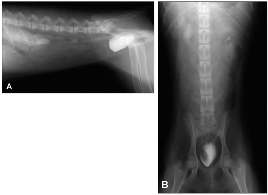
On physical examination, extensive urine staining of the coat was evident in the perineum and caudal aspect of the hind limbs. No other abnormalities were found on physical examination. Abdominal radiography, CBC, serum biochemical analysis, and urinalysis were performed. Bacterial cystitis was the only abnormality found, and aerobic bacterial culture of urine and susceptibility testing were performed to select an appropriate antimicrobial for treatment. Excretory urography was performed (Figure 1).
Figure 1.
Right lateral (A) and ventrodorsal (B) excretory urographic views of a 6-month-old sexually intact female Labrador Retriever–Standard Poodle crossbred dog evaluated for incontinence.
Diagnostic Imaging Findings and Interpretation
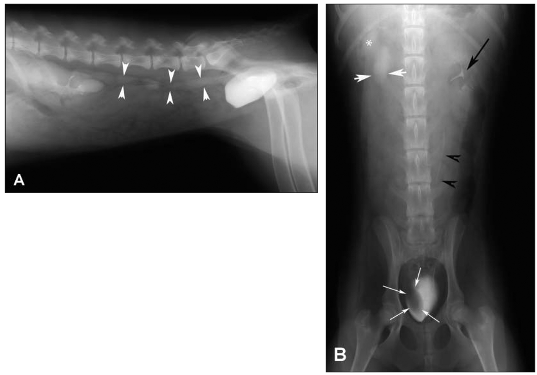
No abnormalities were identified on survey abdominal radiography (images not shown). During the pyelogram phase of excretory urography, the left renal pelvis is normal in appearance (Figure 2). A small volume of contrast medium is identifiable along the left ureter indicating patency, normal diameter, and normal function (peristalsis is shown by intermittent regions lacking contrast that vary between images). The right renal pelvis is distended and poorly defined in part because of poor collecting system opacification. The right ureter is dilated and slightly tortuous, and opacification is less than within the left ureter. There is a filling defect in the right side of the urinary bladder lumen in the region of the right ureteral orifice. These findings are most consistent with a right-sided intraluminal ureterocele with secondary hydronephrosis. A urinary bladder mass with subsequent ureteral obstruction cannot be ruled out on the basis of these findings alone; however, the history of incontinence in a young, otherwise healthy dog supports the diagnosis of an ectopic ureterocele.
Figure 2.
Same excretory urographic images as in Figure 1. Notice that the right renal pelvis is dilated (white asterisk), the right ureter is dilated (white arrowheads), and there is a filling defect within the right side of the urinary bladder lumen (white arrows). The left renal pelvis (black arrow) and left ureter (black arrowheads) appear normal.
Comments
Exploratory surgery confirmed the suspected diagnosis; a large ureterocele was present within the urinary bladder lumen at the location of the right ureteral orifice. The dilation continued caudally within the submucosa of the trigone and terminated in the pelvic urethra. Severe right-sided hydroureter and hydronephrosis were apparent. Given the severity of hydronephrosis, evidence of a poorly functioning kidney on excretory urography, and history of chronic urinary tract infection, a right-sided nephroureterectomy was performed. The ureterocele and ectopic portion of the right ureter were removed, and urethral-trigonal reconstruction was performed. The dog was urinating normally within 2 days after surgery and was considered continent within 2 weeks.
A ureterocele is a cystic dilatation of the submucosal segment of the intravesicular portion of the ureter. It can be orthotopic (entirely intravesicular) or ectopic if any portion of the ureterocele is situated within the urinary bladder neck or urethra. Ureteroceles are often visualized by use of excretory urography as either a positive contrast so-called cobra head dilatation within the urinary bladder or a negative filling defect (with lack of contrast) within the urinary bladder associated with impaired renal function.1 Other diagnostic tests that are useful (particularly when the ureterocele is filled with contrast on excretory urography) include ultrasonography, cystoscopy, contrast-enhanced computed tomography, and urodynamic evaluation.2,3 Additional diagnostic testing may have been helpful in this dog to further characterize the anatomy of the ureterocele and ectopic ureter as well as to evaluate the functional status of both kidneys; however, funding was not available.
Incontinence associated with ectopic ureters and ureteroceles may result from the ectopic position of the ureteral orifice, disruption of the urethral sphincter mechanism, or both. Removal of the urethral portion of the ectopic ureter has been recommended to reduce complications of surgery, including persistent urinary tract infection and incontinence.1,4 In the dog of this report, the ectopic portion of the ureter was removed; the ureter was extremely dilated, occupying > 50% of the urethral diameter at the level of the urinary bladder neck. Complete urinary continence was achieved in this particular dog without further complications.
References
- 1.McLoughlin MA, Hauptman JG, Spaulding K. Canine ureteroceles: a case report and literature review. J Am Anim Hosp Assoc. 1989;25:699–706. [Google Scholar]
- 2.Sutherland-Smith J, Jerram RM, Walker AM, et al. Ectopic ureters and ureteroceles in dogs: presentation, cause, and diagnosis. Compend Contin Educ Pract Vet. 2004;26:303–310. [Google Scholar]
- 3.Samii VF, McLoughlin MA, Mattoon JS, et al. Digital fluoroscopic excretory urography, digital fluoroscopic urethrography, helical computed tomography, and cystoscopy in 24 dogs with suspected ureteral ectopia. J Vet Intern Med. 2004;18:271–281. doi: 10.1892/0891-6640(2004)18<271:dfeudf>2.0.co;2. [DOI] [PubMed] [Google Scholar]
- 4.McLoughlin MA, Chew DJ. Diagnosis and surgical management of ectopic ureters. Clin Tech Small Anim Pract. 2000;15:17–24. doi: 10.1053/svms.2000.7302. [DOI] [PubMed] [Google Scholar]


