Abstract
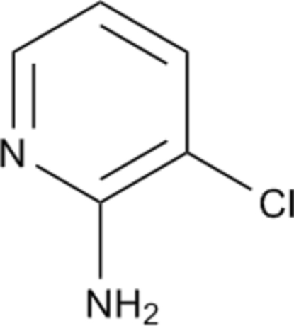
In the title compound, C5H5ClN2, a by-product in the synthesis of ethyl 2-(3-chloropyridin-2-yl)-5-oxopyrazolidine-3-carboxylate, the amine groups form intermolecular hydrogen-bonding associations with pyridine N-atom acceptors, giving centrosymmetric cyclic dimers. Short intermolecular Cl⋯Cl interactions [3.278 (3) Å] also occur.
Related literature
The title compound was isolated as a by-product in the preparation of ethyl 2-(3-chloropyridin-2-yl)-5-oxopyrazolidine-3-carboxylate, an intermediate in the synthesis of the insecticide chlorantraniliprole (systematic name 3-bromo-N-[4-chloro-2-methyl-6-[(methylamino)carbonyl]phenyl]-1-(3-chloro-2-pyridinyl)-1H-pyrazole-5-carboxamide), see: Lahm et al. (2005 ▶). For related structures, see: Chao et al. (1975 ▶); Anagnostis & Turnbull (1998 ▶); Hemamalini & Fun (2010 ▶).
Experimental
Crystal data
C5H5ClN2
M r = 128.56
Monoclinic,

a = 11.149 (8) Å
b = 5.453 (4) Å
c = 9.844 (7) Å
β = 90.581 (12)°
V = 598.5 (7) Å3
Z = 4
Mo Kα radiation
μ = 0.52 mm−1
T = 296 K
0.38 × 0.32 × 0.22 mm
Data collection
Bruker SMART CCD area-detector diffractometer
Absorption correction: multi-scan (SADABS; Bruker, 2001 ▶) T min = 0.827, T max = 0.894
2778 measured reflections
1057 independent reflections
867 reflections with I > 2σ(I)
R int = 0.048
Refinement
R[F 2 > 2σ(F 2)] = 0.059
wR(F 2) = 0.182
S = 1.05
1057 reflections
73 parameters
H-atom parameters constrained
Δρmax = 0.57 e Å−3
Δρmin = −0.31 e Å−3
Data collection: SMART (Bruker, 2001 ▶); cell refinement: SAINT (Bruker, 2001 ▶); data reduction: SAINT; program(s) used to solve structure: SHELXS97 (Sheldrick, 2008 ▶); program(s) used to refine structure: SHELXL97 (Sheldrick, 2008 ▶); molecular graphics: SHELXTL (Sheldrick, 2008 ▶); software used to prepare material for publication: SHELXTL.
Supplementary Material
Crystal structure: contains datablocks I, global. DOI: 10.1107/S1600536811013432/zs2107sup1.cif
Structure factors: contains datablocks I. DOI: 10.1107/S1600536811013432/zs2107Isup2.hkl
Additional supplementary materials: crystallographic information; 3D view; checkCIF report
Table 1. Hydrogen-bond geometry (Å, °).
| D—H⋯A | D—H | H⋯A | D⋯A | D—H⋯A |
|---|---|---|---|---|
| N2—H2A⋯N1i | 0.86 | 2.22 | 3.051 (5) | 162 |
Symmetry code: (i)  .
.
supplementary crystallographic information
Comment
The structures of salts of the halo-substituted aminopyridine, such as 2-amino-5-chloropyridine-fumaric acid (Hemamalini & Fun, 2010), 2-amino-3,5-dichloropyridinium chloride monohydrate (Anagnostis & Turnbull, 1998), are known but the the structure of 2-amino-3-chloropyridine is not known. This compound, C5H5Cl1N2 (I) was isolated as a by-product in the preparation of ethyl 2-(3-chloropyridin-2-yl)-5-oxopyrazolidine-3-carboxylate, an important intermediate in the synthesis of the insecticide chlorantraniliprole (3-bromo-N-[4-chloro-2-methyl-6-[(methylamino) carbonyl]phenyl]-1-(3-chloro-2-pyridinyl)-1H-pyrazole-5-carboxamide) (Lahm et al., 2005). In the structure of (I) (Fig. 1), intermolecular amine N—H···Npyridine hydrogen-bonding interactions (Table 1) give centrosymmetric cyclic dimers (Fig. 2), similar to those found in the structure of 2-aminopyridine (Chao et al., 1975). In (I) there is an intramolecular N—H···Cl interaction [3.001 (3) Å] while in the crystal structure there are also short Cl···Clii interactions [3.278 (3) Å] [symmetry code: (ii) -x + 2, -y, -z + 1].
Experimental
Sodium ethoxide (3.48 g, 50.4 mmol) and 150 ml of absolute ethanol was heated to reflux, after wich 6.80 g (47.4 mmol) of 3-chloro-2-hydrazinylpyridine was added and the mixture was allowed to reflux for 5 minutes. The slurry was then treated dropwise with 9.79 g (56.9 mmol) of diethyl maleate over a period of 5 minutes and the resulting solution was held at reflux for 10 minutes. After cooling to 338 K, the reaction mixture was treated with 5.0 ml (87.3 mmol) of glacial acetic acid. The mixture was diluted with 60 ml water and then cooled to room temperature, giving a precipitate which was isolated via filtration, and separated by column chromatography on silica gel (eluent: ethyl acetate/petroleum ether, 1:5). The title compound was obtained as a yellow solid (0.60 g, 8%) and recyrstallized from dichloromethane to afford colorless single crystals suitable for X-ray diffraction. Anal.: Calc. for C5H5Cl1N2: C, 46.47; H, 3.84; Cl, 27.96; N, 21.85%. Found: C, 46.71; H, 3.99; Cl, 27.58; N, 21.79. 1H NMR(CDCl3): 5.02(s,2H, NH2), 6.62(dd,1H, pyridine-H), 7.48(dd, 1H, pyridine-H), 7.98 (dd, 1H, pyridine-H).
Refinement
Although all H atoms were visible in difference maps, they were placed in geometrically calculated positions, with N—H and C—H = o.86 and 0.93 Å respectively, and included in the final refinement in the riding model approximation, with Uiso(H) = 1.2Ueq(C).
Figures
Fig. 1.
The molecular structure of (I), showing atom numbering scheme and 30% probability displacement ellipsoids.
Fig. 2.
The packing of (I) in ther unit cell viewed down b, showing hydrogen-bonding interactions as dashed lines.
Crystal data
| C5H5ClN2 | F(000) = 264 |
| Mr = 128.56 | Dx = 1.427 Mg m−3 |
| Monoclinic, P21/c | Mo Kα radiation, λ = 0.71073 Å |
| Hall symbol: -P 2ybc | Cell parameters from 1473 reflections |
| a = 11.149 (8) Å | θ = 3.7–27.2° |
| b = 5.453 (4) Å | µ = 0.52 mm−1 |
| c = 9.844 (7) Å | T = 296 K |
| β = 90.581 (12)° | Block, yellow |
| V = 598.5 (7) Å3 | 0.38 × 0.32 × 0.22 mm |
| Z = 4 |
Data collection
| Bruker SMART CCD area-detector diffractometer | 1057 independent reflections |
| Radiation source: fine-focus sealed tube | 867 reflections with I > 2σ(I) |
| graphite | Rint = 0.048 |
| φ and ω scans | θmax = 25.0°, θmin = 1.8° |
| Absorption correction: multi-scan (SADABS; Bruker, 2001) | h = −13→11 |
| Tmin = 0.827, Tmax = 0.894 | k = −6→6 |
| 2778 measured reflections | l = −8→11 |
Refinement
| Refinement on F2 | Primary atom site location: structure-invariant direct methods |
| Least-squares matrix: full | Secondary atom site location: difference Fourier map |
| R[F2 > 2σ(F2)] = 0.059 | Hydrogen site location: inferred from neighbouring sites |
| wR(F2) = 0.182 | H-atom parameters constrained |
| S = 1.05 | w = 1/[σ2(Fo2) + (0.1147P)2 + 0.2179P] where P = (Fo2 + 2Fc2)/3 |
| 1057 reflections | (Δ/σ)max < 0.001 |
| 73 parameters | Δρmax = 0.57 e Å−3 |
| 0 restraints | Δρmin = −0.31 e Å−3 |
Special details
| Geometry. All e.s.d.'s (except the e.s.d. in the dihedral angle between two l.s. planes) are estimated using the full covariance matrix. The cell e.s.d.'s are taken into account individually in the estimation of e.s.d.'s in distances, angles and torsion angles; correlations between e.s.d.'s in cell parameters are only used when they are defined by crystal symmetry. An approximate (isotropic) treatment of cell e.s.d.'s is used for estimating e.s.d.'s involving l.s. planes. |
| Refinement. Refinement of F2 against ALL reflections. The weighted R-factor wR and goodness of fit S are based on F2, conventional R-factors R are based on F, with F set to zero for negative F2. The threshold expression of F2 > σ(F2) is used only for calculating R-factors(gt) etc. and is not relevant to the choice of reflections for refinement. R-factors based on F2 are statistically about twice as large as those based on F, and R- factors based on ALL data will be even larger. |
Fractional atomic coordinates and isotropic or equivalent isotropic displacement parameters (Å2)
| x | y | z | Uiso*/Ueq | ||
| Cl1 | 0.89884 (8) | 0.20978 (18) | 0.46576 (10) | 0.0821 (5) | |
| N1 | 0.6085 (2) | 0.5920 (5) | 0.3676 (2) | 0.0597 (7) | |
| N2 | 0.6357 (3) | 0.2720 (5) | 0.5172 (3) | 0.0716 (8) | |
| H2A | 0.5613 | 0.2836 | 0.5385 | 0.086* | |
| H2B | 0.6804 | 0.1628 | 0.5553 | 0.086* | |
| C1 | 0.6825 (2) | 0.4252 (5) | 0.4237 (3) | 0.0505 (7) | |
| C2 | 0.8035 (2) | 0.4167 (5) | 0.3855 (3) | 0.0535 (7) | |
| C3 | 0.8465 (3) | 0.5728 (6) | 0.2897 (3) | 0.0635 (8) | |
| H3 | 0.9266 | 0.5667 | 0.2645 | 0.076* | |
| C4 | 0.7692 (3) | 0.7404 (7) | 0.2306 (3) | 0.0725 (10) | |
| H4 | 0.7955 | 0.8481 | 0.1640 | 0.087* | |
| C5 | 0.6520 (3) | 0.7431 (6) | 0.2735 (4) | 0.0698 (9) | |
| H5 | 0.6000 | 0.8571 | 0.2345 | 0.084* |
Atomic displacement parameters (Å2)
| U11 | U22 | U33 | U12 | U13 | U23 | |
| Cl1 | 0.0717 (7) | 0.0833 (7) | 0.0913 (8) | 0.0322 (4) | 0.0101 (5) | 0.0145 (4) |
| N1 | 0.0545 (13) | 0.0566 (14) | 0.0681 (15) | 0.0068 (11) | 0.0018 (10) | 0.0047 (11) |
| N2 | 0.0674 (16) | 0.0602 (16) | 0.088 (2) | 0.0131 (12) | 0.0194 (14) | 0.0196 (13) |
| C1 | 0.0572 (14) | 0.0411 (13) | 0.0533 (15) | 0.0036 (11) | 0.0026 (11) | −0.0039 (11) |
| C2 | 0.0558 (15) | 0.0510 (15) | 0.0537 (15) | 0.0099 (11) | 0.0019 (11) | −0.0057 (12) |
| C3 | 0.0561 (15) | 0.076 (2) | 0.0583 (17) | −0.0019 (14) | 0.0070 (13) | 0.0002 (14) |
| C4 | 0.077 (2) | 0.074 (2) | 0.067 (2) | −0.0049 (15) | 0.0047 (17) | 0.0178 (15) |
| C5 | 0.073 (2) | 0.0617 (19) | 0.074 (2) | 0.0057 (14) | −0.0054 (16) | 0.0155 (15) |
Geometric parameters (Å, °)
| Cl1—C2 | 1.735 (3) | C2—C3 | 1.361 (4) |
| N1—C5 | 1.334 (4) | C3—C4 | 1.380 (4) |
| N1—C1 | 1.344 (4) | C3—H3 | 0.9300 |
| N2—C1 | 1.351 (4) | C4—C5 | 1.378 (5) |
| N2—H2A | 0.8600 | C4—H4 | 0.9300 |
| N2—H2B | 0.8600 | C5—H5 | 0.9300 |
| C1—C2 | 1.405 (4) | ||
| C5—N1—C1 | 118.5 (3) | C2—C3—C4 | 118.9 (3) |
| C1—N2—H2A | 120.0 | C2—C3—H3 | 120.6 |
| C1—N2—H2B | 120.0 | C4—C3—H3 | 120.6 |
| H2A—N2—H2B | 120.0 | C5—C4—C3 | 117.9 (3) |
| N1—C1—N2 | 117.3 (3) | C5—C4—H4 | 121.0 |
| N1—C1—C2 | 120.0 (2) | C3—C4—H4 | 121.0 |
| N2—C1—C2 | 122.7 (2) | N1—C5—C4 | 124.0 (3) |
| C3—C2—C1 | 120.7 (3) | N1—C5—H5 | 118.0 |
| C3—C2—Cl1 | 120.2 (2) | C4—C5—H5 | 118.0 |
| C1—C2—Cl1 | 119.0 (2) | ||
| C5—N1—C1—N2 | −179.0 (3) | C1—C2—C3—C4 | 0.1 (5) |
| C5—N1—C1—C2 | 1.5 (4) | Cl1—C2—C3—C4 | −178.0 (2) |
| N1—C1—C2—C3 | −1.3 (4) | C2—C3—C4—C5 | 0.9 (5) |
| N2—C1—C2—C3 | 179.2 (3) | C1—N1—C5—C4 | −0.6 (5) |
| N1—C1—C2—Cl1 | 176.8 (2) | C3—C4—C5—N1 | −0.7 (5) |
| N2—C1—C2—Cl1 | −2.6 (4) |
Hydrogen-bond geometry (Å, °)
| D—H···A | D—H | H···A | D···A | D—H···A |
| N2—H2A···N1i | 0.86 | 2.22 | 3.051 (5) | 162 |
| N2—H2B···Cl1 | 0.86 | 2.61 | 3.001 (4) | 109 |
Symmetry codes: (i) −x+1, −y+1, −z+1.
Footnotes
Supplementary data and figures for this paper are available from the IUCr electronic archives (Reference: ZS2107).
References
- Anagnostis, J. & Turnbull, M. M. (1998). Acta Cryst. C54, 681–683.
- Bruker (2001). SMART, SAINT and SADABS Bruker AXS Inc., Madison, Wisconsin, USA.
- Chao, M., Schemp, E. & Rosenstein, R. D. (1975). Acta Cryst. B31, 2922–2924.
- Hemamalini, M. & Fun, H.-K. (2010). Acta Cryst. E66, o1416–o1417. [DOI] [PMC free article] [PubMed]
- Lahm, G. P., Selby, T. P. & Freudenberger, J. H. (2005). Bioorg. Med. Chem. Lett. 15, 4898–4906. [DOI] [PubMed]
- Sheldrick, G. M. (2008). Acta Cryst. A64, 112–122. [DOI] [PubMed]
Associated Data
This section collects any data citations, data availability statements, or supplementary materials included in this article.
Supplementary Materials
Crystal structure: contains datablocks I, global. DOI: 10.1107/S1600536811013432/zs2107sup1.cif
Structure factors: contains datablocks I. DOI: 10.1107/S1600536811013432/zs2107Isup2.hkl
Additional supplementary materials: crystallographic information; 3D view; checkCIF report


