

Synopsis
This review provides an overview of the assays that are used for measuring escape and avoidance behavior in zebrafish, with a specific focus on zebrafish larvae during the first week of development. Zebrafish larvae display a startle response when exposed to tactile, acoustic, or visual stimuli and will avoid dark areas, moving objects, conspecifics, and open spaces. Emotional states such as fear and anxiety may be induced when larvae are exposed to stimuli that they would normally escape from or avoid. While these emotional states may differ between species and change during development, much can be learned about human fear and anxiety using zebrafish as a model system. The molecular mechanisms of fear and anxiety are highly conserved in vertebrates and are present during early zebrafish development. Larvae during the first week of development display elevated cortisol levels in response to stress and are sensitive to the same anxiolytics that are used for the management of anxiety in humans. Zebrafish larvae are well suited for high-throughput analyses of behavior, and automated systems have been developed for imaging and analyzing the behavior of zebrafish larvae in multiwell plates. These high-throughput analyses will not only provide a wealth of information on the genes and environmental factors that influence escape and avoidance behaviors and the emotional states that may accompany them, but will also facilitate the discovery of novel pharmaceuticals that may be used in the management of anxiety disorders in humans.
Keywords: zebrafish, Danio rerio, behavior, escape, avoidance, fear, anxiety, high-throughput imaging, small molecule screen
Escape, avoidance, fear, and anxiety
In humans, emotional states like fear and anxiety function to avert situations that may be harmful to our health or overall well-being. For example, anxiety can make someone avoid an area with dangerous animals or be more careful crossing a road. However, when deployed excessively or inappropriately, these ancient aids to survival can become counter-productive or contribute to clinically defined anxiety disorders, including generalized anxiety disorder, obsessive-compulsive disorder, panic disorder, post-traumatic stress disorder, and phobias (Craske et al., 2009; Craske and Waters, 2005; Hovatta and Barlow, 2008; LeDoux, 2007; Phelps and LeDoux, 2005). The biology of anxiety is complex, as it involves several regions in the brain, a network of neural connections, and multiple signaling pathways (Denver, 2009; Gerlai, 2010; Maren, 2007). Several of these signaling pathways converge into the activation of the adrenal glands, which are hormone secreting glands on top of the kidneys. The cortex of the adrenal gland produces corticosteroids including cortisol, a common vertebrate stress hormone that regulates a broad range of systems, including glucose metabolism, ionic homeostasis, immune function, and behavior (Alsop and Vijayan, 2009; Denver, 2009). The medulla of the adrenal gland produces the catecholamines epinephrine (adrenaline) and norepinephrine (noradrenaline), which are key signals in the ‘fight-or-flight’ response (Wortsman, 2002).
In this review, we use the term ‘escape’ to describe movements away from an imminent threat (Fig 1A). The term ‘avoidance’ (Fig 1B) is used to describe movements away from an anticipated threat (active avoidance) and movements that maintain a distance from an anticipated threat (passive avoidance). Aversive stimuli may also trigger the emotions of fear and anxiety (Craske et al., 2009; Craske and Waters, 2005; Gerlai, 2010; Hovatta and Barlow, 2008; LeDoux, 2007; Phelps and LeDoux, 2005). We use the term ‘fear’ to describe an internal state characterized by changes in physiology which occur when an organism is confronted with an impending threat or physically present danger. The term ‘anxiety’ is used more restrictively to describe an internal state characterized by changes in physiology which occur when an organism anticipates a threat, often defined clinically as a non-specific threat. Fear and anxiety are thought to be important for the motivation of avoidance behaviors.
Fig 1.

Diagram of escape and avoidance behavior in zebrafish. A) Escape: zebrafish are exposed to an imminent threat and swim away to safety. B) Avoidance: zebrafish anticipate a potential threat and swim away to safety. The fish may dive to the bottom of the tank, swim to a sheltered area, or join a shoal of fish. Once the fish relax, they start to explore their surroundings. In an experimental setup, the threatening stimulus can be adjusted and the tank is typically imaged to determine the exact location and movements of the fish. Thus, quantitative data can be obtained for escape, avoidance, and exploratory behaviors. Emotional states, such as fear, anxiety, and relaxation are more difficult to measure. In general, fear is associated with an imminent threat and anxiety with an anticipated and usually nonspecific threat. While these emotional states likely differ between species and change during development, much can be learned about human fear and anxiety using zebrafish as a model system. The molecular mechanisms of fear and anxiety are highly conserved in vertebrates and are present during early development. In addition, zebrafish escape and avoidance behaviors are modulated by the same anxiolytics that are used in the treatment of human anxiety disorders.
Escape and avoidance behaviors, and the associated emotions of fear and anxiety, can be observed in all vertebrates and probably evolved hundreds of millions of years ago to evade predators or avert other potential dangers (Denver, 2009; LeDoux, 2007; Phelps and LeDoux, 2005; Salas et al., 2006). For example, the capacity to synthesize corticosteroids, such as cortisol, appears to be unique to vertebrates and likely arose prior to the origin of the jawed vertebrates (Denver, 2009). The conservation of molecular mechanisms involved in physiological responses to danger over hundreds of millions of years suggests that various vertebrate model systems can be used to better understand fear and anxiety in humans.
Zebrafish as a model system to study escape and avoidance behavior
Zebrafish have been used extensively to study the genetics of early vertebrate development. The small tropical fish can be maintained at low cost and a modest colony of fish can produce hundreds of fertilized eggs daily (Westerfield, 2007). The embryos develop externally, are nearly transparent, and are accessible to genetic and experimental manipulations. Development is extremely rapid; in just 24 hours, a fertilized egg develops into a larva with eyes, a beating heart, and tail movements (Kimmel et al., 1995). In contrast, a human embryo at 24 hours of development has just entered the two-cell stage. Large scale mutagenesis screens have revealed a range of genes that are important for development of the zebrafish brain (Driever et al., 1996; Granato et al., 1996; Haffter and Nusslein-Volhard, 1996; Schier et al., 1996) and identified genes can be knocked down by injecting antisense morpholinos (Heasman, 2002). While zebrafish is widely used for developmental studies, it has only recently emerged as model system to study behavior (Brennan, 2011). The majority of studies are focused on behavior in adult zebrafish. However, some behaviors develop early and may be observed in larval zebrafish during the first week of development. Zebrafish larvae are ideally suited for large-scale analyses of vertebrate behavior. The behavior of zebrafish larvae can be monitored in multiwell plates, which allows for a systematic screening of the genes, pharmaceuticals, and environmental toxicants that influence behavior. In addition, there is a wealth of information on the signaling pathways that control neural development and many of the molecular tools that have been developed for studying development can also be used to examine brain function. For medium to high-throughput analyses of behavior, it is critical to use robust assays that can be automated. The following sections discuss the available assays for measuring escape and avoidance behavior in zebrafish, starting with adult zebrafish.
Escape and avoidance behavior in adult zebrafish
Assays for measuring escape and avoidance behavior in adult zebrafish make use of various aversive stimuli, including natural threats, novel environments, light, colors, shapes, and open areas.
1) Natural threats
Adult zebrafish respond strongly to threatening stimuli that are likely encountered in nature. Perhaps the most direct natural threat is the presence of a sympatric predator (Bass and Gerlai, 2008; Gerlai, 2010). Another powerful stimulus is the alarm substance released by injured fish, signaling that something harmful may be close by (Egan et al., 2009; Hall and Suboski, 1995; Jesuthasan and Mathuru, 2008; Parra et al., 2009). Zebrafish may respond to such threatening stimuli by escape and avoidance behaviors, which may include diving, freezing, jumping, erratic movements, or shoaling. Zebrafish also respond to an animated image of a predator fish on a computer screen, providing precise control over the stimulus (Gerlai et al., 2009b). The computer-generated stimuli, together with digital recordings of the response, open the door to automated analyses of anxiety.
2) Novel environments
When adult zebrafish are placed in a novel tank, they dive to the bottom of the tank and gradually explore higher levels of the tank in a 5 minute period. This behavior is quantitatively measured in the novel tank diving test (Bencan and Levin, 2008; Bencan et al., 2009; Levin et al., 2007; Wong et al., 2010). Anxiolytic drugs attenuate the diving behavior (Bencan et al., 2009; Levin, 2011). In addition, discontinuation of alcohol or morphine induced anxiety-like behavior in the novel tank diving test indicating that zebrafish may be used to study withdrawal syndromes (Cachat et al., 2010; Stewart et al., 2011).
3) Light, colors, and shapes
Avoidance behavior and anxiety have also been studied in adult zebrafish using tanks with various colors and shapes. Zebrafish prefer dark areas over light areas (scototaxis). They also avoid dark spots on a white background (Ninkovic and Bally-Cuif, 2006), a dark line on a rotating drum (Fleisch and Neuhauss, 2006; Li and Dowling, 1997), and prefer blue over red in a T-maze (Colwill et al., 2005). A rectangular tank with a light and dark compartment is typically used to examine scototaxis-linked anxiety in adult zebrafish (Maximino et al., 2010a; Maximino et al., 2010b). In nature, scototaxis may contribute to predator avoidance, i.e. adult zebrafish stand out clearly when swimming over a light background and are therefore easier to catch. Zebrafish display anxiety-like behavior when confined to the white background that they would normally avoid. In such confinement experiments, it was found that freezing behavior is a particularly viable measure of anxiety (Blaser et al., 2010).
4) Open areas
The preference for an edge vs. an open area (thigmotaxis) is used in rodents as a measure of anxiety (Komada et al., 2008). Thigmotaxis may also be used as an indicator of anxiety in zebrafish. Adult zebrafish tend to swim along the walls in a circular tank, but spend more time in the center of the tank after injection with fluoromethylhistidine, which lowers histamine levels in the brain (Peitsaro et al., 2003). When control fish, treated with saline, are reintroduced to the tanks 3 days later, they spend more time in the center of the tank than they did on the first day, suggesting that the fish remember the environment and are less anxious. A similar thigmotaxis assay has been used to examine zebrafish with reversed left-right asymmetries in the brain (Dadda et al., 2010). These fish are bolder when inspecting a predator, lose their preference for a clockwise swimming direction, and spend less time in the periphery of an open field. Possibly, the preference for the periphery of the tank is similar to the preference for the bottom of the tank in the novel tank diving test, i.e. in both cases the fish avoid an open area. In conclusion, various assays are available for measuring escape and avoidance behavior in adult zebrafish. However, can these assays be used to better understand fear and anxiety in humans?
Validation of behavioral assays
As briefly mentioned in the examples above, the behavioral assays can be validated using known anxiolytic or anxiogenic drugs (Table 1). This validation is important as it confirms the premise of the assay and examines if the assay can be used for the discovery of novel drugs with medical applications in humans (Gerlai, 2010). For example, zebrafish behavior has been examined after treatment with - or withdrawal from - alcohol (Echevarria et al., 2011; Egan et al., 2009; Fernandes and Gerlai, 2009; Gerlai et al., 2009a; Gerlai et al., 2006; Loucks and Carvan, 2004), cocaine (Darland and Dowling, 2001; Lopez Patino et al., 2008a; Lopez Patino et al., 2008b), nicotine and nicotine antagonists (Bencan and Levin, 2008; Levin et al., 2007), fluoxetine (Prozac) (Egan et al., 2009), Buspirone (Bencan et al., 2009), diazepam (Valium) (Bencan et al., 2009; Lopez Patino et al., 2008a), and caffeine (Egan et al., 2009). In addition to the use of anxiolytic and anxiogenic drugs, endogenous hormones can be measured immediately following the assay. For example, cortisol levels in zebrafish increase after direct or visual contact with a predator (Barcellos et al., 2007) and also after withdrawal from alcohol or morphine (Cachat et al., 2010). Conversely, cortisol levels are reduced by treatment with fluoxetine in the novel tank diving test (Egan et al., 2009). The cortisol measurements show that zebrafish display a physiological stress response when exposed to aversive stimuli (Clark et al., 2011), and indicate that zebrafish and humans share common pathways that regulate anxiety.
Table 1.
Anxiolytic drugs and their effects on zebrafish behavior
| Drug | Trade name |
Type | Effect on zebrafish behavior | Age | Refs |
|---|---|---|---|---|---|
| 1. Anti-anxiety medication (a) | |||||
| Diazepam | Valium | BZD | Increased exploratory behavior in the novel tank diving test Decreased activity in the photomotor response assay | Adult Embryo | (Bencan et al., 2009) (Kokel et al., 2010) |
| Buspirone | Buspar | AZA | Increased exploratory behavior in the novel tank diving test Altered rest-wake profiles: increased rest | Adult Larvae | (Bencan et al., 2009) (Rihel et al., 2010) |
| 2. Antidepressants that are used for treatment of anxiety disorders (a) | |||||
| Fluoxetine | Prozac | SSRI | Increased exploratory behavior in the novel tank diving test Decrease in spontaneous swimming activity Altered rest-wake profiles: increased rest | Adult Larvae Larvae | (Egan et al., 2009; Wong et al., 2010) (Airhart et al., 2007) (Rihel et al., 2010) |
| Imipramine | Tofranil | TCA | Altered rest-wake profiles | Larvae | (Rihel et al., 2010) |
| Phenelzine | Nardil | MAOI | Altered rest-wake profiles | Larvae | (Rihel et al., 2010) |
| 3. Medication to control physical symptoms of anxiety (a) | |||||
| Propranolol | Inderal | BB | Decreased activity in the photomotor response assay | Embryo | (Kokel et al., 2010) |
| 4. Other drugs with anxiolytic properties | |||||
| Nicotine | - | ACR | Increased exploratory behavior in the novel tank diving test Altered rest-wake profiles | Adult Larvae | (Bencan and Levin, 2008; Levin et al., 2007) (Rihel et al., 2010) |
| Alcohol | - | - | Increased exploratory behavior in the novel tank diving test | Adult | (Egan et al., 2009; Wong et al., 2010) |
| Morphine | - | ORA | Increased exploratory behavior in the novel tank diving test | Adult | (Wong et al., 2010) |
| Anxiogenic drugs or exposures (induce anxiety) | |||||
| Caffeine | - | ARI | Freezing, erratic swimming, reduced habituation, and reduced exploratory behavior in the novel tank diving test | Adult | (Egan et al., 2009; Wong et al., 2010) |
| Drug withdrawal | - | - | Reduced exploratory behavior in the novel tank diving test Hyperactivity and stereotypy | Adult Adult | (Cachat et al., 2010) (Lopez Patino et al., 2008a; Lopez Patino et al., 2008b) |
The table shows examples of anxiolytic and anxiogenic drugs and their effects on zebrafish behavior.
National Institute of Mental Health (www.nimh.nih.gov/health/publications/mental-health-medications). BZD = benzodiazepines; bind to GABA receptors. AZA = azapirones; bind to serotonin receptors. SSRI = selective serotonin reuptake inhibitor. TCA = tricyclic antidepressants; inhibit the reuptake of serotonin and norepinephrine and bind to various receptors and ion channels. MAOI = monoamine oxidase inhibitors; suppress the breakdown of the neurotransmitters dopamine, serotonin, and norepinephrine. BB= beta blockers; inhibit the action of epinephrine and norepinephrine on beta-adrenergic receptors. ACR = nicotinic acetylcholine receptor activator. ORA = opioid receptor activator. ARI = adenosine receptor inhibitor.
Unfortunately, cortisol levels cannot be measured in vivo during the behavioral experiments. For behavioral studies it would be interesting to monitor hormone levels, neural activity, or other physiological parameters in real time. While there are technical challenges to such in vivo measurements, we propose that two anxiety-linked physiological parameters might be measured in free-swimming fish during the behavioral assays, i.e. the heart rate and skin color.
Heart rate
To date, the heart rate has not been imaged in free-swimming zebrafish. However, recent innovations in imaging technologies may make this possible in the future. The main challenges for imaging the heart rate in free-swimming adult zebrafish are: 1) that the heart is covered by opaque tissues, and 2) that imaging systems either lack the speed or resolution to monitor the beating heart in a large field of view. The first issue has recently been resolved in the transparent zebrafish line Casper (Wenner, 2009) and the second issue may be resolved by innovations in imaging technologies and data storage in the near future. Alternatively, the heart beat may be monitored in an electrocardiogram or ECG (Milan et al., 2006; Sun et al., 2009; Yu et al., 2010), or by photocardiography, which makes use of near-infrared light that is detected by a phototransistor located outside the body (Yoshida et al., 2009). However, these more invasive techniques may be incompatible with the analysis of normal behavior in free-swimming fish. Once available, imaging techniques that monitor the heart beat in free-swimming adult fish could be used to complement existing behavioral assays and indicate the level of fear or anxiety in response to threatening stimuli.
Skin color
The skin of zebrafish can be lighter or darker depending on the distribution of the melanosomes, which are black melanin-containing pigment granules within the pigment cells, the melanophores (Hirata et al., 2005; Logan et al., 2006). When the melanosomes aggregate the skin is lighter and when the melanosomes disperse the skin is darker. In zebrafish, this phenomenon plays a role in background adaptation. The aggregation of the melanosomes is regulated by melanin-concentrating and melanocyte-stimulating hormones (Logan et al., 2006), and by the alpha2-adrenoceptors, which are known to mediate functions of epinephrine and norepinephrine signaling (Iwashita et al., 2006; Ruuskanen et al., 2005). For example, dexmedetomidine, an alpha2-adrenoceptor agonist, induces a lighter skin color, an effect that is antagonized by atipamezole, an alpha2-adrenoceptor antagonist (Ruuskanen et al., 2005). Similarly, the melanosomes aggregate when zebrafish are placed in water containing epinephrine (Iwashita et al., 2006). The latter study also showed that melanosomes disperse when fish are placed in water containing yohimbine, an alpha2-adrenoceptor antagonist. This dispersal is observed regardless of background color, indicating that this antagonist blocks the endogenous aggregation signal. Since epinephrine and norepinephrine are key signals in the ‘fight or flight’ response, skin color might be useful as a real-time indicator of fear and anxiety in free-swimming zebrafish.
Some behaviors such as shoaling, defending spawning sites, and mating develop later in life and may only be observed in juvenile or adult zebrafish (Miller and Gerlai, 2011). However, a broad range of behaviors can be observed in zebrafish larvae during early stages of development. There is significant interest in using zebrafish larvae in behavioral assays, since the larvae are ideally suited for high-throughput applications.
The development of behavior in zebrafish larvae
Zebrafish larvae hatch from their chorion between 2 and 3 days post-fertilization (dpf) and are 3.1 – 3.5 mm long at this time (Kimmel et al., 1995). Hatched larvae initially lie on their side on the bottom of the tank and occasionally dart forward or circle around. The larvae display a startle response when exposed to abrupt stimuli, including touch, sound, water flow, or light (Burgess and Granato, 2007b; Emran et al., 2008; Fetcho et al., 2008; McHenry et al., 2009; McLean and Fetcho, 2009). At 4 dpf, the larvae are approximately 4 mm long and either lay on the bottom of the tank, or hang heads up on the side of the tank. The swim bladder gradually inflates (Lindsey et al., 2010) and the larvae start swimming around between 4 and 5 dpf. At the same time, larvae will follow moving objects with their eyes, a behavior that is referred to as the optokinetic response or OKR (Brockerhoff, 2006; Maurer et al., 2011). At 5 dpf, the larvae have excellent vision and are actively hunting for food (Neuhauss, 2003). The eyes include four types of cone cells, which give the larvae tetrachromatic color vision ranging from ultraviolet to red (Fleisch and Neuhauss, 2006). The larvae display a spontaneous locomotor repertoire that includes routine turns and slow scoots. In addition, the larvae display a more vigorous C-start and burst swimming to escape from threatening stimuli (Budick and O'Malley, 2000). Zebrafish larvae will swim in the same direction as a pattern of moving stripes, a behavior that is called the optomotor response (OMR). This response depends on input from green and red cones, and can be observed at 6 dpf, but is more robust at 7 dpf (Fleisch and Neuhauss, 2006; Orger and Baier, 2005; Orger et al., 2008; Portugues and Engert, 2009). At 5, 6, and 7 dpf, larvae display diurnal rhythms in activity, i.e. the larvae are more active during the day than they are at night (Prober et al., 2006). These circadian rhythms are regulated by hypocretin expression and by the synthesis and secretion of melatonin, a sleep-inducing hormone produced by the pineal gland in the brain (Appelbaum and Gothilf, 2006; Appelbaum et al., 2009; Cahill, 1996; Kazimi and Cahill, 1999; Zhdanova, 2011; Zhdanova et al., 2001). Zebrafish larvae display left-right asymmetries in brain morphology, which have been linked to asymmetric behavior (Barth et al., 2005; Creton, 2009; Dadda et al., 2010; Facchin et al., 2009; McManus, 2005), boldness, and exploratory behavior (Dadda et al., 2010; Facchin et al., 2008). For example, larvae at 7 dpf prefer a clockwise orientation in a two fish assay, in which the larvae look at each other with their right eye (Creton, 2009). Thus, zebrafish larvae display a range of interesting behaviors during the first week of development. Their sensory and motor systems mature quickly and equip them with the ability to detect and avoid threatening and unpleasant stimuli in their environment.
Escape and avoidance behavior in zebrafish larvae
There are a number of stimuli that have been shown to evoke escape and avoidance behaviors in zebrafish larvae during the first week of development. The properties of these stimuli and their use in behavioral assays to examine fear and anxiety are discussed below. The stimuli include 1) tactile stimuli, acoustic stimuli, and water flow, 2) abrupt changes in light intensity, 3) a gradient of light, 4) moving objects including a bouncing ball and other zebrafish larvae, and 5) open areas.
1) Tactile stimuli, acoustic stimuli, and water flow
Zebrafish larvae display a startle or escape response that can be evoked by gently touching the head or tail. The startle response can be observed at 2 dpf and has been used in a large-scale genetic screen for motility defects (Granato et al., 1996). This screen identified 166 mutants with specific defects in embryonic motility comprising at least 48 genes. At 4 dpf, tactile stimuli induce a rapid escape response away from the stimulus. While the swim frequencies are similar at 2 and 4 dpf (ranging from 20 to 85 Hz), the 4 dpf larvae display a more refined escape response by limiting head movements at lower swim frequencies (McLean and Fetcho, 2009; McLean et al., 2008). The neural mechanisms have been studied in detail and include sensory input that is relayed by the Mauthner cells, and other reticulospinal interneurons, to contralateral motor neurons in the spinal cord (Gahtan et al., 2002; Kohashi and Oda, 2008; Sillar, 2009). The primary motor neurons that drive the fast escape response develop first, and the neurons that drive slower swimming movements are added at more ventral locations in later development (McLean et al., 2007; McLean and Fetcho, 2009). The escape response can also be triggered by water flow at 3–4 dpf (Froehlicher et al., 2009; Kohashi and Oda, 2008) or by acoustic stimuli at 5 and 6 dpf (Burgess and Granato, 2007b; Zeddies and Fay, 2005). Water flow is sensed by the mechanosensory hair cells in the lateral line system and may contribute to a rapid escape when a predator strikes (McHenry et al., 2009).
2) Abrupt changes in light intensity
Zebrafish larvae display a startle response when exposed to a sudden change in light intensity. The startle response is characterized by a brief period of elevated activity and can be induced when the light is abruptly turned off at 3 dpf (Easter and Nicola, 1996) or when the light is abruptly turned on or off at 4 or 5 dpf (Emran et al., 2008). When the light is turned off at 6 dpf, locomotion initially increases to a maximum at 4 minutes and then decreases to a low level at 20 minutes (MacPhail et al., 2009). In contrast, when the light is turned on at 6 dpf, locomotion gradually increases to a stable level at 20 minutes. Acute exposures to 1% or 2% alcohol increased activity in the 6 dpf larvae, while exposure to 4% alcohol decreased activity, suggesting that larval locomotion may be used to examine drugs with anxiolytic properties (MacPhail et al., 2009). To separate anxiolytic effects from defects in vision or defects in muscle contractions, investigators often show that zebrafish with reduced anxiety still have normal functioning sensory and motor systems. Alternatively, it is possible to record multiple behavioral parameters (profiles) and examine which parameters, or combination of parameters, are linked to anxiolytics, but not to other psychoactive compounds. Such multi-parameter investigations have been particularly fruitful in large-scale screens. A recent study shows that the startle response can be induced by a pulse of light at 30 hpf when the embryos are still inside the chorion (Kokel et al., 2010). This pulse of light induces a 5 second period of increased activity, while a second light pulse 10 seconds later does not. This assay was used in a large-scale small molecule screen that showed specific behavioral profiles in response to known anxiolytics such as diazepam. Cluster analysis of the behavioral profiles, combined with structural analyses of the clustered compounds, revealed novel monoamine oxidase inhibitors that could have therapeutic potential for the treatment of depression and anxiety. Thus, zebrafish embryos, at very early stages in development, may be used for the discovery of novel anxiolytics.
3) A gradient of light
Zebrafish larvae at 7 dpf are phototactic, i.e. they prefer a lighter section of the tank over a darker section of the tank (Brockerhoff et al., 1995). The larvae thus display an opposite response to light than adult fish, which prefer darker sections in a tank. Assays for measuring phototaxis were recently optimized in 5–7 dpf larvae by dimming a uniform field of illumination, followed by a 10-fold weaker spot of light (Burgess et al., 2010; Mueller and Neuhauss, 2010). The larvae display a routine turn towards the light followed by a series of slow swims (scoots) in the direction of the light source. The larvae show a maximal aggregation close to the spot light after approximately 60 seconds.
4) Moving objects, including a bouncing ball and other zebrafish larvae
Zebrafish larvae at 6 dpf will swim away from a computer-generated ‘bouncing ball’ (Colwill and Creton, 2010; Creton, 2009). The bouncing ball is created in PowerPoint, and consists of a 2.5 cm diameter disc moving left and right on a computer screen. The computer screen sits flat on a horizontal surface and a dish with zebrafish larvae is placed on top of the screen. Zebrafish larvae swim away from the bouncing ball (escape or active avoidance) and remain at a safe distance from the bouncing ball (passive avoidance). Possibly, the bouncing ball mimics a predator or the shadow of a predator. The dish with zebrafish larvae is imaged by a high-resolution camera and the location and orientation of the larvae is examined by automated image analysis. Advantages of this system are its flexibility for programming stimuli and its suitability for high-throughput applications. The same imaging system was used to measure the interaction of two zebrafish larvae in a 12-well plate (Fig 2). Zebrafish larvae at 7 dpf prefer to swim in opposite quadrants, indicating that the larvae avoid each other’s company (Creton, 2009).
Fig 2.
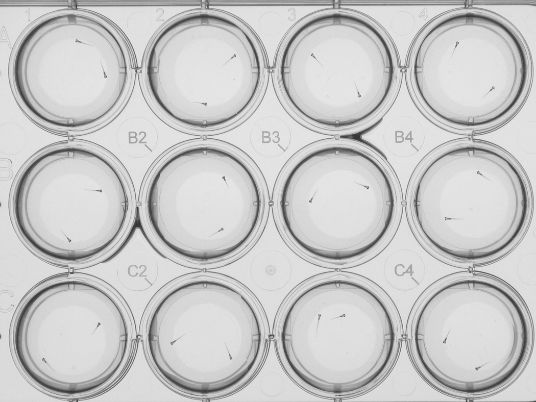
Zebrafish larvae imaged in a 12-well plate at 7 days post-fertilization. The larvae were imaged using a high-resolution imaging system that allows for the automated analysis of larval locations and orientations. For a description of the imaging system see (Colwill and Creton, 2010; Creton, 2009).
5) Open areas
Zebrafish larvae are thigmotactic, i.e. they prefer the edge over the center when imaged in multiwell plates (Colwill and Creton, 2011)). In nature, open areas may expose zebrafish larvae to predators and this exposure could induce an anxiety-like state in the larvae, similar to the anxiogenic effects of open areas in adult zebrafish (Peitsaro et al., 2003) and rodents (Komada et al., 2008). Since the location of larvae can be measured in high-throughput applications, this behavior may be used for the discovery of novel drugs with anxiolytic properties.
Validation of the behavioral assays in zebrafish larvae
The behavioral assays in zebrafish larvae can be validated using drugs with anxiolytic properties, including fluoxetine (Prozac) (Airhart et al., 2007; Rihel et al., 2010), alpha 2-adrenergic receptor agonists, such as clonidine (Rihel et al., 2010), diazepam (Valium), and beta-blockers (Kokel et al., 2010). The main classes of medical anxiolytics are shown in Table 1, each with a few examples of the observed effects on zebrafish behavior. Overall, larval behavior may be altered by hundreds or even thousands of compounds as shown by behavioral analyses in small molecule screens (Kokel et al., 2010; Rihel et al., 2010). In such high-throughput screens, the observed behavioral profiles can be clustered to link specific behaviors with known anxiolytics. The cluster analysis is an elegant approach for identifying which behaviors are affected by anxiolytics and not by other psychoactive compounds, such as sedatives or mood stabilizers. Moreover, the cluster analysis can reveal novel compounds with anxiolytic properties, and thus has value in drug discovery. In addition to the pharmacological validation, it is possible to evaluate the level of anxiety in the behavioral assays by measuring endogenous hormone levels. Interestingly, zebrafish produce cortisol under control of the hypothalamus–pituitary–interrenal (HPI) axis during early development (Alsop and Vijayan, 2009). Corticotrope deficiency leads to lowered cortisol levels in 6 dpf zebrafish larvae (Dickmeis et al., 2007). In addition, cortisol concentrations increase approximately 2-fold when 4 dpf zebrafish larvae are exposed to rapid swirling (Alsop and Vijayan, 2008, 2009) or osmotic stress (Alderman and Bernier, 2009). While the cortisol levels can only be measured in homogenized fish, other parameters might be measured in real time in living free-swimming larvae. A particularly promising approach is the use of fluorescent or bioluminescent zebrafish lines that emit blue, green, or red light in response to specific signals (Naumann et al., 2010; Wyart and Del Bene, 2011). In addition, it may be possible to image the heart beat and skin color, as discussed above for adult zebrafish.
Heart beat
The beating heart is visible in zebrafish larvae and image analysis software can be used to examine specific parameters of the heart beat. Norepinephrine added to the water increased the wall motion amplitude in 4 day old larvae, suggesting that larvae are sensitive to this hormone at early stages in development (Denvir et al., 2008). In 5–7 dpf larvae, the heart rate can be modulated by mild electric shocks (Mann et al., 2010), which first induce a slow heart rate (bradycardia) followed by a fast heart rate (tachycardia). The observed tachycardia can be blocked by the beta-blocker propranolol, suggesting that adrenergic signals control the heartbeat in larval fish (Mann et al., 2010). Apart from imaging the heart beat by transmitted light microscopy, it is also possible to image the beating heart in thin optical sections using advanced fluorescence imaging techniques. For example, transgenic zebrafish that express DsRed in the blood cells and myocardium and EGFP in the endocardium have been imaged in vivo by selective plane illumination microscopy (SPIM), providing a high-resolution view of the tissues that constitute the heart (Huisken and Stainier, 2009). In each of the examples above, the heartbeat was imaged in immobilized larvae. While technically challenging, it would be interesting to image the heart beat during behavioral assays in free-swimming larvae. Such experiments could provide new insights in the physiology of fear and anxiety associated with escape and avoidance behaviors.
Skin color
Skin color might be used as another measure of anxiety. As discussed above, adult zebrafish display a lighter color in response to epinephrine or a white background. Zebrafish larvae at 5–7 dpf also adjust their pigment in response to the background, which can be used as a first hint to potential blindness in large-scale screens (Balm and Groneveld, 1998; Fleisch and Neuhauss, 2006; Neuhauss, 2003; Peng et al., 2009). The changes in color may in part be regulated by epinephrine / alpha2-adenoceptor signaling, similar to the regulation of skin color adult fish (Iwashita et al., 2006; Ruuskanen et al., 2005). Interestingly, when larvae are exposed to alcohol, the melanosomes disperse throughout the cytoplasm resulting in a dark appearance (Peng et al., 2009). We propose that such changes in color could be measured in real time in free-swimming larvae to correlate specific swimming behaviors with elevated levels of anxiety.
Automated imaging of escape and avoidance behaviors in zebrafish larvae
Elegant imaging systems have been developed for the automated analysis of behavior in zebrafish larvae. Some of these systems are now commercially available. For example, both Noldus and ViewPoint have developed efficient imaging systems for the automated analysis of larval activity in 96-well plates. The automated imaging system developed by Noldus Information Technology includes a DanioVision observation chamber and EthoVision software (see http://www.noldus.com). The EthoVision software has for example been used to isolate seizure-resistant zebrafish larvae in a large-scale mutagenesis screen (Baraban et al., 2007) and to examine the effects of lighting conditions and alcohol on larval activity (MacPhail et al., 2009). The automated imaging system developed by ViewPoint includes a ZebraBox imaging system and ZebraLab software (see http://www.viewpointlifesciences.com). This system has been used for measuring circadian rhythms in zebrafish larvae (Prober et al., 2006) and for a high-throughput small molecule screen to identify drugs that alter sleep-wake behaviors of zebrafish larvae (Rihel et al., 2010). For practical information on how to use this system for measuring larval locomotor activity, see the video published by Emran et al. (Emran et al., 2008). The Noldus and ViewPoint systems are both equipped with infrared illumination for imaging in the dark and can thus be used to examine the effect of light-dark cycles in a multiwell format. This feature also enables both systems to be used to monitor changes in sleep patterns that may co-occur with chronic anxiety and stress. The imaging systems record larval activity in all 96 wells simultaneously, using one larva per well. Future developments may make it possible to automatically track multiple larvae in a single well, to acquire information on larval orientation, and to provide local stimuli in the multiwell plates. These innovations would pave the way for automated analyses of social behaviors and more detailed analyses of the swimming patterns in response to specific threats and stimuli previously associated with these threats.
Various laboratories have developed custom-built systems for automated measurements of larval location, orientation, and bending angles. For example, Burgess and Granato developed an imaging system with a high-speed 1000 frames per second camera and custom-written kinematic analysis software, to identify the position, orientation, and curvature of 6–7 dpf larvae during the startle response (Burgess and Granato, 2007a, b). Approximately 30 larvae were imaged simultaneously for a 0.4 – 1 second period in a 6 cm dish. The images are acquired at a sufficient resolution (0.25 megapixels) to identify the orientation of the tail, trunk, and head segments by automated image analysis. A high-speed 1000 frames per second camera has also been used to examine the escape response of individual larvae triggered by touching the tail (McLean et al., 2008). In this case, the bending at different places along the body was automatically quantified by custom-written software that tracked three regions along the midline of the 4–5 dpf zebrafish larvae. Thus, both systems are geared towards brief periods of highspeed imaging to quantify parameters of the fast escape response. On the other side of the spectrum is a high-resolution imaging system that monitors zebrafish larvae in time-lapse mode using a 9 megapixel camera (Colwill and Creton, 2010; Creton, 2009). The spatial resolution of this imaging system makes it possible to measure the location and orientation of zebrafish larvae in multiwell plates (Fig 2). In addition, threatening stimuli can be presented to the larvae on an LCD screen, which makes this system well-suited for studying escape and avoidance behaviors. An alternative approach to such high-resolution recordings is to use a microscope and image one well at a time. Such microscopes can be equipped with a motorized stage to automatically move from one well to the next. This approach was used successfully in a largescale screen for small molecules that affect a light-induced startle response in zebrafish embryos (Kokel et al., 2010).
Conclusions and future directions
This is an exciting era for studying zebrafish behavior. The genetic and neural networks that influence behavior are being uncovered at an unprecedented pace. In addition, there have been significant innovations in imaging technologies that can be used for automated analyses of behavior. These advanced imaging technologies are likely to further improve in the near future. For example, innovations in camera hardware and digital storage media will make it possible to image at higher speeds and at higher resolution. The automated analysis of large movie files will likely be faster, and will include more options for analyzing behaviors other than activity. Such innovations will particularly pay off when imaging the behavior of zebrafish larvae in multiwell plates. For example, it should be possible to automate the analysis of social behavior, learning, and memory in a multiwell plate format. The high-resolution movies may contain information beyond the location of the fish. For example, they may reveal rapid changes in skin color and changes in heart beat frequencies that are associated with specific behaviors. Moreover, it may be possible to acquire information on a range of neural and physiological parameters by imaging fluorescent and luminescent indicators. For studies on fear and anxiety it would be major step forward to image the concentrations of cortisol and epinephrine in real time in free swimming fish. Overall, high-throughput analyses of escape and avoidance behavior in zebrafish will likely lead to the identification of novel drugs that can be used in the management of human anxiety disorders. In addition, high-throughput analyses may be used to better understand the genetic and environmental factors that cause anxiety disorders.
Acknowledgements
This work was supported by the Eunice Kennedy Shriver National Institute of Child Health and Human Development (NICHD, R01HD060647) and the National Institute of Environmental Health Sciences (NIEHS, R03ES017755).
References
- Airhart MJ, Lee DH, Wilson TD, Miller BE, Miller MN, Skalko RG. Movement disorders and neurochemical changes in zebrafish larvae after bath exposure to fluoxetine (PROZAC) Neurotoxicol Teratol. 2007;29:652–664. doi: 10.1016/j.ntt.2007.07.005. [DOI] [PubMed] [Google Scholar]
- Alderman SL, Bernier NJ. Ontogeny of the corticotropin-releasing factor system in zebrafish. Gen Comp Endocrinol. 2009;164:61–69. doi: 10.1016/j.ygcen.2009.04.007. [DOI] [PubMed] [Google Scholar]
- Alsop D, Vijayan MM. Development of the corticosteroid stress axis and receptor expression in zebrafish. Am J Physiol Regul Integr Comp Physiol. 2008;294:R711–R719. doi: 10.1152/ajpregu.00671.2007. [DOI] [PubMed] [Google Scholar]
- Alsop D, Vijayan MM. Molecular programming of the corticosteroid stress axis during zebrafish development. Comp Biochem Physiol A Mol Integr Physiol. 2009;153:49–54. doi: 10.1016/j.cbpa.2008.12.008. [DOI] [PubMed] [Google Scholar]
- Appelbaum L, Gothilf Y. Mechanism of pineal-specific gene expression: the role of E-box and photoreceptor conserved elements. Mol Cell Endocrinol. 2006;252:27–33. doi: 10.1016/j.mce.2006.03.021. [DOI] [PubMed] [Google Scholar]
- Appelbaum L, Wang GX, Maro GS, Mori R, Tovin A, Marin W, Yokogawa T, Kawakami K, Smith SJ, Gothilf Y, et al. Sleep-wake regulation and hypocretin-melatonin interaction in zebrafish. Proc Natl Acad Sci U S A. 2009;106:21942–21947. doi: 10.1073/pnas.906637106. [DOI] [PMC free article] [PubMed] [Google Scholar]
- Balm PH, Groneveld D. The melanin-concentrating hormone system in fish. Ann N Y Acad Sci. 1998;839:205–209. doi: 10.1111/j.1749-6632.1998.tb10760.x. [DOI] [PubMed] [Google Scholar]
- Baraban SC, Dinday MT, Castro PA, Chege S, Guyenet S, Taylor MR. A large-scale mutagenesis screen to identify seizure-resistant zebrafish. Epilepsia. 2007;48:1151–1157. doi: 10.1111/j.1528-1167.2007.01075.x. [DOI] [PMC free article] [PubMed] [Google Scholar]
- Barcellos LJG, Ritter F, Kreutz LC, Quevedo RM, da Silva LB, Bedin AC, Finco J, Cericato L. Whole-body cortisol increases after direct and visual contact with a predator in zebrafish, Danio rerio. Aquaculture. 2007;272:774–778. [Google Scholar]
- Barth KA, Miklosi A, Watkins J, Bianco IH, Wilson SW, Andrew RJ. fsizebrafish show concordant reversal of laterality of viscera, neuroanatomy, and a subset of behavioral responses. Curr Biol. 2005;15:844–850. doi: 10.1016/j.cub.2005.03.047. [DOI] [PMC free article] [PubMed] [Google Scholar]
- Bass SL, Gerlai R. Zebrafish (Danio rerio) responds differentially to stimulus fish: the effects of sympatric and allopatric predators and harmless fish. Behav Brain Res. 2008;186:107–117. doi: 10.1016/j.bbr.2007.07.037. [DOI] [PubMed] [Google Scholar]
- Bencan Z, Levin ED. The role of alpha7 and alpha4beta2 nicotinic receptors in the nicotine-induced anxiolytic effect in zebrafish. Physiol Behav. 2008;95:408–412. doi: 10.1016/j.physbeh.2008.07.009. [DOI] [PMC free article] [PubMed] [Google Scholar]
- Bencan Z, Sledge D, Levin ED. Buspirone, chlordiazepoxide and diazepam effects in a zebrafish model of anxiety. Pharmacol Biochem Behav. 2009;94:75–80. doi: 10.1016/j.pbb.2009.07.009. [DOI] [PMC free article] [PubMed] [Google Scholar]
- Blaser RE, Chadwick L, McGinnis GC. Behavioral measures of anxiety in zebrafish (Danio rerio) Behav Brain Res. 2010;208:56–62. doi: 10.1016/j.bbr.2009.11.009. [DOI] [PubMed] [Google Scholar]
- Brennan CH. Zebrafish behavioural assays of translational relevance for neurobehavioural disease. Reviews in the Neurosciences. 2011;22:37–48. doi: 10.1515/RNS.2011.006. [DOI] [PubMed] [Google Scholar]
- Brockerhoff SE. Measuring the optokinetic response of zebrafish larvae. Nat Protoc. 2006;1:2448–2451. doi: 10.1038/nprot.2006.255. [DOI] [PubMed] [Google Scholar]
- Brockerhoff SE, Hurley JB, Janssen-Bienhold U, Neuhauss SC, Driever W, Dowling JE. A behavioral screen for isolating zebrafish mutants with visual system defects. Proc Natl Acad Sci U S A. 1995;92:10545–10549. doi: 10.1073/pnas.92.23.10545. [DOI] [PMC free article] [PubMed] [Google Scholar]
- Budick SA, O'Malley DM. Locomotor repertoire of the larval zebrafish: swimming, turning and prey capture. J Exp Biol. 2000;203:2565–2579. doi: 10.1242/jeb.203.17.2565. [DOI] [PubMed] [Google Scholar]
- Burgess HA, Granato M. Modulation of locomotor activity in larval zebrafish during light adaptation. J Exp Biol. 2007a;210:2526–2539. doi: 10.1242/jeb.003939. [DOI] [PubMed] [Google Scholar]
- Burgess HA, Granato M. Sensorimotor gating in larval zebrafish. J Neurosci. 2007b;27:4984–4994. doi: 10.1523/JNEUROSCI.0615-07.2007. [DOI] [PMC free article] [PubMed] [Google Scholar]
- Burgess HA, Schoch H, Granato M. Distinct retinal pathways drive spatial orientation behaviors in zebrafish navigation. Curr Biol. 2010;20:381–386. doi: 10.1016/j.cub.2010.01.022. [DOI] [PMC free article] [PubMed] [Google Scholar]
- Cachat J, Canavello P, Elegante M, Bartels B, Hart P, Bergner C, Egan R, Duncan A, Tien D, Chung A, et al. Modeling withdrawal syndrome in zebrafish. Behav Brain Res. 2010;208:371–376. doi: 10.1016/j.bbr.2009.12.004. [DOI] [PubMed] [Google Scholar]
- Cahill GM. Circadian regulation of melatonin production in cultured zebrafish pineal and retina. Brain Res. 1996;708:177–181. doi: 10.1016/0006-8993(95)01365-2. [DOI] [PubMed] [Google Scholar]
- Clark KJ, Boczek NJ, Ekker SC. Stressing zebrafish for behavioral genetics. Reviews in the Neurosciences. 2011;22:49–62. doi: 10.1515/RNS.2011.007. [DOI] [PMC free article] [PubMed] [Google Scholar]
- Colwill RM, Creton R. Automated imaging of avoidance behavior in larval zebrafish. In: Kalueff AV, Cachat JM, editors. Neuromethods. 1st Edition. Vol 51. Zebrafish Neurobehavioral Protocols Humana Press, Springer Protocols; 2010. ISBN: 978-1-60761-952-9. [Google Scholar]
- Colwill RM, Creton R. Locomotor behaviors in zebrafish (Danio rerio) larvae. Behav Processes. 2011;86:222–229. doi: 10.1016/j.beproc.2010.12.003. [DOI] [PMC free article] [PubMed] [Google Scholar]
- Colwill RM, Raymond MP, Ferreira L, Escudero H. Visual discrimination learning in zebrafish (Danio rerio) Behav Processes. 2005;70:19–31. doi: 10.1016/j.beproc.2005.03.001. [DOI] [PubMed] [Google Scholar]
- Craske MG, Rauch SL, Ursano R, Prenoveau J, Pine DS, Zinbarg RE. What is an anxiety disorder? Depress Anxiety. 2009;26:1066–1085. doi: 10.1002/da.20633. [DOI] [PubMed] [Google Scholar]
- Craske MG, Waters AM. Panic disorder, phobias, and generalized anxiety disorder. Annu Rev Clin Psychol. 2005;1:197–225. doi: 10.1146/annurev.clinpsy.1.102803.143857. [DOI] [PubMed] [Google Scholar]
- Creton R. Automated analysis of behavior in zebrafish larvae. Behav Brain Res. 2009;203:127–136. doi: 10.1016/j.bbr.2009.04.030. [DOI] [PubMed] [Google Scholar]
- Dadda M, Domenichini A, Piffer L, Argenton F, Bisazza A. Early differences in epithalamic left-right asymmetry influence lateralization and personality of adult zebrafish. Behav Brain Res. 2010;206:208–215. doi: 10.1016/j.bbr.2009.09.019. [DOI] [PubMed] [Google Scholar]
- Darland T, Dowling JE. Behavioral screening for cocaine sensitivity in mutagenized zebrafish. Proc Natl Acad Sci U S A. 2001;98:11691–11696. doi: 10.1073/pnas.191380698. [DOI] [PMC free article] [PubMed] [Google Scholar]
- Denver RJ. Structural and functional evolution of vertebrate neuroendocrine stress systems. Ann N Y Acad Sci. 2009;1163:1–16. doi: 10.1111/j.1749-6632.2009.04433.x. [DOI] [PubMed] [Google Scholar]
- Denvir MA, Tucker CS, Mullins JJ. Systolic and diastolic ventricular function in zebrafish embryos: influence of norepenephrine, MS-222 and temperature. BMC Biotechnol. 2008;8:21. doi: 10.1186/1472-6750-8-21. [DOI] [PMC free article] [PubMed] [Google Scholar]
- Dickmeis T, Lahiri K, Nica G, Vallone D, Santoriello C, Neumann CJ, Hammerschmidt M, Foulkes NS. Glucocorticoids play a key role in circadian cell cycle rhythms. PLoS Biol. 2007;5:e78. doi: 10.1371/journal.pbio.0050078. [DOI] [PMC free article] [PubMed] [Google Scholar]
- Driever W, Solnica-Krezel L, Schier AF, Neuhauss SC, Malicki J, Stemple DL, Stainier DY, Zwartkruis F, Abdelilah S, Rangini Z, et al. A genetic screen for mutations affecting embryogenesis in zebrafish. Development. 1996;123:37–46. doi: 10.1242/dev.123.1.37. [DOI] [PubMed] [Google Scholar]
- Easter SS, Jr, Nicola GN. The development of vision in the zebrafish (Danio rerio) Dev Biol. 1996;180:646–663. doi: 10.1006/dbio.1996.0335. [DOI] [PubMed] [Google Scholar]
- Echevarria DJ, Toms CN, Jouandot DJ. Alcohol induced behavior change in zebrafish models. Reviews in the Neurosciences. 2011;22:85–93. doi: 10.1515/RNS.2011.010. [DOI] [PubMed] [Google Scholar]
- Egan RJ, Bergner CL, Hart PC, Cachat JM, Canavello PR, Elegante MF, Elkhayat SI, Bartels BK, Tien AK, Tien DH, et al. Understanding behavioral and physiological phenotypes of stress and anxiety in zebrafish. Behav Brain Res. 2009;205:38–44. doi: 10.1016/j.bbr.2009.06.022. [DOI] [PMC free article] [PubMed] [Google Scholar]
- Emran F, Rihel J, Dowling JE. A behavioral assay to measure responsiveness of zebrafish to changes in light intensities. J Vis Exp. 2008 doi: 10.3791/923. [DOI] [PMC free article] [PubMed] [Google Scholar]
- Facchin L, Argenton F, Bisazza A. Lines of Danio rerio selected for opposite behavioural lateralization show differences in anatomical left-right asymmetries. Behav Brain Res. 2009;197:157–165. doi: 10.1016/j.bbr.2008.08.033. [DOI] [PubMed] [Google Scholar]
- Facchin L, Burgess HA, Siddiqi M, Granato M, Halpern ME. Determining the function of zebrafish epithalamic asymmetry. Philos Trans R Soc Lond B Biol Sci. 2008 doi: 10.1098/rstb.2008.0234. [DOI] [PMC free article] [PubMed] [Google Scholar]
- Fernandes Y, Gerlai R. Long-term behavioral changes in response to early developmental exposure to ethanol in zebrafish. Alcohol Clin Exp Res. 2009;33:601–609. doi: 10.1111/j.1530-0277.2008.00874.x. [DOI] [PMC free article] [PubMed] [Google Scholar]
- Fetcho JR, Higashijima S, McLean DL. Zebrafish and motor control over the last decade. Brain Res Rev. 2008;57:86–93. doi: 10.1016/j.brainresrev.2007.06.018. [DOI] [PMC free article] [PubMed] [Google Scholar]
- Fleisch VC, Neuhauss SC. Visual behavior in zebrafish. Zebrafish. 2006;3:191–201. doi: 10.1089/zeb.2006.3.191. [DOI] [PubMed] [Google Scholar]
- Froehlicher M, Liedtke A, Groh KJ, Neuhauss SC, Segner H, Eggen RI. Zebrafish (Danio rerio) neuromast: promising biological endpoint linking developmental and toxicological studies. Aquat Toxicol. 2009;95:307–319. doi: 10.1016/j.aquatox.2009.04.007. [DOI] [PubMed] [Google Scholar]
- Gahtan E, Sankrithi N, Campos JB, O'Malley DM. Evidence for a widespread brain stem escape network in larval zebrafish. J Neurophysiol. 2002;87:608–614. doi: 10.1152/jn.00596.2001. [DOI] [PubMed] [Google Scholar]
- Gerlai R. Zebrafish antipredatory responses: a future for translational research? Behav Brain Res. 2010;207:223–231. doi: 10.1016/j.bbr.2009.10.008. [DOI] [PMC free article] [PubMed] [Google Scholar]
- Gerlai R, Chatterjee D, Pereira T, Sawashima T, Krishnannair R. Acute and chronic alcohol dose: population differences in behavior and neurochemistry of zebrafish. Genes Brain Behav. 2009a doi: 10.1111/j.1601-183X.2009.00488.x. [DOI] [PMC free article] [PubMed] [Google Scholar]
- Gerlai R, Fernandes Y, Pereira T. Zebrafish (Danio rerio) responds to the animated image of a predator: towards the development of an automated aversive task. Behav Brain Res. 2009b;201:318–324. doi: 10.1016/j.bbr.2009.03.003. [DOI] [PMC free article] [PubMed] [Google Scholar]
- Gerlai R, Lee V, Blaser R. Effects of acute and chronic ethanol exposure on the behavior of adult zebrafish (Danio rerio) Pharmacol Biochem Behav. 2006;85:752–761. doi: 10.1016/j.pbb.2006.11.010. [DOI] [PMC free article] [PubMed] [Google Scholar]
- Granato M, van Eeden FJ, Schach U, Trowe T, Brand M, Furutani-Seiki M, Haffter P, Hammerschmidt M, Heisenberg CP, Jiang YJ, et al. Genes controlling and mediating locomotion behavior of the zebrafish embryo and larva. Development. 1996;123:399–413. doi: 10.1242/dev.123.1.399. [DOI] [PubMed] [Google Scholar]
- Haffter P, Nusslein-Volhard C. Large scale genetics in a small vertebrate, the zebrafish. Int J Dev Biol. 1996;40:221–227. [PubMed] [Google Scholar]
- Hall D, Suboski MD. Visual and olfactory stimuli in learned release of alarm reactions by zebra danio fish (Brachydanio rerio) Neurobiol Learn Mem. 1995;63:229–240. doi: 10.1006/nlme.1995.1027. [DOI] [PubMed] [Google Scholar]
- Heasman J. Morpholino oligos: making sense of antisense? Dev Biol. 2002;243:209–214. doi: 10.1006/dbio.2001.0565. [DOI] [PubMed] [Google Scholar]
- Hirata M, Nakamura K, Kondo S. Pigment cell distributions in different tissues of the zebrafish, with special reference to the striped pigment pattern. Dev Dyn. 2005;234:293–300. doi: 10.1002/dvdy.20513. [DOI] [PubMed] [Google Scholar]
- Hovatta I, Barlow C. Molecular genetics of anxiety in mice and men. Ann Med. 2008;40:92–109. doi: 10.1080/07853890701747096. [DOI] [PubMed] [Google Scholar]
- Huisken J, Stainier DY. Selective plane illumination microscopy techniques in developmental biology. Development. 2009;136:1963–1975. doi: 10.1242/dev.022426. [DOI] [PMC free article] [PubMed] [Google Scholar]
- Iwashita M, Watanabe M, Ishii M, Chen T, Johnson SL, Kurachi Y, Okada N, Kondo S. Pigment pattern in jaguar/obelix zebrafish is caused by a Kir7.1 mutation: implications for the regulation of melanosome movement. PLoS Genet. 2006;2:e197. doi: 10.1371/journal.pgen.0020197. [DOI] [PMC free article] [PubMed] [Google Scholar]
- Jesuthasan SJ, Mathuru AS. The alarm response in zebrafish: innate fear in a vertebrate genetic model. J Neurogenet. 2008;22:211–228. doi: 10.1080/01677060802298475. [DOI] [PubMed] [Google Scholar]
- Kazimi N, Cahill GM. Development of a circadian melatonin rhythm in embryonic zebrafish. Brain Res Dev Brain Res. 1999;117:47–52. doi: 10.1016/s0165-3806(99)00096-6. [DOI] [PubMed] [Google Scholar]
- Kimmel CB, Ballard WW, Kimmel SR, Ullmann B, Schilling TF. Stages of embryonic development of the zebrafish. Dev Dyn. 1995;203:253–310. doi: 10.1002/aja.1002030302. [DOI] [PubMed] [Google Scholar]
- Kohashi T, Oda Y. Initiation of Mauthner- or non-Mauthner-mediated fast escape evoked by different modes of sensory input. J Neurosci. 2008;28:10641–10653. doi: 10.1523/JNEUROSCI.1435-08.2008. [DOI] [PMC free article] [PubMed] [Google Scholar]
- Kokel D, Bryan J, Laggner C, White R, Cheung CY, Mateus R, Healey D, Kim S, Werdich AA, Haggarty SJ, et al. Rapid behavior-based identification of neuroactive small molecules in the zebrafish. Nat Chem Biol. 2010;6:231–237. doi: 10.1038/nchembio.307. [DOI] [PMC free article] [PubMed] [Google Scholar]
- Komada M, Takao K, Miyakawa T. Elevated plus maze for mice. J Vis Exp. 2008 doi: 10.3791/1088. [DOI] [PMC free article] [PubMed] [Google Scholar]
- LeDoux J. The amygdala. Curr Biol. 2007;17:R868–R874. doi: 10.1016/j.cub.2007.08.005. [DOI] [PubMed] [Google Scholar]
- Levin ED. Zebrafish assessment of cognitive improvement and anxiolysis: filling the gap between in vitro and rodent models for drug development. Reviews in the Neurosciences. 2011 doi: 10.1515/RNS.2011.009. in press. [DOI] [PMC free article] [PubMed] [Google Scholar]
- Levin ED, Bencan Z, Cerutti DT. Anxiolytic effects of nicotine in zebrafish. Physiol Behav. 2007;90:54–58. doi: 10.1016/j.physbeh.2006.08.026. [DOI] [PubMed] [Google Scholar]
- Li L, Dowling JE. A dominant form of inherited retinal degeneration caused by a non-photoreceptor cell-specific mutation. Proc Natl Acad Sci U S A. 1997;94:11645–11650. doi: 10.1073/pnas.94.21.11645. [DOI] [PMC free article] [PubMed] [Google Scholar]
- Lindsey BW, Smith FM, Croll RP. From inflation to flotation: contribution of the swimbladder to whole-body density and swimming depth during development of the zebrafish (Danio rerio) Zebrafish. 2010;7:85–96. doi: 10.1089/zeb.2009.0616. [DOI] [PubMed] [Google Scholar]
- Logan DW, Burn SF, Jackson IJ. Regulation of pigmentation in zebrafish melanophores. Pigment Cell Res. 2006;19:206–213. doi: 10.1111/j.1600-0749.2006.00307.x. [DOI] [PubMed] [Google Scholar]
- Lopez Patino MA, Yu L, Cabral H, Zhdanova IV. Anxiogenic effects of cocaine withdrawal in zebrafish. Physiol Behav. 2008a;93:160–171. doi: 10.1016/j.physbeh.2007.08.013. [DOI] [PubMed] [Google Scholar]
- Lopez Patino MA, Yu L, Yamamoto BK, Zhdanova IV. Gender differences in zebrafish responses to cocaine withdrawal. Physiol Behav. 2008b;95:36–47. doi: 10.1016/j.physbeh.2008.03.021. [DOI] [PMC free article] [PubMed] [Google Scholar]
- Loucks E, Carvan MJ., 3rd Strain-dependent effects of developmental ethanol exposure in zebrafish. Neurotoxicol Teratol. 2004;26:745–755. doi: 10.1016/j.ntt.2004.06.017. [DOI] [PubMed] [Google Scholar]
- MacPhail RC, Brooks J, Hunter DL, Padnos B, Irons TD, Padilla S. Locomotion in larval zebrafish: Influence of time of day, lighting and ethanol. Neurotoxicology. 2009;30:52–58. doi: 10.1016/j.neuro.2008.09.011. [DOI] [PubMed] [Google Scholar]
- Mann KD, Hoyt C, Feldman S, Blunt L, Raymond A, Page-McCaw PS. Cardiac response to startle stimuli in larval zebrafish: sympathetic and parasympathetic components. Am J Physiol Regul Integr Comp Physiol. 2010;298:1288–1297. doi: 10.1152/ajpregu.00302.2009. [DOI] [PubMed] [Google Scholar]
- Maren S. Neuroscience. The threatened brain. Science. 2007;317:1043–1044. doi: 10.1126/science.1147797. [DOI] [PubMed] [Google Scholar]
- Maurer CM, Ying-Yu H, Neuhauss SCF. Application of zebrafish oculomotor behavior to model human disorders. Reviews in the Neurosciences. 2011 doi: 10.1515/RNS.2011.003. in press. [DOI] [PubMed] [Google Scholar]
- Maximino C, de Brito TM, Colmanetti R, Pontes AA, de Castro HM, de Lacerda RI, Morato S, Gouveia A., Jr Parametric analyses of anxiety in zebrafish scototaxis. Behav Brain Res. 2010a;210:1–7. doi: 10.1016/j.bbr.2010.01.031. [DOI] [PubMed] [Google Scholar]
- Maximino C, Marques de Brito T, Dias CA, Gouveia A, Jr, Morato S. Scototaxis as anxiety-like behavior in fish. Nat Protoc. 2010b;5:209–216. doi: 10.1038/nprot.2009.225. [DOI] [PubMed] [Google Scholar]
- McHenry MJ, Feitl KE, Strother JA, Van Trump WJ. Larval zebrafish rapidly sense the water flow of a predator's strike. Biol Lett. 2009;5:477–479. doi: 10.1098/rsbl.2009.0048. [DOI] [PMC free article] [PubMed] [Google Scholar]
- McLean DL, Fan J, Higashijima S, Hale ME, Fetcho JR. A topographic map of recruitment in spinal cord. Nature. 2007;446:71–75. doi: 10.1038/nature05588. [DOI] [PubMed] [Google Scholar]
- McLean DL, Fetcho JR. Spinal interneurons differentiate sequentially from those driving the fastest swimming movements in larval zebrafish to those driving the slowest ones. J Neurosci. 2009;29:13566–13577. doi: 10.1523/JNEUROSCI.3277-09.2009. [DOI] [PMC free article] [PubMed] [Google Scholar]
- McLean DL, Masino MA, Koh IY, Lindquist WB, Fetcho JR. Continuous shifts in the active set of spinal interneurons during changes in locomotor speed. Nat Neurosci. 2008;11:1419–1429. doi: 10.1038/nn.2225. [DOI] [PMC free article] [PubMed] [Google Scholar]
- McManus C. Reversed bodies, reversed brains, and (some) reversed behaviors: of zebrafish and men. Dev Cell. 2005;8:796–797. doi: 10.1016/j.devcel.2005.05.006. [DOI] [PubMed] [Google Scholar]
- Milan DJ, Jones IL, Ellinor PT, MacRae CA. In vivo recording of adult zebrafish electrocardiogram and assessment of drug-induced QT prolongation. Am J Physiol Heart Circ Physiol. 2006;291:H269–H273. doi: 10.1152/ajpheart.00960.2005. [DOI] [PubMed] [Google Scholar]
- Miller NY, Gerlai R. Shoaling in zebrafish: what we don't know. Reviews in the Neurosciences. 2011;22:17–25. doi: 10.1515/RNS.2011.004. [DOI] [PubMed] [Google Scholar]
- Mueller KP, Neuhauss SC. Behavioral neurobiology: how larval fish orient towards the light. Curr Biol. 2010;20:R159–R161. doi: 10.1016/j.cub.2009.12.028. [DOI] [PubMed] [Google Scholar]
- Naumann EA, Kampff AR, Prober DA, Schier AF, Engert F. Monitoring neural activity with bioluminescence during natural behavior. Nat Neurosci. 2010;13:513–520. doi: 10.1038/nn.2518. [DOI] [PMC free article] [PubMed] [Google Scholar]
- Neuhauss SC. Behavioral genetic approaches to visual system development and function in zebrafish. J Neurobiol. 2003;54:148–160. doi: 10.1002/neu.10165. [DOI] [PubMed] [Google Scholar]
- Ninkovic J, Bally-Cuif L. The zebrafish as a model system for assessing the reinforcing properties of drugs of abuse. Methods. 2006;39:262–274. doi: 10.1016/j.ymeth.2005.12.007. [DOI] [PubMed] [Google Scholar]
- Orger MB, Baier H. Channeling of red and green cone inputs to the zebrafish optomotor response. Vis Neurosci. 2005;22:275–281. doi: 10.1017/S0952523805223039. [DOI] [PubMed] [Google Scholar]
- Orger MB, Kampff AR, Severi KE, Bollmann JH, Engert F. Control of visually guided behavior by distinct populations of spinal projection neurons. Nat Neurosci. 2008;11:327–333. doi: 10.1038/nn2048. [DOI] [PMC free article] [PubMed] [Google Scholar]
- Parra KV, Adrian JC, Jr, Gerlai R. The synthetic substance hypoxanthine 3-N-oxide elicits alarm reactions in zebrafish (Danio rerio) Behav Brain Res. 2009;205:336–341. doi: 10.1016/j.bbr.2009.06.037. [DOI] [PMC free article] [PubMed] [Google Scholar]
- Peitsaro N, Kaslin J, Anichtchik OV, Panula P. Modulation of the histaminergic system and behaviour by alpha-fluoromethylhistidine in zebrafish. J Neurochem. 2003;86:432–441. doi: 10.1046/j.1471-4159.2003.01850.x. [DOI] [PubMed] [Google Scholar]
- Peng J, Wagle M, Mueller T, Mathur P, Lockwood BL, Bretaud S, Guo S. Ethanol-modulated camouflage response screen in zebrafish uncovers a novel role for cAMP and extracellular signal-regulated kinase signaling in behavioral sensitivity to ethanol. J Neurosci. 2009;29:8408–8418. doi: 10.1523/JNEUROSCI.0714-09.2009. [DOI] [PMC free article] [PubMed] [Google Scholar]
- Phelps EA, LeDoux JE. Contributions of the amygdala to emotion processing: from animal models to human behavior. Neuron. 2005;48:175–187. doi: 10.1016/j.neuron.2005.09.025. [DOI] [PubMed] [Google Scholar]
- Portugues R, Engert F. The neural basis of visual behaviors in the larval zebrafish. Curr Opin Neurobiol. 2009;19:644–647. doi: 10.1016/j.conb.2009.10.007. [DOI] [PMC free article] [PubMed] [Google Scholar]
- Prober DA, Rihel J, Onah AA, Sung RJ, Schier AF. Hypocretin/orexin overexpression induces an insomnia-like phenotype in zebrafish. J Neurosci. 2006;26:13400–13410. doi: 10.1523/JNEUROSCI.4332-06.2006. [DOI] [PMC free article] [PubMed] [Google Scholar]
- Rihel J, Prober DA, Arvanites A, Lam K, Zimmerman S, Jang S, Haggarty SJ, Kokel D, Rubin LL, Peterson RT, et al. Zebrafish behavioral profiling links drugs to biological targets and rest/wake regulation. Science. 2010;327:348–351. doi: 10.1126/science.1183090. [DOI] [PMC free article] [PubMed] [Google Scholar]
- Ruuskanen JO, Peitsaro N, Kaslin JV, Panula P, Scheinin M. Expression and function of alpha-adrenoceptors in zebrafish: drug effects, mRNA and receptor distributions. J Neurochem. 2005;94:1559–1569. doi: 10.1111/j.1471-4159.2005.03305.x. [DOI] [PubMed] [Google Scholar]
- Salas C, Broglio C, Duran E, Gomez A, Ocana FM, Jimenez-Moya F, Rodriguez F. Neuropsychology of learning and memory in teleost fish. Zebrafish. 2006;3:157–171. doi: 10.1089/zeb.2006.3.157. [DOI] [PubMed] [Google Scholar]
- Schier AF, Neuhauss SC, Harvey M, Malicki J, Solnica-Krezel L, Stainier DY, Zwartkruis F, Abdelilah S, Stemple DL, Rangini Z, et al. Mutations affecting the development of the embryonic zebrafish brain. Development. 1996;123:165–178. doi: 10.1242/dev.123.1.165. [DOI] [PubMed] [Google Scholar]
- Sillar KT. Escape behaviour: reciprocal inhibition ensures effective escape trajectory. Curr Biol. 2009;19:R697–R699. doi: 10.1016/j.cub.2009.07.015. [DOI] [PubMed] [Google Scholar]
- Stewart A, Wong K, Cachat J, Gaikwad S, Kyzar E, Wu N, Hart P, Piet V, Utterback E, Elegante M, et al. Zebrafish models to study drug abuse-related phenotypes. Reviews in the Neurosciences. 2011;22:95–105. doi: 10.1515/RNS.2011.011. [DOI] [PubMed] [Google Scholar]
- Sun P, Zhang Y, Yu F, Parks E, Lyman A, Wu Q, Ai L, Hu CH, Zhou Q, Shung K, et al. Micro-electrocardiograms to study post-ventricular amputation of zebrafish heart. Ann Biomed Eng. 2009;37:890–901. doi: 10.1007/s10439-009-9668-3. [DOI] [PMC free article] [PubMed] [Google Scholar]
- Wenner M. The most transparent research. Nat Med. 2009;15:1106–1109. doi: 10.1038/nm1009-1106. [DOI] [PubMed] [Google Scholar]
- Westerfield M. A guide for the laboratory use of zebrafish (Danio rerio) 5th Edition. Eugene: University of Oregon Press; 2007. THE ZEBRAFISH BOOK. [Google Scholar]
- Wong K, Elegante M, Bartels B, Elkhayat S, Tien D, Roy S, Goodspeed J, Suciu C, Tan J, Grimes C, et al. Analyzing habituation responses to novelty in zebrafish (Danio rerio) Behav Brain Res. 2010;208:450–457. doi: 10.1016/j.bbr.2009.12.023. [DOI] [PubMed] [Google Scholar]
- Wortsman J. Role of epinephrine in acute stress. Endocrinol Metab Clin North Am. 2002;31:79–106. doi: 10.1016/s0889-8529(01)00024-x. [DOI] [PubMed] [Google Scholar]
- Wyart C, Del Bene F. Let there be light: zebrafish neurobiology and the optogenetic revolution. Reviews in the Neurosciences. 2011;22:121–130. doi: 10.1515/RNS.2011.013. [DOI] [PubMed] [Google Scholar]
- Yoshida M, Hirano R, Shima T. Photocardiography: a novel method for monitoring cardiac activity in fish. Zoolog Sci. 2009;26:356–361. doi: 10.2108/zsj.26.356. [DOI] [PubMed] [Google Scholar]
- Yu F, Li R, Parks E, Takabe W, Hsiai TK. Electrocardiogram signals to assess zebrafish heart regeneration: implication of long QT intervals. Ann Biomed Eng. 2010;38:2346–2357. doi: 10.1007/s10439-010-9993-6. [DOI] [PMC free article] [PubMed] [Google Scholar]
- Zeddies DG, Fay RR. Development of the acoustically evoked behavioral response in zebrafish to pure tones. J Exp Biol. 2005;208:1363–1372. doi: 10.1242/jeb.01534. [DOI] [PubMed] [Google Scholar]
- Zhdanova IV. Sleep and its regulation in zebrafish. Reviews in the Neurosciences. 2011;22:27–36. doi: 10.1515/RNS.2011.005. [DOI] [PubMed] [Google Scholar]
- Zhdanova IV, Wang SY, Leclair OU, Danilova NP. Melatonin promotes sleep-like state in zebrafish. Brain Res. 2001;903:263–268. doi: 10.1016/s0006-8993(01)02444-1. [DOI] [PubMed] [Google Scholar]