


Abstract
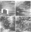
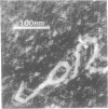
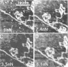
We have developed a chemical treatment for the mica surface which allows biopolymers to be held in place for atomic force microscopy, even under water, using conventional, untreated force sensing tips. We illustrate the procedure with images of lambda DNA and fd phage. The phage adheres well enough to permit in situ imaging of the adsorption process in water. These experiments yield a mean length for the phage of 883 +/- 72 nm. This compares with a measured length of 883 +/- 33 nm when the phage are imaged after drying following adsorption from water, showing that the effect of dehydration is quite small. Adhesion forces between the force sensing tip and the substrate and the sensing tip and the biomolecules are very different in the three media (air, water and propanol). The apparent height of the phage and the width and height of the DNA depends upon these adhesion forces quite strongly. In contrast, changing the Hookean spring force exerted by the scanning tip makes little difference. These results suggest that the chemical factors involved in adhesion can dominate atomic force images and that the composition of the scanning tip is at least as important a factor as its geometry.
Full text
PDF
Images in this article
Selected References
These references are in PubMed. This may not be the complete list of references from this article.
- Arnold G. E., Day L. A., Dunker A. K. Tryptophan contributions to the unusual circular dichroism of fd bacteriophage. Biochemistry. 1992 Sep 1;31(34):7948–7956. doi: 10.1021/bi00149a028. [DOI] [PubMed] [Google Scholar]
- Bustamante C., Vesenka J., Tang C. L., Rees W., Guthold M., Keller R. Circular DNA molecules imaged in air by scanning force microscopy. Biochemistry. 1992 Jan 14;31(1):22–26. doi: 10.1021/bi00116a005. [DOI] [PubMed] [Google Scholar]
- Crewe A. V., Wall J. A scanning microscope with 5 A resolution. J Mol Biol. 1970 Mar;48(3):375–393. doi: 10.1016/0022-2836(70)90052-5. [DOI] [PubMed] [Google Scholar]
- Dunker A. K., Klausner R. D., Marvin D. A., Wiseman R. L. Letter: Filamentous bacterial viruses. X. X-ray diffraction studies of the R4-protein mutant. J Mol Biol. 1974 Jan 5;82(1):115–117. doi: 10.1016/0022-2836(74)90579-8. [DOI] [PubMed] [Google Scholar]
- Frank H., Day L. A. Electron microscopic observations on fd bacteriophage, its alkali denaturation products and its DNA. Virology. 1970 Sep;42(1):144–154. doi: 10.1016/0042-6822(70)90247-3. [DOI] [PubMed] [Google Scholar]
- Hansma H. G., Sinsheimer R. L., Li M. Q., Hansma P. K. Atomic force microscopy of single- and double-stranded DNA. Nucleic Acids Res. 1992 Jul 25;20(14):3585–3590. doi: 10.1093/nar/20.14.3585. [DOI] [PMC free article] [PubMed] [Google Scholar]
- Hansma H. G., Vesenka J., Siegerist C., Kelderman G., Morrett H., Sinsheimer R. L., Elings V., Bustamante C., Hansma P. K. Reproducible imaging and dissection of plasmid DNA under liquid with the atomic force microscope. Science. 1992 May 22;256(5060):1180–1184. doi: 10.1126/science.256.5060.1180. [DOI] [PubMed] [Google Scholar]
- Lindsay S. M., Nagahara L. A., Thundat T., Knipping U., Rill R. L., Drake B., Prater C. B., Weisenhorn A. L., Gould S. A., Hansma P. K. STM and AFM images of nucleosome DNA under water. J Biomol Struct Dyn. 1989 Oct;7(2):279–287. doi: 10.1080/07391102.1989.10507771. [DOI] [PubMed] [Google Scholar]
- Lindsay S. M., Tao N. J., DeRose J. A., Oden P. I., Lyubchenko YuL, Harrington R. E., Shlyakhtenko L. Potentiostatic deposition of DNA for scanning probe microscopy. Biophys J. 1992 Jun;61(6):1570–1584. doi: 10.1016/S0006-3495(92)81961-6. [DOI] [PMC free article] [PubMed] [Google Scholar]
- Lyubchenko Y. L., Gall A. A., Shlyakhtenko L. S., Harrington R. E., Jacobs B. L., Oden P. I., Lindsay S. M. Atomic force microscopy imaging of double stranded DNA and RNA. J Biomol Struct Dyn. 1992 Dec;10(3):589–606. doi: 10.1080/07391102.1992.10508670. [DOI] [PubMed] [Google Scholar]
- Lyubchenko Y. L., Jacobs B. L., Lindsay S. M. Atomic force microscopy of reovirus dsRNA: a routine technique for length measurements. Nucleic Acids Res. 1992 Aug 11;20(15):3983–3986. doi: 10.1093/nar/20.15.3983. [DOI] [PMC free article] [PubMed] [Google Scholar]
- Thundat T., Allison D. P., Warmack R. J., Ferrell T. L. Imaging isolated strands of DNA molecules by atomic force microscopy. Ultramicroscopy. 1992 Jul;42-44(Pt B):1101–1106. doi: 10.1016/0304-3991(92)90409-d. [DOI] [PubMed] [Google Scholar]
- Vesenka J., Guthold M., Tang C. L., Keller D., Delaine E., Bustamante C. Substrate preparation for reliable imaging of DNA molecules with the scanning force microscope. Ultramicroscopy. 1992 Jul;42-44(Pt B):1243–1249. doi: 10.1016/0304-3991(92)90430-r. [DOI] [PubMed] [Google Scholar]
- Weisenhorn A. L., Gaub H. E., Hansma H. G., Sinsheimer R. L., Kelderman G. L., Hansma P. K. Imaging single-stranded DNA, antigen-antibody reaction and polymerized Langmuir-Blodgett films with an atomic force microscope. Scanning Microsc. 1990 Sep;4(3):511–516. [PubMed] [Google Scholar]
- Yang J., Takeyasu K., Shao Z. Atomic force microscopy of DNA molecules. FEBS Lett. 1992 Apr 20;301(2):173–176. doi: 10.1016/0014-5793(92)81241-d. [DOI] [PubMed] [Google Scholar]