Abstract
The fusion (F) proteins of Newcastle disease virus (NDV) and Nipah virus (NiV) are both triggered by binding to receptors, mediated in both viruses by a second protein, the attachment protein. However, the hemagglutinin-neuraminidase (HN) attachment protein of NDV recognizes sialic acid receptors, whereas the NiV G attachment protein recognizes ephrinB2/B3 as receptors. Chimeric proteins composed of domains from the two attachment proteins have been evaluated for fusion-promoting activity with each F protein. Chimeras having NiV G-derived globular domains and NDV HN-derived stalks, transmembranes, and cytoplasmic tails are efficiently expressed, bind ephrinB2, and trigger NDV F to promote fusion in Vero cells. Thus, the NDV F protein can be triggered by binding to the NiV receptor, indicating that an aspect of the triggering cascade induced by the binding of HN to sialic acid is conserved in the binding of NiV G to ephrinB2. However, the fusion cascade for triggering NiV F by the G protein and that of triggering NDV F by the chimeras can be distinguished by differential exposure of a receptor-induced conformational epitope. The enhanced exposure of this epitope marks the triggering of NiV F by NiV G but not the triggering of NDV F by the chimeras. Thus, the triggering cascade for NiV G-F fusion may be more complex than that of NDV HN and F. This is consistent with the finding that reciprocal chimeras having NDV HN-derived heads and NiV G-derived stalks, transmembranes, and tails do not trigger either F protein for fusion, despite efficient cell surface expression and receptor binding.
Keywords: Fusion Protein, Receptors, Viral Protein, Virus Entry, Virus, Paramyxovirus
Introduction
The Paramyxoviridae is a family of enveloped, negative-stranded RNA viruses that includes human parainfluenza virus types 1–4, measles virus, mumps virus, Newcastle disease virus (NDV),3 Sendai virus, parainfluenza virus 5, respiratory syncytial virus, and the recently emerged henipaviruses, Nipah (NiV) and Hendra (1). The latter two viruses are unique among the paramyxoviruses in being able to cause severe encephalitis and high mortality rates in both animals and humans (2). On the basis of their highly infectious nature and virulence, they are listed as NIAID, National Institutes of Health Group I Emerging Pathogens. NDV is an avian pathogen that is also being used as a vaccine vector (3) and oncolytic agent because of its ability to selectively kill tumor cells (reviewed in Ref. 4).
Paramyxoviruses gain entry into cells by promoting the direct fusion of the viral and cellular membranes. However, these viruses are unusual in that the receptor binding and fusion-promoting activities reside on two different spike structures (1). For most paramyxoviruses, this distribution of functions requires a mechanism by which the two processes can be linked for the promotion of fusion. This is accomplished by a virus-specific interaction between the two proteins that makes it possible for receptor binding to trigger activation of the homologous fusion protein (reviewed in Refs. 1, 5).
Paramyxoviruses can be divided into two classes with respect to the types of receptors their attachment proteins recognize. Viruses, including NDV, that have a hemagglutinin-neuraminidase (HN) attachment protein bind to sialic acid-containing proteins and lipids on the cell surface and possess neuraminidase (NA) activity (1). Other viruses in the family, including measles virus and the henipaviruses, recognize specific protein receptors. The NiV attachment protein (called G) recognizes ephrinB2 and B3 as receptors (6–8) and exhibits neither hemagglutinating nor NA activity (9, 10).
The ectodomains of the attachment proteins of all paramyxoviruses consist of a membrane-proximal stalk that supports a terminal globular head in which resides the receptor binding site (1). The evaluation of chimeric attachment proteins composed of stalks and heads derived from the HN proteins of different members of the family has established that the stalk region of HN determines specificity for the homologous fusion (F) protein (11–13). Subsequently, a domain was identified in the stalk of NDV HN (14) that may mediate the interaction with F. An analogous domain was identified in the stalk of the measles virus attachment protein (15, 16). However, the F-interactive site(s) on NiV/Hendra virus G remain(s) to be identified.
We have constructed and evaluated a series of chimeric attachment proteins composed of stalks and globular heads derived from the NDV HN and NiV G proteins. Our results reveal that it is possible to trigger the NDV F protein with chimeras that bind to the NiV receptor. This is the first demonstration of the triggering of a paramyxovirus F protein by binding to a different class of receptor. This indicates that an aspect of the NDV HN triggering cascade is conserved in the binding of NiV G to its receptor. However, the triggering of NDV F by these chimeras does not require a step that appears to be a requirement for the triggering of NiV F by the NiV G protein. Also, none of several NiV G stalk-NDV HN head chimeras is capable of triggering NiV F, despite efficient cell surface expression and receptor binding. Together, these findings suggest that the triggering cascade for NiV F is more complex than that for NDV F. We speculate that triggering of NiV F by the G protein may involve another contribution from the head region, distinct from its receptor binding activity.
EXPERIMENTAL PROCEDURES
Recombinant Plasmids
The preparation of pCAGGS expression vectors for the HN and F proteins of NDV strain Australia-Victoria has been described previously (17). The HN and F genes were generously provided by Trudy Morrison and Robert Lamb, respectively. The NiV G gene was inserted into pCAGGS between the ClaI and StuI sites, and the NiV F gene was inserted between EcoRI and SacI.
Construction of Chimeric Attachment Protein Genes
All chimeras were constructed in pBluescript SK(+) (Stratagene, La Jolla, CA) and facilitated by the introduction of HindIII sites at the desired positions in both NDV HN and NiV G using the QuikChange site-directed mutagenesis kit (Stratagene). The segments specific for the N-terminal stalk regions of the proteins were exchanged. In each chimera, there is a lysine-leucine insertion between the two segments necessary for the introduction of the HindIII site.
Cells, Transfections, and Quantitation of Cell Surface Expression
Vero or PK13 (ephrinB2- and B3-deficient) cells (ATCC) were used for experiments involving chimeras with HN-derived stalks and G-derived heads (HN-G chimeras). BHK-21F cells (generous gift of Rebecca Dutch) were used for experiments involving the reciprocal chimeras (G-HN). For all experiments, cells were seeded in six-well plates at 2 × 10⁵ cells/well 1 day prior to transfection. WT and chimeric proteins were expressed using the Lipofectamine 2000 transfection reagent (Invitrogen) and 1 μg of each DNA per well, according to protocols provided by the company. All assays were performed at 48 h post-transfection. Cell surface expression was quantified by flow cytometry (performed by the University of Massachusetts Medical Center Flow Cytometry Core Laboratory) using a NiV G-specific polyclonal antiserum (806) (18) for HN-G chimeras and a mixture of conformation-specific mAbs for NDV HN, including HN1b, HN2a, HN3c, HN4a, and HN23a (19–22) for the G-HN chimeras. Secondary antibodies were obtained from KPL Laboratories (Gaithersburg, MD).
EphrinB2 Binding Assay
The ability of HN-G chimeras to bind ephrinB2 was determined by a modification of the procedure described by Negrete et al. (7). PK13 cells were transfected as above. The medium was removed, and the monolayers were incubated for 1 h at room temperature with 2 μg of soluble ephrinB2-human Fc protein (ephrinB2-Fc) (R&D Systems, Minneapolis, MN). Binding of ephrinB2 was quantified by flow cytometry.
Receptor Binding Enhancement (RBE) Assay
The effect of receptor binding on the recognition of the HN-G chimeras by mAbs was determined by preincubating a monolayer of PK13 cells expressing the chimeras with soluble ephrinB2 and then quantifying the binding of the respective antibody by flow cytometry.
Hemadsorption (HAd) and NA Assays
The receptor binding activity of G-HN chimeras was assayed by their ability to adsorb guinea pig erythrocytes (Bio-Link Laboratories, Liverpool, NY). NA activity was determined colorimetrically using sialyllactose (Accurate Chemical & Scientific Co., Westbury, NY) as a substrate. Both protocols have been described previously (23).
Content-mixing Assay for Fusion
The ability of each chimera to complement the F proteins in the promotion of cell-cell fusion was quantitated using a modification of a content-mixing assay (24), which measures β-galactosidase activity in target cells following fusion induced by the glycoprotein-expressing effector cells. Effector Vero or BHK-21F cells were transfected with 1 μg each of G/HN/chimera DNA and the NiV/NDV F DNA, as well as 1 μg of pCAGT7 DNA (25) (generous gift of Eric Lazear). The following day, another set of monolayers (target) was infected with WT vaccinia virus (multiplicity of infection of 1 for Vero cells and 10 for BHK-21F cells) and transfected with 1 μg of pG1NT7β-galactosidase (26). Five hours later, the cells were trypsinized, and equal numbers of the two cell populations were combined and incubated overnight. The next day, the extent of fusion was quantitated colorimetrically.
Immunoprecipitation
Proteins were immunoprecipitated with either antiserum 806 (HN-G chimeras) or with a mixture of conformation-specific anti-HN mAbs (G-HN chimeras) as described previously (23) and analyzed by SDS-PAGE in the presence or absence of β-mercaptoethanol. Rainbow markers were obtained from GE Healthcare.
Biotinylation
The cell surface expression of chimera 214G-HN124 was examined by biotinylation and Western blot. Cell surface proteins were biotinylated with membrane-impermeable sulfo-NHS-SS-biotin (Pierce) for 30 min in the cold with gentle agitation. Cells were lysed, and proteins were immunoprecipitated as described above, boiled in 10% SDS, diluted 50-fold with TN buffer (50 mm Tris-HCl (pH 8.0), 150 mm sodium chloride) and reprecipitated with streptavidin-agarose beads (Pierce). Proteins were separated on SDS-PAGE and analyzed by Western blot, using mAb 14f, which recognizes a linear epitope in NDV HN (22).
Sucrose Gradients
The oligomeric structure of the HN-G chimeras was analyzed by sucrose gradient sedimentation analysis (27).
RESULTS
The NiV and NDV Attachment Proteins Do Not Complement the Heterologous F Proteins in the Promotion of Fusion
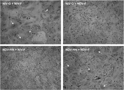
To determine whether the NiV and NDV attachment and fusion proteins can complement each other in the promotion of fusion, heterologous combinations of the two proteins were coexpressed in Vero cells, and the monolayers were stained and examined for syncytium formation (Fig. 1). Although extensive syncytium formation was visible in the NiV G/NiV F-coexpressing cells and, although to a lesser extent, in the NDV HN/NDV F-coexpressing cells, no fusion was detected in cells coexpressing either heterologous combination of the attachment and fusion proteins. Similar results were obtained using BHK-21F cells (data not shown). Thus, NDV HN was unable to complement NiV F, and NiV G was unable to complement NDV F in the promotion of fusion.
FIGURE 1.
The NiV and NDV attachment proteins do not complement the heterologous F proteins in the promotion of fusion. At 48 h post-transfection, Vero cell monolayers expressing NiV G and NiV F, NiV G and NDV F, NDV HN and NiV F, or NDV HN and NDV F were fixed with methanol and stained with Giemsa stain. Syncytia are indicated by white arrows.
Construction of Chimeric Attachment Proteins Composed of N-terminal Segments from the NDV HN Protein and C-terminal Segments Derived from the NiV G Protein
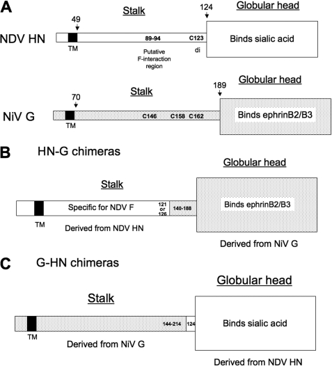
Because NDV HN and NiV G recognize different types of receptors, we wanted to determine whether attachment to the heterologous receptor is capable of triggering the promotion of fusion by the NDV F and NiV F proteins. To test this possibility, chimeric attachment proteins composed of stalks from one of the attachment proteins and globular domains from the heterologous attachment protein were constructed. There is a 24% amino acid homology between the NDV HN and NiV G proteins. A schematic diagram of the structures of the two proteins is shown in Fig. 2A.
FIGURE 2.
Alignment of NDV HN and NiV G and strategy for the design of chimeras. A, the residues in the ectodomains of the NDV HN and NiV G proteins that make up the membrane-proximal stalk and C-terminal globular head of each protein are indicated. In NDV HN, the head, which binds sialic acid-containing receptors, begins at residue 124. The cysteine at position 123 (Cys-123) that mediates disulfide-linked dimer formation is indicated. A putative F-interactive domain has been identified in the stalk at residues 89–94. In NiV G, the globular head, which includes the ephrinB2/B3 binding site, begins at residue 189. Cysteines at positions 146, 158 and 162 (Cys-146, Cys-158, and Cys-166, respectively) mediate disulfide-linked dimer and tetramer formation. B, strategy for the design of HN-G chimeras. In these chimeras, the N-terminal segment extends to either residue 121 or 126 of NDV HN, and the C-terminal NiV G-derived segment begins at varying positions in the stalk and base of the head of G, ranging from position 140 to 188. C, strategy for the design of G-HN chimeras. In these chimeras, the N-terminal NiV G-derived segment is of varying length ranging from 144 to 214 residues. In each case, the C-terminal NDV HN-derived segment begins at HN residue 124.
In the ectodomain of NDV HN, the transition from the stalk region to the head occurs between residues 123 and 124 (28), with the cysteine at position 123 mediating disulfide-linked dimer formation in some strains, including AV (29–31). The specificity of NDV HN for its homologous F protein has been shown to be determined by the stalk region of the protein (11), and a putative interaction site has been identified at stalk residues 89–94 (14). Two sialic acid binding sites have been identified in the globular head (28, 32).
In the NiV G ectodomain, the stalk is thought to encompass residues 71–188, with residue 189 being the membrane-proximal residue in the globular domain (33). There are three cysteines in the stalk at positions 146, 158, and 162, which are thought to mediate disulfide-linked oligomer formation. Domains in the globular head region mediate binding to receptors ephrinB2 and B3 (33, 34). The domain(s) in NiV G that mediate the interaction with the homologous F protein has not been identified.
The strategy for the construction of chimeras (HN-G) having NDV HN-derived stalks and NiV-derived heads is shown in Fig. 2B. The switch point in the chimeras is either at NDV HN residue 121 or residue 126 so that a chimera may or may not include the cysteine at position 123 of that protein. The globular segment derived from NiV G may begin at any of several residues between NiV G residues 140 and 188 so that the chimeras may include none, two, or all three of the cysteines in the membrane-distal end of the stalk of NiV G. The nomenclature for this series of chimeras indicates the most C-terminal HN-derived residue and the most N-terminal NiV G-derived residue. Thus, chimera 121HN-G149 has the N-terminal 121 residues from NDV HN and a C-terminal segment that begins at NiV G residue 149.
The HN-G Attachment Protein Chimeras Are Efficiently Expressed at the Cell Surface and Retain the Ability to Bind to EphrinB2
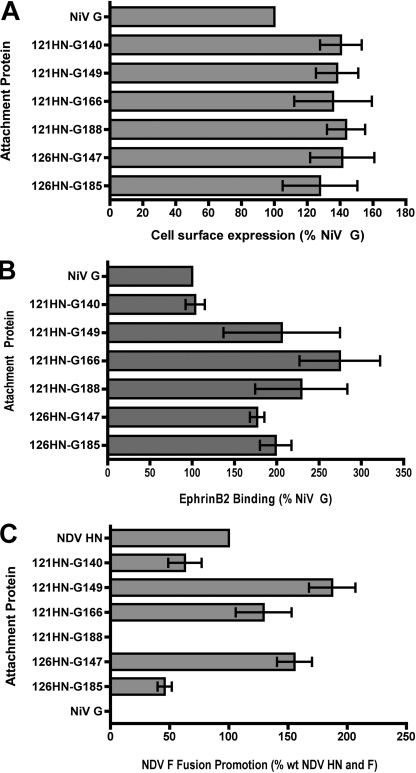
To determine whether chimeras having NDV HN-derived stalks and G-derived terminal globular heads are transported to the cell surface, they were expressed in Vero cells, and the amount of each protein present at the cell surface was determined by flow cytometry using polyclonal antiserum to the G protein (Fig. 3A). All six of the chimeras tested were expressed more efficiently than the WT NiV G protein, which is set at 100%. The amounts of expression vary from 128% (chimera 126HN-G185) to 141% of WT (chimera 121HN-G188). Thus, the substitution of the stalk of HN for that of NiV G did not impair the ability of the attachment protein to be transported to the cell surface and may have actually enhanced it relative to that of the NiV G protein. However, we cannot exclude the possibility that the presence of the HN-derived stalk may have increased the ability of antiserum to recognize the globular domain of G.
FIGURE 3.
Functional characterization of HN-G chimeras. A, cell surface expression of the HN-G chimeras. Expression at the surface of Vero cells was quantitated by flow cytometry using a polyclonal antiserum (806-term) specific for NiV G. Data are corrected for background obtained with vector alone and normalized to the value obtained with NiV G, which is set at 100%. Data represent mean ± S.D. n = 6. B, ability of the chimeras to bind ephrinB2. The chimeras or NiV G were expressed in PK13 cells, and at 48 h post-transfection the monolayers were incubated at room temperature with 2 μg of ephrinB2-Fc. After washing, binding was detected by flow cytometry and corrected for background obtained with vector alone. Data are expressed relative to the binding obtained with NiV G, which is set at 100%. Data represent mean ± S.D. n = 6. C, ability of the chimeras to complement NDV F in the promotion of fusion. Vero cells coexpressing NDV F and either a chimera, NDV HN, or NiV G were mixed with target cells overnight at 37 °C. The extent of fusion was then quantified in the content-mixing assay with data obtained with cells expressing vector and NDV F as background. Data are expressed relative to that obtained with NDV HN and F, which is set at 100%. Data represent mean ± S.D. n = 6.
We next wondered whether this series of chimeras retained the ability to bind to NiV receptors. This was tested by expressing the proteins in PK13 cells, which lack NiV receptors, and treating the monolayers with soluble ephrinB2 (Fig. 3B). Again, data are expressed relative to WT NiV G, which is set at 100%. One chimera, 121HN-G140 bound ephrinB2-human Fc to an extent similar to the WT protein. However, each of the other chimeras binds it significantly more efficiently than the WT NiV G protein, ranging from nearly 2-fold to almost 3-fold greater. Although these binding levels are not corrected for differences in expression, it is not likely that the increased receptor binding is due solely to the increased expression, because the latter is increased by less than 50% for each of the chimeras. Thus, in general, the replacement of the NiV G stalk with that from NDV HN significantly enhances the ability of the globular domain of G to bind ephrinB2.
All of the HN-G chimera were tested for the ability to bind sialic acid receptors by assaying their hemadsorption activity using guinea pig erythrocytes. None of the chimeras exhibited detectable activity in this assay (data not shown). As controls, NDV HN exhibited extensive HAd activity, and NiV G was negative.
Several of the HN-G Chimeras Are Capable of Complementing NDV F in the Promotion of Fusion
To determine whether it is possible to trigger NDV F to promote fusion by binding to the NiV receptor, each member of the HN-G series of chimeras was tested for its ability to complement NDV F in the promotion of fusion in Vero cells (Fig. 3C). In this regard, the chimeras segregate into three groups. Surprisingly, three of the chimeras were capable of complementing NDV F more efficiently than WT HN. This was especially true of chimera 121HN-G149, which promoted fusion almost twice as efficiently as WT HN (187.3% of WT), but chimeras 126HN-G147 and 121HN-G166 also promoted fusion with NDV F more efficiently than WT HN. Two other chimeras, 121HN-G140 and 126HN-G185, promoted fusion with NDV F less efficiently than WT HN, 62.9% and 45.8% of WT, respectively. Finally, one chimera, 121HN-G188, did not complement NDV F for fusion at a detectable level, even though it is both expressed and binds ephrinB2 efficiently. As a control, NiV G did not complement NDV F for fusion at a detectable level. Also, none of the chimeras complemented the NiV F protein in fusion promotion (data not shown). Thus, several chimeras that bound ephrinB2 rather than sialic acid as receptor were capable of triggering NDV F to promote fusion.
Oligomeric Structure of the HN-G Chimeras
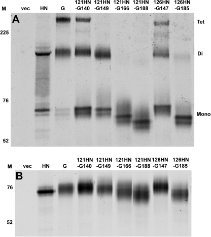
The oligomeric structure of the chimeras was analyzed by SDS-PAGE under non-reducing conditions and by sucrose gradient sedimentation. For SDS-PAGE, the attachment proteins were expressed in Vero cells, radioactively labeled, and chased to the surface for 90 min in medium. Cells were lysed in the presence of N-ethylmaleimide, and the chimeric proteins were immunoprecipitated using antiserum 806 and resolved by SDS-PAGE under non-reducing conditions (Fig. 4A). For comparison, the WT NDV-AV HN and NiV G proteins were also treated in the same way, although HN was immunoprecipitated with a mixture of HN-specific mAbs. The WT HN protein migrated as a mixture of monomers and dimers. The dimer is disulfide-linked through the cysteine at position 123 in the stalk (29–31). The HN protein from this strain of NDV also possesses a cysteine residue in its cytoplasmic domain that can mediate disulfide-linked tetramer formation upon cell lysis (31). However, formation of this bond is prevented by the inclusion of an alkylating agent in the lysis buffer. Thus, NDV-AV HN is a non-disulfide-linked tetramer of disulfide-linked dimers (27).
FIGURE 4.
SDS-PAGE analysis of HN-G chimeras. Each of the chimeras, as well as NDV HN and NiV G, were expressed in Vero cells, radiolabeled, and chased to the surface by incubation with medium for 90 min. Cells were lysed, and the chimeras and NiV G were immunoprecipitated with NiV G-specific antiserum. NDV HN was immunoprecipitated with a mixture of conformation-specific mAbs to that protein. Proteins were resolved by SDS-PAGE in the absence (A) or presence (B) of β-mercaptoethanol. The numbers in the lanes marked M indicate the migration rates of markers in kilodaltons. Tet, di, and mono indicate tetramers, dimers, and monomers, respectively.
The NiV G protein migrated in the gel as a mixture of disulfide-linked dimers and tetramers with a lesser amount of monomer (Fig. 4A). The intermolecular disulfide bonds responsible for these oligomers are thought to be mediated by one or more of the cysteines at positions 146, 158, and 162 in the stalk region of NiV G. The first four chimeras in the series, in which the HN-derived segment terminates at HN residue 121, do not include NDV HN residue Cys-123 and migrated as a mixture of monomers, dimers, and tetramers, depending on the point at which the NiV G-derived segment begins. Chimeras 121HN-G166 and 121HN-G188 ran solely as monomers, consistent with their having none of the stalk cysteines from either HN or G. Finally, chimeras 126HN-G147 and 126HN-G185 both ran partially as dimers in non-reduced SDS-PAGE, although the former also exhibited tetramers on the gel (Fig. 4A). This is consistent with the presence of the cysteine in HN at position 123 that mediates disulfide-linked dimer formation in that protein. As expected, when the chimeras were electrophoresed in the presence of a reducing agent, they all ran exclusively as monomers, albeit with slightly different migration rates because of their different sizes (Fig. 4B).
It is important to note that one of the chimeras (121HN-G166) that lacks all of the stalk cysteines from both proteins and ran exclusively as a monomer in non-reducing SDS-PAGE (Fig. 4A) still promoted fusion with NDV F almost 30% more efficiently than WT HN (Fig. 3C), although, when normalized for expression and ephrinB2 binding, fusion was actually lower than that of WT HN.
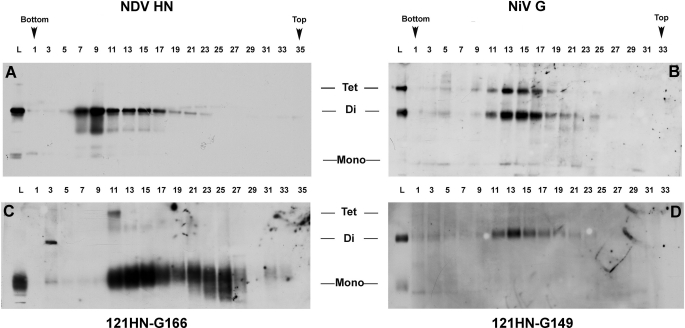
Because the SDS-PAGE migration pattern does not reflect the native oligomeric state, we compared the sucrose gradient sedimentation rate of this chimera to those of the WT NDV HN and NiV G proteins (Fig. 5). First of all, although both of the latter two proteins are tetrameric, NDV HN sedimented at a faster rate than NiV G. This likely means that the globular heads of the NDV tetramers are more tightly folded than those of NiV G. Most importantly, the sedimentation pattern of chimera 121HN-G166 (Fig. 5C) is noticeably different from that of either parent protein. Although a major population cosedimented with NiV G in fractions 11–17, there was a significant amount of a slower-sedimenting species in fractions 19–27. This suggests that some of the protein exists as dimers or possibly even monomers.
FIGURE 5.
Sucrose gradient sedimentation analysis of chimeras compared with NDV HN and NiV G. Vero cells expressing NDV HN (A), NiV G (B), chimera 121HN-G166 (C), and chimera 121HN-G149 (D) were lysed, and the lysates were layered onto continuous 7.5 to 22.5% sucrose gradients in MNT buffer (20 mm morpholino-ethanesulfonic acid, 30 mm Tris, 100 mm sodium chloride (pH 5.0)) plus 0.05% dodecyl-β-d-maltoside. The proteins in odd-numbered fractions were subjected to trichloroacetic acid precipitation, SDS-PAGE (no β-mercaptoethanol), and Western blot analysis. The chimeras and NiV G were both detected using antiserum 806, and HN was detected with mAb 14f. Tet, di, and mono indicate tetramers, dimers, and monomers, respectively. The lane marked L contains the lysate.
For another chimera, 121HN-G149, only monomers and dimers were detected in non-reducing SDS-PAGE (Fig. 4A). However, this chimera sedimented in the gradients at a rate very similar to that of NiV G, albeit without the disulfide-linked tetramers visible in the gradient gel (Fig. 5D). This suggests that, although the dimers of this chimera are not disulfide-linked, the protein is still tetrameric.
The HN-G Chimeras Do Not Exhibit the RBE of an mAb That Appears to Detect Part of the Triggering Cascade in the NiV G Protein
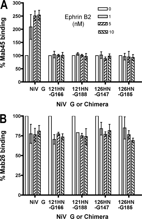
Mab45 is a conformational monoclonal antibody whose binding to the NiV G protein is enhanced upon NiV G binding to ephrinB2 (18). This enhancement represents increased binding to a domain thought to reside near the base of the globular domain distinct from the receptor binding site. This increased binding is postulated to be part of the fusion cascade in NiV G that results in the triggering of NiV F.
To determine whether the triggering of the NDV F protein by the HN-G chimeras also involves this mAb45 RBE, we tested several chimeras for this phenomenon (Fig. 6A). The binding of mAb45 to the chimeras expressed at the surface of PK13 cells was quantified by flow cytometry both in the absence of soluble ephrinB2 and in the presence of concentrations varying from 1 to 5 to 10 nm. As a control, the WT NiV G protein exhibited the RBE with a 2.5-fold increase in binding of the antibody at the highest concentration of receptor relative to that in its absence, as previously observed by Aguilar et al. (18). Four chimeras were also tested in this assay, two that are highly fusogenic (121HN-G166 and 126HN-G147), one that is weakly fusogenic (126HN-G185), and one that is fusion-null (121HN-G188). As shown in Fig. 6A, none of the chimeras exhibited the mAb45 RBE. There was no significant difference in the level of binding of the antibody in the absence or presence of any concentration of soluble receptor tested. Thus, the mAb45 RBE, which appears to reflect a step in the triggering cascade for NiV fusion, is not required for the chimeras to trigger the NDV F protein. This result distinguishes the mechanism of triggering of NiV F by the binding of NiV G to ephrinB2 from that by which the binding of the chimeras to ephrinB2 triggers NDV F.
FIGURE 6.
Four of the HN-G chimeras do not exhibit the enhanced binding of mAb45 in the presence of soluble ephrinB2 that is exhibited by the WT NiV G protein. Anti-NiV G mAb45 (A) or mAb26 (B) binding to WT NiV G and chimeras 121HN-G166 (highly fusogenic), 121HN-G188 (nonfusogenic), 126HN-G147 (highly fusogenic), and 126HN-G185 (weakly fusogenic) expressed at the surface of PK13 cells in the absence of receptor or in the presence of 1, 5, or 10 nm soluble ephrinB2 protein at room temperature, as detected by flow cytometry. Data for each protein are expressed relative to that of the protein expressed in the absence of receptor, which is set at 100%. Data represent mean ± S.D. n = 3.
As a control, the same chimeras were tested for the relatively modest decrease in the binding of a second antibody mAb26, observed with the WT NiV G protein (Fig. 6B). The epitope recognized by this antibody is thought to reside near the surface of the globular head close to the receptor binding site (18). As one would expect, all four of the chimeras (even the one that does not promote fusion) exhibited a decrease in the binding of this antibody very similar to that exhibited by the WT G protein. Thus, the loss of mAb45 RBE by the chimeras is not a general phenomenon of mAbs specific for the G protein.
Several Chimeras with Stalks Derived from the NiV G Protein and Globular Heads Derived from NDV HN Are Expressed, Are Competent to bind Sialic Acid-containing Receptors, but Do Not Trigger NiV F to Promote Fusion
Having successfully demonstrated that binding to the NiV receptor can trigger the NDV F protein, we wondered if the reverse would also be true. So, we prepared several chimeras (G-HN) in which the N-terminal segment with varying lengths of the stalk are derived from the NiV G protein, and the C-terminal globular head is derived from NDV HN.
The strategy for the construction of this set of chimeras is shown in Fig. 2C. The NiV G-derived stalk segment of the chimeras extends to either residue 144, 166, 179, 182, or 214 in NiV G, and the NDV HN-derived C-terminal segment begins at residue 124. The nomenclature for this series of chimeras indicates the most C-terminal G-derived residue and the most N-terminal NDV HN-derived residue. As an example, chimera 144G-HN124 has the N-terminal 144 residues from NiV G and a C-terminal segment that begins at NDV HN residue 124. Thus, none of the chimeras includes the cysteine at residue 123 in NDV HN that mediates disulfide-linked dimer formation. All of the G-HN chimeras, except 144G-HN124, include all the cysteines in the stalk of NiV G that mediate disulfide-linked oligomer formation.
The 214G-124HN chimera was constructed so that it contains a domain that was previously shown to be critical for the binding of mAb45 and may include the epitope recognized by the antibody (18). The mAb45 domain likely maps in the β6S4/β1H1 region defined by amino acids 195–211 at the base of the globular head. This is the only chimera in which the switch point falls within the head region of one of the parent proteins.
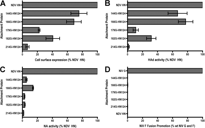
The functional activities of the chimeras were assayed after expression in BHK-21F cells. The level of cell surface expression of the G-HN chimeras was determined by flow cytometry using a mixture of conformation-specific anti-HN mAbs. Chimeras 144G-HN124 and 166G-HN124 were expressed at the cell surface (Fig. 7A) and hemadsorbed guinea pig erythrocytes at 60–80% of the level of WT HN (Fig. 7B). Chimeras 179G-HN124 and 182G-HN124 were expressed considerably less efficiently, at 22.3% and 40.5% of WT HN, respectively (Fig. 7A). Their HAd activities were also reduced with 179G-HN124 binding only about 10% of WT HN and 182G-HN124 only at 31.6% of the WT protein (Fig. 7B). Chimera 214G-HN124 was only weakly expressed (<10% of WT HN) (Fig. 7A) and exhibited barely detectable HAd activity (Fig. 7B).
FIGURE 7.
Functional characterization of G-HN chimeras. A, cell surface expression of the G-HN chimeras. Expression at the surface of BHK-21F cells was quantitated by flow cytometry using a mixture of conformation-specific mAbs specific for NDV HN. Data are corrected for background obtained with vector alone and normalized to the value obtained with HN, which is set at 100%. Data represent mean ± S.D. n = 6. B, ability of the chimeras to hemadsorb guinea pig erythrocytes. The chimeras or NDV HN were expressed in BHK-21F cells, and at 48 h post-transfection the monolayers were incubated at room temperature with a suspension of erythrocytes. After washing away unadsorbed erythrocytes, HAd activity was quantitated by lysing bound erythrocytes and determining the absorption of hemoglobin at 540 nm. Data are corrected for background obtained with vector alone and expressed relative to the binding obtained with NDV HN, which is set at 100%. Data represent mean ± S.D. n = 8. C, NA activity of the chimeras. The ability of the chimeras expressed at the surface of BHK-21F cells to cleave sialic acid from neuraminlactose was determined by a colorimetric NA assay. Data are corrected for background obtained with vector alone and expressed relative to the binding obtained with NDV HN, which is set at 100%. Data represent mean ± S.D. n = 3. D, BHK-21F cells coexpressing NiV F and either a chimera, NDV HN, or NiV G, were mixed with target cells overnight at 37 °C. The extent of fusion was then quantitated in the content-mixing assay with data obtained with cells expressing vector and NiV F as background. Data are expressed relative to that obtained with NiV G and F, which is set at 100%. Data represent mean ± S.D. n = 6.
Because the NA active site of the NDV HN protein resides in its globular domain, we also tested each of the chimeras for this activity (Fig. 7C). They all exhibited markedly reduced NA activity relative to the WT HN protein, ranging from as low as 1.7% (chimera 214G-HN124) to as much as 14.1% (chimera 166G-HN124).
Each of the G-HN chimeras was then tested for its ability to complement the NiV F protein in the promotion of fusion of BHK-21F cells using the content-mixing assay. As shown in Fig. 7D, none of the chimeras was able to complement NiV to a significant extent above vector background. (They also failed to complement NDV F.) Similar results were obtained in Vero cells (data not shown). Thus, chimeras that include NiV G-derived N-terminal segments of varying lengths and ostensibly intact NDV HN-derived globular domains were unable to trigger the NiV F protein to promote fusion, despite retention of extensive receptor binding activity.
Oligomeric Structure of the HN-G Chimeras
The oligomeric structure of the HN-G chimeras was analyzed by SDS-PAGE under non-reducing conditions identical to those in Fig. 4A. For SDS-PAGE, the chimeras were expressed in BHK-21F cells, radioactively labeled, and chased to the surface. Cells were lysed in the presence of N-ethylmaleimide, and the chimeric proteins were immunoprecipitated using a mixture of conformation-specific anti-HN mAbs and resolved by SDS-PAGE under non-reducing conditions (supplemental Fig. 1A). All of the chimeras, except 144G-HN124, migrated as a mixture of monomers, dimers and tetramers, similar to the control NiV G protein immunoprecipitated with 806-term antiserum. Chimera 144G-HN124 migrated almost exclusively as monomers, consistent with its lack of stalk cysteines from either protein. As before (Fig. 4A), the WT HN protein migrated as a mixture of monomers and dimers, although it is likely predominantly tetrameric. When the chimeras were electrophoresed in the presence of a reducing agent, they all ran exclusively as monomers, but with slightly different migration rates because of their different compositions (supplemental Fig. 1B).
Chimera 214G-HN124 is interesting in that it was detected only minimally at the cell surface by flow cytometry (Fig. 7A) but was immunoprecipitated quite efficiently, although the same antiserum (806) was used in both cases. To confirm that this chimera is not transported to the surface, cells expressing the chimera were biotinylated with a membrane-impermeable biotinylating agent, the proteins were immunoprecipitated with anti-HN mAbs and then with streptavidin beads, followed by SDS-PAGE and Western blotting with mAb 14f. Chimera 214G-HN124 was not detected in this assay, whereas the control WT HN and chimera 166G-HN124 were detected quite efficiently (data not shown). This confirms that chimera 214G-HN124 is partially folded but remains inside the cell.
DISCUSSION
We show here that the NDV F protein can be triggered to promote fusion by chimeric attachment proteins that bind to NiV receptors via a NiV G-derived globular head. This not only confirms that the F-specific domain of NDV HN resides in its stalk region (11) but also demonstrates that this domain can be induced to trigger the conversion of the NDV F protein to its fusion-active conformation by binding to a different class of receptor. Moreover, fusion is observed with several different chimeras having NiV G-derived C-terminal segments beginning at varying positions N-terminal to the beginning of the globular domain in the G protein. Thus, in these chimeras, the fusion cascade is apparently seamlessly transmitted through the interface between the NiV G-derived head and NDV HN-derived stalk segments. This means that an aspect of the fusion cascade triggered when NDV HN binds to its sialic acid-containing receptor is conserved in the binding of NiV G to ephrinB2/B3.
The ability to trigger NDV F by binding to a protein receptor may be facilitated by the fact that the sialic acid binding site on the head of HN and the ephrinB2/B3 binding sites on the head of G colocalize with each other at the top center of the β-sheet propeller (35). One can envision how binding to receptors, although they may be different, at similar topological sites might trigger comparable changes in two proteins with similar overall structures.
The lone HN-G chimera tested that is not able to complement NDV F for fusion is 121HN-G188 (Fig. 3C). Evidently, the signal that induces the triggering of NDV F is not transmitted in this chimera. Noteworthy in this regard is the relatively reduced level of fusion promoted by chimera 126HN-G185, which has only three additional NiV G-derived residues. It may be that the switch point in chimeras 121HN-G188 is too close to the stalk-head interface in NiV G.
Although most HN-G chimeras are capable of complementing NDV F for fusion, the triggering of NDV F and NiV F by the globular head of NiV G can, nonetheless, be distinguished in at least two ways. First, the RBE of mAb45 that appears to be an integral part of the triggering cascade for NiV F is not exhibited by any of the chimeras in their triggering of NDV F. Although all of the chimeras bind mAb45, none of them exhibits the RBE of the antibody. Second, the requirement for intermolecular disulfide bonds in the membrane-distal end of the stalk of G for the triggering of NiV and NDV F is also different. A chimera (121HN-G166) lacking stalk cysteines (migrating as a monomer in non-reduced SDS-PAGE) (Fig. 4A) is capable of efficiently triggering the NDV F protein to promote fusion (Fig. 3C). This is a marked departure from the properties of NiV G proteins in which individual stalk cysteines at positions 146, 158, or 162 are mutated to serine.4 These mutated proteins are fusion-null, despite efficient cell surface expression and intact ephrinB2 binding activity. Thus, stalk intermolecular disulfide bonds are not required for ephrinB2 binding to trigger NDV F, although they are required for it to trigger NiV F. These two phenotypic differences separate the mechanism of triggering NiV F and NDV F by binding to the NiV ephrin receptors.
The fact that specific conformational changes in the head of NiV G upon receptor binding are apparently required for the triggering of NiV F but not NDV F suggests that the triggering cascade in NiV may be more complex than that in NDV. It may be that, contrary to the mechanism of triggering the NDV F protein, triggering of NiV F requires more of a contribution from the globular head of G than merely receptor binding. This is consistent with the idea that the mAb45 RBE constitutes a receptor-induced activation site and that the epitope recognized by this mAb may reside near the base of the globular head of G (18).
In addition, this scenario is also consistent with the inability of the G-HN chimeras to trigger the NiV F protein, despite efficient cell surface expression and HAd activity for at least two of them. (Although the G-HN chimeras have relatively low NA levels, these levels have been shown to be sufficient for the promotion of fusion (36).) Moreover, whereas point mutations in a putative F-interactive site in the stalk of NDV HN that eliminate fusion also eliminate the HN-F interaction (14), fusion-null point mutations in the stalk NiV G stalk retain their ability to interact with NiV F, at least intracellularly (37). Indeed, unlike NDV HN, no mutation has been identified to date in the stalk of NiV G that eliminates G-F complex formation.
A great deal of evidence exists to suggest that NDV and NiV use different mechanisms to trigger fusion (reviewed in Refs. 5, 38, 39). Although in both cases binding to receptor serves as the trigger for fusion, the two viruses appear to use different strategies to regulate the process. Most mutated NDV HN proteins that lack receptor binding activity also fail to interact with the homologous F protein at the cell surface (17). Also, the extent of HN-F complex formation at the cell surface is proportional to the amount of fusion promoted (14). This is consistent with HN and F interacting at the cell surface triggered by receptor binding, a mode of fusion that has been called the association or provocateur (40) model.
For the henipaviruses, the relationship between receptor binding and G-F complex formation appears to be quite different. The amount of fusion promoted by NiV G and mutated F proteins is inversely proportional to the avidity of the G-F complex (41, 42), and decreased receptor binding activity results in increased detection of G-F complexes (37). In addition, receptor binding itself has been reported to result in decreased avidities of NiV F-G interactions (18). This is consistent with NiV fusion being promoted by dissociation of a preformed protein complex. F-G dissociation is thus thought to be triggered by the binding of G to receptors, a mode of fusion called the dissociation or clamp (40) model.
Taking these findings and the data presented here together, one might speculate about the intriguing possibility that the organization of the HN-F and G-F complexes may be different. Whereas the NDV F protein is activated through a transient interaction with the HN protein that is mediated completely by the stalk domain of the latter, the NiV F protein may be “clamped” in its prefusion, metastable conformation by an interaction that could be bidentate, requiring domains in both the stalk and head regions of NiV G. F would presumably be released from G upon the binding of the latter to receptors, thus freeing F to undergo the extensive conformational change required for fusion. Though this is a complex issue, it should be possible to begin to address it, as well as the mechanistic bases for the clamp versus provocateur modes of fusion promotion, using these two sets of attachment protein chimeras.
Supplementary Material
This work was supported, in whole or in part, by National Institutes of Health Grant AI49268 (to R. M. I.), a subproject of U19 Grant AI057319 (to the University of Massachusetts Center for Translational Research on Human Immunology and Biodefense), and AI069317 (to B. L.). This work was also supported by Subproject Award U54 AI065359 from the Pacific Southwest Regional Center for Excellence in Biodefense and Emerging Infectious Diseases.

The on-line version of this article (available at http://www.jbc.org) contains supplemental Fig. 1.
O. Negrete, personal communication.
- NDV
- Newcastle disease virus
- NiV
- Nipah virus
- HN
- hemagglutin-neuraminidase
- NA
- neuraminidase
- G
- Nipah virus attachment protein
- F
- fusion
- RBE
- receptor binding enhancement
- HAd
- hemadsorption
- AV
- Australia-Victoria.
REFERENCES
- 1. Lamb R. A., Parks G. D. (2007) Fields Virology, 5th Ed., pp. 1449–1496, Wolters Kluwer/Lippincott Williams and Wilkins, Philadelphia [Google Scholar]
- 2. Murray K., Selleck P., Hooper P., Hyatt A., Gould A., Gleeson L., Westbury H., Hiley L., Selvey L., Rodwell B. (1995) Science 268, 94–97 [DOI] [PubMed] [Google Scholar]
- 3. Bukreyev A., Collins P. L. (2008) Curr. Opin. Mol. Ther. 10, 46–55 [PubMed] [Google Scholar]
- 4. Schirrmacher V., Fournier P. (2009) Methods Mol. Biol. 542, 565–605 [DOI] [PMC free article] [PubMed] [Google Scholar]
- 5. Iorio R. M., Melanson V. R., Mahon P. J. (2009) Future Virol. 4, 335–351 [DOI] [PMC free article] [PubMed] [Google Scholar]
- 6. Bonaparte M. I., Dimitrov A. S., Bossart K. N., Crameri G., Mungall B. A., Bishop K. A., Choudhry V., Dimitrov D. S., Wang L. F., Eaton B. T., Broder C. C. (2005) Proc. Natl. Acad. Sci. U.S.A. 102, 10652–10657 [DOI] [PMC free article] [PubMed] [Google Scholar]
- 7. Negrete O. A., Levroney E. L., Aguilar H. C., Bertolotti-Ciarlet A., Nazarian R., Tajyar S., Lee B. (2005) Nature 436, 401–405 [DOI] [PubMed] [Google Scholar]
- 8. Negrete O. A., Wolf M. C., Aguilar H. C., Enterlein S., Wang W., Mühlberger E., Su S. V., Bertolotti-Ciarlet A., Flick R., Lee B. (2006) PLoS Pathog. 2, e7. [DOI] [PMC free article] [PubMed] [Google Scholar]
- 9. Yu M., Hansson E., Langedijk J. P., Eaton B. T., Wang L. F. (1998) Virology 251, 227–233 [DOI] [PubMed] [Google Scholar]
- 10. Harcourt B. H., Tamin A., Ksiazek T. G., Rollin P. E., Anderson L. J., Bellini W. J., Rota P. A. (2000) Virology 271, 334–349 [DOI] [PubMed] [Google Scholar]
- 11. Deng R., Wang Z., Mirza A. M., Iorio R. M. (1995) Virology 209, 457–469 [DOI] [PubMed] [Google Scholar]
- 12. Tsurudome M., Kawano M., Yuasa T., Tabata N., Nishio M., Komada H., Ito Y. (1995) Virology 213, 190–203 [DOI] [PubMed] [Google Scholar]
- 13. Tanabayashi K., Compans R. W. (1996) J. Virol. 70, 6112–6118 [DOI] [PMC free article] [PubMed] [Google Scholar]
- 14. Melanson V. R., Iorio R. M. (2004) J. Virol. 78, 13053–13061 [DOI] [PMC free article] [PubMed] [Google Scholar]
- 15. Lee J. K., Prussia A., Paal T., White L. K., Snyder J. P., Plemper R. K. (2008) J. Biol. Chem. 283, 16561–16572 [DOI] [PMC free article] [PubMed] [Google Scholar]
- 16. Paal T., Brindley M. A., St. Clair C., Prussia A., Gaus D., Krumm S. A., Snyder J. P., Plemper R. K. (2009) J. Virol. 83, 10480–10493 [DOI] [PMC free article] [PubMed] [Google Scholar]
- 17. Li J., Quinlan E., Mirza A., Iorio R. M. (2004) J. Virol. 78, 5299–5310 [DOI] [PMC free article] [PubMed] [Google Scholar]
- 18. Aguilar H. C., Ataman Z. A., Aspericueta V., Fang A. Q., Stroud M., Negrete O. A., Kammerer R. A., Lee B. (2009) J. Biol. Chem. 284, 1628–1635 [DOI] [PMC free article] [PubMed] [Google Scholar]
- 19. Iorio R. M., Borgman J. B., Glickman R. L., Bratt M. A. (1986) J. Gen. Virol. 67, 1393–1403 [DOI] [PubMed] [Google Scholar]
- 20. Iorio R. M., Bratt M. A. (1983) J. Virol. 48, 440–450 [DOI] [PMC free article] [PubMed] [Google Scholar]
- 21. Iorio R. M., Glickman R. L., Riel A. M., Sheehan J. P., Bratt M. A. (1989) Virus Res. 13, 245–261 [DOI] [PubMed] [Google Scholar]
- 22. Iorio R. M., Syddall R. J., Sheehan J. P., Bratt M. A., Glickman R. L., Riel A. M. (1991) J. Virol. 65, 4999–5006 [DOI] [PMC free article] [PubMed] [Google Scholar]
- 23. Mahon P. J., Mirza A. M., Musich T. A., Iorio R. M. (2008) J. Virol. 82, 10386–10396 [DOI] [PMC free article] [PubMed] [Google Scholar]
- 24. Corey E. A., Mirza A. M., Levandowsky E., Iorio R. M. (2003) J. Virol. 77, 6913–6922 [DOI] [PMC free article] [PubMed] [Google Scholar]
- 25. Okuma K., Nakamura M., Nakano S., Niho Y., Matsuura Y. (1999) Virology 254, 235–244 [DOI] [PubMed] [Google Scholar]
- 26. Nussbaum O., Broder C. C., Berger E. A. (1994) J. Virol. 68, 5411–5422 [DOI] [PMC free article] [PubMed] [Google Scholar]
- 27. Melanson V. R., Iorio R. M. (2006) J. Virol. 80, 623–633 [DOI] [PMC free article] [PubMed] [Google Scholar]
- 28. Crennell S., Takimoto T., Portner A., Taylor G. (2000) Nat. Struct. Biol. 7, 1068–1074 [DOI] [PubMed] [Google Scholar]
- 29. Sheehan J. P., Iorio R. M., Syddall R. J., Glickman R. L., Bratt M. A. (1987) Virology 161, 603–606 [DOI] [PubMed] [Google Scholar]
- 30. Mirza A. M., Sheehan J. P., Hardy L. W., Glickman R. L., Iorio R. M. (1993) J. Biol. Chem. 268, 21425–21431 [PubMed] [Google Scholar]
- 31. McGinnes L. W., Morrison T. G. (1994) Virology 200, 470–483 [DOI] [PubMed] [Google Scholar]
- 32. Zaitsev V., von Itzstein M., Groves D., Kiefel M., Takimoto T., Portner A., Taylor G. (2004) J. Virol. 78, 3733–3741 [DOI] [PMC free article] [PubMed] [Google Scholar]
- 33. Bowden T. A., Aricescu A. R., Gilbert R. J., Grimes J. M., Jones E. Y., Stuart D. I. (2008) Nat. Struct. Mol. Biol. 15, 567–572 [DOI] [PubMed] [Google Scholar]
- 34. Xu K., Rajashankar K. R., Chan Y. P., Himanen J. P., Broder C. C., Nikolov D. B. (2008) Proc. Natl. Acad. Sci. U.S.A. 105, 9953–9958 [DOI] [PMC free article] [PubMed] [Google Scholar]
- 35. Bowden T. A., Crispin M., Jones E. Y., Stuart D. I. (2010) Biochem. Soc. Trans. 38, 1349–1355 [DOI] [PMC free article] [PubMed] [Google Scholar]
- 36. Mirza A. M., Deng R., Iorio R. M. (1994) J. Virol. 68, 5093–5099 [DOI] [PMC free article] [PubMed] [Google Scholar]
- 37. Bishop K. A., Stantchev T. S., Hickey A. C., Khetawat D., Bossart K. N., Krasnoperov V., Gill P., Feng Y. R., Wang L., Eaton B. T., Wang L. F., Broder C. C. (2007) J. Virol. 81, 5893–5901 [DOI] [PMC free article] [PubMed] [Google Scholar]
- 38. Iorio R. M., Mahon P. J. (2008) Trends Microbiol. 16, 135–137 [DOI] [PMC free article] [PubMed] [Google Scholar]
- 39. Smith E. C., Popa A., Chang A., Masante C., Dutch R. E. (2009) FEBS J. 276, 7217–7227 [DOI] [PMC free article] [PubMed] [Google Scholar]
- 40. Connolly S. A., Leser G. P., Jardetzky T. S., Lamb R. A. (2009) J. Virol. 83, 10857–10868 [DOI] [PMC free article] [PubMed] [Google Scholar]
- 41. Aguilar H. C., Matreyek K. A., Filone C. M., Hashimi S. T., Levroney E. L., Negrete O. A., Bertolotti-Ciarlet A., Choi D. Y., McHardy I., Fulcher J. A., Su S. V., Wolf M. C., Kohatsu L., Baum L. G., Lee B. (2006) J. Virol. 80, 4878–4889 [DOI] [PMC free article] [PubMed] [Google Scholar]
- 42. Aguilar H. C., Matreyek K. A., Choi D. Y., Filone C. M., Young S., Lee B. (2007) J. Virol. 81, 4520–4532 [DOI] [PMC free article] [PubMed] [Google Scholar]
Associated Data
This section collects any data citations, data availability statements, or supplementary materials included in this article.


