Abstract
Community-associated methicillin-resistant Staphylococcus aureus accounts for a large portion of the increased staphylococcal disease incidence and can cause illness ranging from mild skin infections to rapidly fatal sepsis syndromes. Currently, we have limited understanding of S. aureus-derived mechanisms contributing to bacterial pathogenesis and host inflammation during staphylococcal disease. Herein, we characterize an influential role for the saeR/S two-component gene regulatory system in mediating cytokine induction using mouse models of S. aureus pathogenesis. Invasive S. aureus infection induced the production of localized and systemic pro-inflammatory cytokines, including tumor necrosis factor alpha (TNF-α), interferon gamma (IFN-γ), interleukin (IL)-6 and IL-2. In contrast, mice infected with an isogenic saeR/S deletion mutant demonstrated significantly reduced pro-inflammatory cytokine levels. Additionally, secreted factors influenced by saeR/S elicited pro-inflammatory cytokines in human blood ex vivo. Our study further demonstrated robust saeR/S-mediated IFN-γproduction during both invasive and subcutaneous skin infections. Results also indicated a critical role for saeR/S in promoting bacterial survival and enhancing host mortality during S. aureus peritonitis. Taken together, this study provides insight into specific mechanisms used by S. aureus during staphylococcal disease and characterizes a relationship between a bacterial global regulator of virulence and the production of pro-inflammatory mediators.
Introduction
Staphylococcus aureus predominates as a global cause of bacterial infections, which can range from mild skin irritations to severe life-threatening invasive disease [1]. Increases in reported cases of methicillin-resistant S. aureus infections (MRSA) including community-associated MRSA (CA-MRSA) infections that occur in otherwise healthy individuals, independent of hospital settings [2]–[4], are an important public health concern. Generally presenting with soft tissue infections, S. aureus disease is also associated with such severe conditions as septicemia, necrotizing pneumonia and necrotizing fasciitis [3], [5]–[8]. In 2005, the United States reported over 18,000 deaths resulting from invasive MRSA disease, a number surpassing the annual fatalities associated with HIV/AIDS [9], [10].
Gram-positive bacterial infections account for ∼50% of all reported sepsis cases and are associated with the dysfunctional production of pro-inflammatory cytokines [11]–[14]. Systemic S. aureus infections are associated with the endogenous production of interferon gamma (IFN-γ), tumor necrosis factor alpha (TNF-α) and interleukin (IL)-6 [15], [16]. Additionally, innate immune effectors exhibit severely diminished anti-bacterial function during sepsis and S. aureus infections [17]–[21]. However, studies characterizing pathogen-derived mediators of the host inflammatory response have predominately focused on single toxins and proteins, rendering the inflammatory modulating effects of global virulence regulators undefined.
S. aureus possesses 16 two-component gene-regulatory systems that monitor changing environmental conditions to influence gene transcription [22]–[24]. Numerous studies have indicated a regulatory role for the S. aureus two-component system SaeR/S in the expression of secreted virulence factors [25]–[33]. Previous findings have demonstrated a critical role for saeR/S in evading destruction by neutrophils and enhancing mortality in murine bacteremic models [18], [33]. However, significant gaps in our understanding of how saeR/S contributes to S. aureus pathogenesis exist. To that end, we investigated the influence of saeR/S on pathogen survival and the host response during invasive disease and demonstrated that saeR/S strongly influenced the production of inflammatory cytokines during S. aureus infection. These current findings further support the hypothesis that saeR/S is a critical mediator of pathogenesis during staphylococcal disease.
Results
SaeR/S significantly enhances S. aureus survival, dissemination and mortality during invasive disease
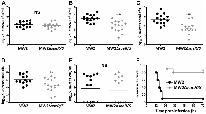
Invasive staphylococcal disease is associated with bacterial persistence and dissemination to deep tissues. To investigate the effects of saeR/S on bacterial survival and dissemination during invasive infection, we used a mouse model of S. aureus peritonitis via intraperitoneal (i.p.) inoculation with wild-type (MW2) or S. aureus with deleted saeR/S (MW2ΔsaeR/S) (figure 1). At 4 hours post-infection, saeR/S did not impact S. aureus survival in the peritoneum, as both MW2- and MW2ΔsaeR/S-infected mice exhibited similar bacterial loads (P>0.05; figure 1A ). However, at 10 hours post-infection, saeR/S-regulated factors significantly increased S. aureus survival in the peritoneum of MW2-infected mice (P<0.0001; figure 1B ). To further investigate the contributions of saeR/S on S. aureus tissue infiltration, we harvested both kidneys and enumerated S. aureus burdens at 10 hours post-infection. Mice infected with MW2ΔsaeR/S had significantly less colony forming units (cfu) in the kidneys (P<0.0001; figure 1C ), suggesting a role for saeR/S in pathogen dissemination. To characterize the influence of saeR/S on S. aureus dissemination from the infectious foci, we exsanguinated mice and harvested hearts for additional S. aureus quantification. Consistent with peritoneal cavity and kidney burdens, the absence of saeR/S significantly reduced bacterial loads in the hearts from MW2ΔsaeR/S-infected mice (P<0.01; figure 1D ). Surprisingly, both groups exhibited similar S. aureus burdens in the blood (P>0.05; figure 1E ). This suggests a transient presence of S. aureus in the blood following peritonitis. Consistent with observations of the influence of saeR/S on murine mortality during S. aureus bacteremia [18], [33], saeR/S-regulated factors significantly enhanced morbidity and mortality in the peritonitis model (1 mouse infected with MW2 survived the 72 hours time course, compared to 8 mice infected with MW2ΔsaeR/S; P<0.001; figure 1F ). These results are congruent with previous observations that saeR/S contributes to host mortality [18], [33] and demonstrates saeR/S is essential for S. aureus survival and dissemination following invasive infection.
Figure 1. SaeR/S promotes bacterial persistence and host mortality in a mouse model of S. aureus peritonitis.
Mice were inoculated i.p. with MW2 or MW2ΔsaeR/S (5×10⁷ cfu) for (A) 4 hours and (B–E) 10 hours. (A) S. aureus burden in the peritoneal exudate, NS. For data in (A), results are from 3 biological replicates of 5 mice/group (n = 15). (B) S. aureus burden in peritoneal exudate, ****P<0.0001. (C) S. aureus burden in the kidneys, ****P<0.0001. (D) S. aureus burden in the heart, **P<0.01. E, S. aureus burden in the blood, NS. For data in (B–E), results are from 2 biological replicates of 7 and 8 mice/group (n = 15). All tissues compared were individually analyzed by t test. (F) Survival curve for mice inoculated (i.p.) with 5×10⁷ cfu of MW2 or MW2ΔsaeR/S, *P<0.001 as determined by logrank test (n = 10/group). Mice receiving PBS had no S. aureus cfu (data not shown). NS = not significant.
SaeR/S promotes pro-inflammatory cytokine gene transcription
Sepsis syndromes are commonly associated with an early rapid induction of pro-inflammatory cytokines [13]. Following i.p. infection with MW2 and MW2ΔsaeR/S, we investigated the influence of saeR/S-regulated factors on the transcriptional regulation of 84 host-derived inflammation-associated genes (Tables 1, S1 and S2). Leukocytes were isolated from the peritoneal cavity at 4 hours post-inoculation, when the bacterial burdens between MW2 and MW2ΔsaeR/S were virtually equal (figure 1A ). Deletion of saeR/S resulted in a down-regulation of key inflammatory cytokines (Tables 1 and S1). Of the 84 assayed genes, 47 were down-regulated ≥3-fold with only 6 genes up-regulated ≥3-fold, MW2ΔsaeR/S relative to MW2 (Tables 1 and S1). Several pro-inflammatory genes commonly expressed during early sepsis were down-regulated in MW2ΔsaeR/S-infected mice compared to MW2-infected mice, including: IFN-γ: -11.27-fold (P≤.05), TNF: -9.37-fold (P>0.10), IL-18: −7.17 (P≤0.01), CD40 ligand: −4.92 (P≤0.01), IL-1β: −4.07 (P≤0.05) and C-reactive protein: −4.43 (P≤0.05) (Tables 1 and S1). Interestingly, transcription of anti-inflammatory cytokine genes were relatively unaffected with the exception of IL1RII, which was up-regulated 21.38-fold, MW2ΔsaeR/S relative to MW2 (Table S1). Both MW2 and MW2ΔsaeR/S-infected mice displayed up-regulation of cytokine transcription compared to PBS-inoculated control mice (Table S2). These results suggest differences in transcriptional activity stem from saeR/S-regulated factors, and indicate a saeR/S-mediated pro-inflammatory cytokine response during S. aureus infection.
Table 1. SaeR/S-mediated factors elicit host pro-inflammatory transcription during invasive disease.
| Gene symbol | Encoded protein | Fold-regulation change | P value |
| ifng | Interferon gamma | −11.27 | ≤0.05 |
| tnfrsf1a | Tumor necrosis factor receptor superfamily, member 1a | −3.03 | ≤0.01 |
| il18 | Interleukin 18 | −7.17 | ≤0.01 |
| cd40lg | CD40 ligand | −4.92 | ≤0.01 |
| il1b | Interleukin-1β | −4.07 | ≤0.05 |
| crp | C-reactive protein | −4.43 | ≤0.05 |
| il11 | Interleukin 11 | −3.65 | ≤0.05 |
NOTE. Genes listed display fold-regulation values of MW2ΔsaeR/S-infected mice relative to MW2-infected mice (3 per group). RNA was collected from all leukocytes isolated from the peritoneum, 4 hours post-infection. Fold-regulation and P values calculated using SA Biosciences™ web-based software utilizing the ΔΔCt method.
SaeR/S-regulated factors promote pro-inflammatory cytokines in the blood during invasive disease
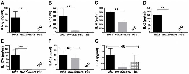
Sepsis and septic shock are associated with the systemic production of inflammatory cytokines [11]–[14]. To determine if the saeR/S-mediated inflammatory response was systemic during S. aureus peritonitis, we infected mice i.p. with 5×10⁷ cfu of MW2 or MW2ΔsaeR/S for 10 hours and measured protein levels of several pro- and anti-inflammatory cytokines in the serum by cytometric bead array (figure 2). Of note, serum IFN-γand TNF levels were significantly reduced in mice infected with MW2ΔsaeR/S (P<0.05 and P<0.01, respectively; figures 2A and 2B ). Additional pro-inflammatory cytokines associated with sepsis syndromes were influenced by saeR/S as serum IL-6 and IL-2 concentrations were significantly lower in MW2ΔsaeR/S-infected mice (P<0.01; figure 2C and 2D ). Serum IL-17A levels were also significantly reduced in mice infected with MW2ΔsaeR/S (P<0.01; figure 2E ). In contrast, IL-4 and IL-10 serum concentrations did not exhibit differences between MW2 and MW2ΔsaeR/S-infected groups (P>0.05; figures 2F and 2G ). These results are consistent with previous sepsis syndrome observations, in which an early pro-inflammatory cytokine response (IFN-γ, TNF, IL-2, and IL-6) was concomitant with a relatively subdued anti-inflammatory cytokine (IL-4 and IL-10) response [13], [14]. The differentially regulated transcriptional activity in the peritoneum (Tables 1 and S1) and the polarized pro-inflammatory cytokine response in the blood (figure 2) indicate that saeR/S-regulated factors promote localized and systemic inflammation during invasive disease.
Figure 2. SaeR/S elicits systemic pro-inflammatory cytokines during invasive S. aureus infection.
Mice were inoculated i.p. with MW2, MW2ΔsaeR/S (5×10⁷ cfu) or PBS for 10 hours and serum cytokine concentrations were measured by cytometric bead array. Serum concentrations (pg/ml) of (A) IFN-γ, (B) TNF, (C) IL-6, (D) IL-2, (E) IL-17A, (F) IL-10 and (G) IL-4. Results displayed are from 5 mice (MW2), 7 mice (MW2ΔsaeR/S) and 2 mice (PBS). *P<0.05 and **P<0.01 compared to cytokine expression in MW2-infected mice for each cytokine investigated as determined by t test. NS = not significant; N/D = not detectable.
Secreted factors, regulated by saeR/S, significantly enhance the production of inflammatory proteins in human whole blood
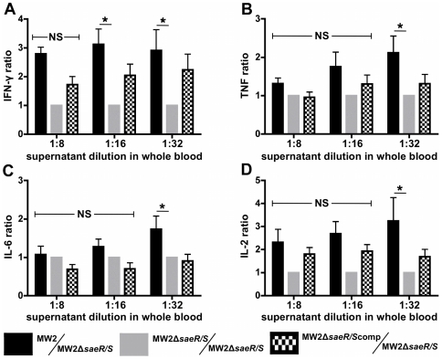
Numerous secreted S. aureus virulence factors are regulated by saeR/S [25]–[33]. To investigate the concerted role of these exoproteins on the production of cytokines in human blood and to confirm the saeR/S-mediated production of IFN-γ, TNF, IL-6, IL-2 and IL-17A observed in mice (figure 2 and Table 1), we treated human whole blood with supernatants from cultures of MW2, MW2ΔsaeR/S and a saeR/S-complemented MW2ΔsaeR/S strain (MW2ΔsaeR/Scomp) and compared plasma cytokine levels (figure 3). The data shown are normalized for each individual donor to account for large variations in the magnitude of cytokine expression between individuals. Consistent with cytokine profiles observed in our mouse studies, MW2-treated human blood produced significantly higher levels of IFN-γand TNF compared to MW2ΔsaeR/S-treated blood (P<0.05; figures 3A and 3B ). Treatment with MW2ΔsaeR/Scomp supernatant restored the observed IFN-γphenotype and partially restored the TNF phenotype (figures 3A and 3B , respectively). IL-2 and IL-6 levels were also significantly elevated in the MW2 treatment groups (P<0.05; figures 3C and 4D , respectively), whereas IL–17A was not significantly influenced by supernatants within treatment groups (data not shown). Control groups (media-treated and no treatment) did not result in substantial production of the assayed proteins (data not shown). These results demonstrate that saeR/S-regulated secreted factors stimulate the production of pro-inflammatory cytokines in human blood.
Figure 3. Secreted factors influenced by saeR/S promote inflammatory cytokines in human whole blood.
S. aureus supernatants were collected at early stationary growth phase. Human whole blood was exposed to diluted S. aureus supernatants from MW2, MW2ΔsaeR/S and MW2ΔsaeR/Scomp strains. Cytokine levels were measured after 3 hours. Results represent the blood plasma concentration ratios of (A) IFN-γ, (B) TNF, (C) IL-6 and (D) IL-2. Ratios represent the concentration of protein measured in MW2, MW2ΔsaeR/S or MW2ΔsaeR/Scomp-treated whole blood to the concentration measured in MW2ΔsaeR/S-treated blood, normalized for each donor. Protein concentrations were measured by cytometric bead array. Results are from 4 separate donors. *P<0.05 versus MW2 as measured by ANOVA with Tukey's post-test. NS = not significant.
Figure 4. SaeR/S significantly increases IFN-γand TNF-α at the site of infection during invasive disease.
Mice were inoculated i.p. with MW2 or MW2ΔsaeR/S (5×10⁷ or 5×10⁶ cfu) for 10 hours. (A) Bacterial burden in peritoneal exudates. Concentrations (pg/ml) of (B) IFN-γand (C) TNF-α in peritoneal exudates. *P<0.05, **P<0.01 and ***P<0.001 compared to MW2 in each panel as determined by t test. Protein levels for uninfected tissue controls were undetectable. Results are from 7 mice/group for high bacterial infection (5×10⁷ cfu), 8 mice/group for 10-fold reduced bacterial infection (5×10⁶ cfu) and 5 mice in the PBS group. N/D = not detectable.
The absence of SaeR/S significantly attenuates production of localized IFN-γ and TNF-α production
To further investigate the role of saeR/S-mediated factors on the production of IFN-γ and TNF-α at the infectious foci, we measured protein levels in the peritoneal exudates following i.p. inoculation with MW2 or MW2ΔsaeR/S. At 10 hours post-infection, IFN-γ was significantly reduced in mice infected with MW2ΔsaeR/S compared to MW2, following i.p. inoculation with 5×10⁷ cfu (P<0.001; figure 4B ). Significant increases in IFN-γ production in MW2 compared to MW2ΔsaeR/S were also observed using a ten-fold decrease in i.p. inoculum (5×10⁶ cfu; P<0.05; figure 4B ). Of note, mice infected with 5×10⁶ MW2 cfu produced significantly higher IFN-γ concentrations compared to mice infected with 5×10⁷ MW2ΔsaeR/S cfu (P<0.01; figure 4B ). This demonstrates that differences in bacterial burden (figure 4A ) do not account for the observed decrease in IFN-γ in MW2ΔsaeR/S-infected mice and that the robust IFN-γ production is saeR/S-mediated. TNF-α protein was also significantly elevated in mice infected with MW2 compared to MW2ΔsaeR/S, for both 5×10⁷ and 5×10⁶ cfu inoculums (P<0.01; figure 2C ). Collectively, these findings demonstrate that saeR/S-regulated factors elicit the production of IFN-γ and TNF-α at the infectious foci during invasive S. aureus infection.
SaeR/S promotes IFN-γ during USA300 skin infection
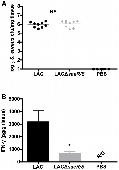
S. aureus pulsed-field gel electrophoresis type USA300 (LAC) is a major cause of CA-MRSA skin infections [34]. To investigate if saeR/S promotes IFN-γ during skin infection and to confirm our previous observation that IFN-γ production is influenced by saeR/S-regulated factors, we infected mice subcutaneously with LAC and an isogenic deletion mutant of saeR/S (LACΔsaeR/S) [33]. At 8 hours post-infection, the deletion of saeR/S did not impact bacterial load at the site of infection (Figure 5A ). These findings are consistent with our previous studies demonstrating saeR/S does not significantly influence abscess size or S. aureus burden during early skin infection (i.e. less than two days) [18], [33]. However, saeR/S did promote a significant increase in IFN-γ in LAC-infected mice compared to LACΔsaeR/S-infected mice (P<0.05; Figure 5B ). IFN-γ concentrations in un-infected mouse skin tissues were undetectable (Figure 5B ). These data are consistent with our observations of enhanced IFN-γ during peritonitis (figure 4) and are in further support that robust IFN-γ production observed during S. aureus disease is saeR/S-mediated.
Figure 5. Skin infection with LAC induces IFN-γ in a saeR/S-dependent manner.
Mice were inoculated subcutaneously with LAC, LACΔsaeR/S (1×10⁷ cfu) or DPBS for 8 hours. Infected tissues were excised and homogenized for bacterial load enumeration and IFN-γ concentration measurements. (A) Bacterial burden at the site of infection. (B) IFN-γ concentration (pg/g tissue) in affected tissues. *P<0.05 compared to MW2 in each panel as determined by t test. Results are from 3 biological replicates of 3 mice/group (n = 9) and 2 mice/PBS group (n = 6). NS = not significant; N/D = not detectable.
Discussion
In the current study, we found that absence of saeR/S significantly decreased the localized and systemic production of pro-inflammatory mediators (i.e. TNF-α, IFN-γ, IL-1β, IL-2, IL-6 and IL-17A) during invasive staphylococcal disease (Tables 1, S1 and figures 2– 4). We also observed that robust IFN-γ production was saeR/S-mediated during both S. aureus peritonitis and superficial skin infections (Table 1 and figures 4 and 5). Significantly elevated systemic pro-inflammatory cytokines coupled with the onset of mortality in wild-type-infected mice strongly indicate that saeR/S is critical for mediating sepsis, a phenotype absent in mutant-infected groups. This conclusion is supported by the ‘cytokine storm’ hypothesis, a phenomena characterized by a rapid pro-inflammatory cytokine response that correlates very strongly with coagulation dysfunction, organ failure and death [35], [36]. Our observation demonstrating both pro-inflammatory transcript abundance and protein concentration as significantly elevated in MW2-infected groups (figures 2– 4 and Tables 1 and S1), when bacterial burdens are virtually equal at the sites of inflammation (figures 1A, 1E and 4A ), suggest pro-inflammatory cytokine production stems from saeR/S-regulated factors. This idea is further supported by our data demonstrating IFN-γ production is significantly reduced in mice infected with MW2ΔsaeR/S compared to MW2, even when the bacterial burden in mutant S. aureus-infected mice is significantly increased (∼10-fold) over wild-type S. aureus-infected mice (figures 4A and 4B ). These data suggest factors regulated by saeR/S are responsible for the robust IFN-γ response observed during S. aureus infection. However, additional studies are needed to define the contribution of other S. aureus global regulators of virulence in mediating the production of IFN-γ and other pro-inflammatory cytokines.
During S. aureus infection, the role of IFN-γ has been disputed with studies demonstrating that this inflammatory mediator plays either protective or deleterious roles. For example, using a surgical wound model, McLoughlin et al [37] reported that in the absence of IFN-γ, a decreased S. aureus burden was observed at the site of infection. Zhao et al [38] observed that monoclonal antibody-neutralization of IFN-γ decreased the frequency and severity of S. aureus-mediated arthritis. Using IFN-γ-deficient mice, Sasaki et al [39] reported a decrease in S. aureus burden and an increase in survival rates using a bacteremic model of infection. Conversely, others have demonstrated that administration of exogenous IFN-γ decreased mouse mortality and reduced bacterial loads following S. aureus bacteremia [38], [40]. Our current findings correlate increased IFN-γ with elevated bacterial burdens and increased morbidity and mortality. However, it is likely that the observed effects of IFN-γ during S. aureus disease are dependent upon multiple factors, including strain of S. aureus studied and type/route of infection. Clearly, additional studies are necessary to characterize the precise role of IFN-γ as a mediator of S. aureus pathogenesis.
S. aureus produces several factors that have been implicated in pro-inflammation, including superantigens and exotoxins [41]. Superantigens non-specifically bind the major histocompatibility complex type II (MHCII) of antigen-presenting cells to T-cell receptors, causing massive T-cell activation and release of pro-inflammatory cytokines [42]. SaeR/S regulates several of these factors (18, 33, 43]. For example, Pantrangi et al [43] showed saeR/S to positively regulate staphylococcal superantigen-like genes ssl5 and ssl8. Staphylococcal enterotoxin C exhibits a superantigenic phenotype and is also regulated by saeR/S [18], [44]. SaeR/S also regulates exotoxin/cytolysin production (i.e. hla and hlg) and these factors elicit pro-inflammatory cytokines [18], [33], [45]. Finally, our previous study showed that saeR/S-regulated factors significantly enhanced the lysis of neutrophils [18]. Cellular lysis promotes the release of host-derived intracellular components and peripheral leukocytes may recognize these danger-associated molecular patterns (DAMPs) to further perpetuate the pro-inflammatory response [46]. Thus, saeR/S regulates a full repertoire of factors capable of eliciting a dysfunctional pro-inflammatory cytokine response, an essential mechanism of sepsis.
In addition to saeR/S, other S. aureus regulatory systems play pivotal roles in virulence and inflammation during infection. Both agr and sarA are essential for full virulence during invasive S. aureus disease [47], [48]. Heyer et al [49] explored the role of agr- and sarA-mediated cytokine production in a murine model of S. aureus pneumonia and reported neither regulator was required for the production of IL-8, a potent pro-inflammatory mediator of neutrophil recruitment. However, both agr and sarA did promote granulocyte-macrophage colony-stimulating factor (GM-CSF), a cytokine implicated in global pro-inflammatory responses during invasive infection [49]. Furthermore, agr has been shown to elicit cytokines and chemokines, important in leukocyte trafficking, from endothelial cells [50]. Taken together with our current report, these studies support the hypothesis that robust host inflammatory responses result from pathogen-derived factors under the influence of global regulators. However, more studies are needed to indentify the specific S. aureus factors responsible for the pro-inflammatory response.
In the current study, we report key pro-inflammatory cytokines, associated with sepsis syndromes, as being induced in response to factors regulated by the S. aureus two-component gene regulatory system, saeR/S. We hypothesize that saeR/S plays a critical role in S. aureus pathogenesis during invasive disease, likely through a synergistic mechanism of innate immune evasion [25] and the initiation of potentially dysfunctional host inflammatory pathways. This study provides a foundation for future work to identify the individual contributions of specific saeR/S-regulated factors to the host inflammatory response during staphylococcal disease.
Materials and Methods
Bacterial strains and culture
S. aureus isolates, pulsed-field gel electrophoresis type USA400 (MW2) and pulsed-field gel electrophoresis type USA300 (LAC), were selected based on clinical relevance [5]–[11], [17], [18], [33], [51]–[54]. S. aureus was cultured in tryptic soy broth (TSB) supplemented with 0.5% glucose and harvested as described elsewhere [17]. MW2, MW2saeR/S and MW2ΔsaeR/Scomp were generated in prior investigations [18]. LAC and LACΔsaeR/S were generated in prior investigations [33].
Mouse infection models
All animal studies were performed in accordance with the National Institutes of Health guidelines and approved by the Animal Care and Use Committee at Montana State University-Bozeman. For the peritonitis model, male and female BALB/c mice (aged 8–10 weeks) were purchased from commercial sources and the Montana State University Animal Resource Center. S. aureus was harvested at mid-exponential growth phase, washed in sterile Dulbecco's phosphate buffered saline (DPBS) and re-suspended in sterile DPBS at a concentration of 5×10⁶ or 5× cells per 100 µl. All mice were inoculated via intraperitoneal route (i.p.) with MW2 or MW2ΔsaeR/S and control mice received sterile DPBS.
Bacterial burdens were determined as follows: to enumerate S. aureus in the peritoneum, the peritoneal cavity was washed with 10 ml sterile HANKs' balanced salt solution using an 18 gauge needle and 10 ml syringe. Exudate was diluted in distilled water (dH20) and plated on tryptic soy agar (TSA) plates for enumeration of colony forming units (cfu). To determine S. aureus load in the blood, mice were anesthetized in isoflurane then exsanguinated via the axillary vessels. Blood was diluted in dH20 and plated on TSA. To determine S. aureus burden in selected organs, hearts and both kidneys were aseptically removed, washed in dH20 and then homogenized in dH2O. Homogenates were diluted in dH2O and plated on TSA. TSA plates were incubated overnight in 37°C; 5% CO2 and cfu counted the following day.
The survival study was performed by inoculating mice i.p. with 5×10⁷ S. aureus. Mice were monitored every 2 hours for 48 hours and then every 4 hours for an additional 24 hours. Mice were euthanized if they became immobile, exhibited labored breathing or were unable to eat or drink. Survival statistics were performed using a log-rank test.
Skin infection models were performed as described elsewhere [18], [33]. Crl;SKH1-hrBR hairless mice (Charles River) were inoculated subcutaneously with 1×10⁷ bacteria. Eight hours post-bacterial inoculation, the infected skin area was excised using a 9 mm diameter “punch.” Tissues were homogenized in sterile DPBS for bacterial enumeration and cytokine measurements by ELISA per the manufacturer's instructions (R&D Systems).
Mouse inflammatory gene expression
To compare host inflammatory transcript levels, mice were inoculated i.p. with 5×10⁷ MW2 or MW2ΔsaeR/S or control DPBS for 4 hours. Peritoneal cavities were washed as described above, and the exudate was centrifuged for 5 min at 600 x g. Pellets were resuspended in RLT lysing buffer (Qiagen) and RNA was purified using RNeasy kits as described by the manufacturer (Qiagen). Contaminating DNA was digested on-column using DNase (Qiagen). Complementary DNA (cDNA) was synthesized using ∼200 ng purified RNA and C-03 RT2 First Strand Kit (SA Biosciences). Detection of cDNA was performed using RT2 Real-time™ SYBR Green/ROX PCR master mix (SA Biosciences). Master mix with cDNA was loaded onto the Mouse Inflammatory Cytokines and Receptors RT2 Profiler™ PCR Array (SA Biosciences). Real-time PCR was performed using a 7500 Fast Real-time PCR system with Fast Real-time PCR system software v1.4.0 (Applied Biosystems). Calculated threshold (Ct) values were uploaded to the manufacturer's website (SA Biosciences; http://www.sabiosciences.com/pcr/arrayanalysis.php) for analysis using the ΔΔCt method. Fold-regulation and P values were calculated by SA Biosciences software (SA Biosciences; http://www.sabiosciences.com). Gene analyses are displayed as fold-regulation values of MW2ΔsaeR/S-infected mice relative to MW2-infected mice (Tables 1 and S1) and MW2 or MW2ΔsaeR/S-infected mice relative to DPBS-treated mice (Table S2).
Mouse cytokine assays
TNF-α and IFN-γ concentrations in peritoneal exudates were measured using sandwich ELISA per the manufacturer's instructions (R&D Systems). Mouse blood was collected as described above, and mouse serum was separated from whole blood by centrifugation. Protein levels of IFN-γ, TNF, IL-6, IL-2, IL-17a, IL-4 and IL-10 were measured using mouse Th1/Th2/Th17 Cytometric Bead Arrays per the manufacturer's instructions (BD Biosciences). All results are displayed as mean concentrations ± SEM (pg/ml).
Cytokine expression in human whole blood
Heparinzed venous human whole blood was collected from healthy individuals in accordance with an approved protocol by the Montana State University Institutional Review Board. Donors provided written consent to participate in the study. Overnight S. aureus cultures were inoculated in TSB at a ratio of 1∶100 and allowed to incubate for 6 hours to early stationary growth phase. At this time, no differences in bacterial growth exist between S. aureus strains (data not shown) [18]. Ten ml of S. aureus cell suspension was pelleted by centrifugation, and 1 ml of supernatant was sterile filtered using 0.22 µm syringe filters. Filtrate was plated to ensure the absence of S. aureus (data not shown). Filtered S. aureus supernatant was incubated with 1 ml human blood at final ratios of 1∶8, 1∶16 and 1∶32 using an end-over-end apparatus (Heto Rotamix RK) at 20 RPM for 3 hours at 37°C and 10% CO2. Plasma was isolated by centrifugation and stored at −80°C until assayed for protein concentration using human Th1/Th2/Th17 Cytometric Bead Arrays per the manufacturer's instructions (BD Biosciences).
Statistical Analyses
All data sets were analyzed using GraphPad Prism, version 4.0c for Macintosh (GraphPad Software, San Diego, CA). All mouse data sets were analyzed using paired t tests (see figure legends). Human blood data sets were analyzed using one-way analysis of variance (ANOVA) with Tukey's post-test. For bar graphs, error bars represent the standard error of the mean. Mouse survival statistics were analyzed using the log-rank test.
Supporting Information
Genes listed display fold-regulation values of MW2ΔsaeR/S-infected mice relative to MW2-infected mice (3 per group). RNA was collected from all cells washed from the peritoneum, 4 hrs post-infection. Fold-regulation values and P values calculated using SA Biosciences™ web-based software utilizing the ΔΔCt method.
(DOC)
Genes listed display fold-regulation values of MW2 and MW2ΔsaeR/S-infected mice relative to PBS-treated mice (3 per group). RNA was collected from all cells washed from the peritoneum, 4 hrs post-infection. Fold-regulation values and P values calculated using SA Biosciences™ web-based software utilizing the ΔΔCt method.
(DOC)
Acknowledgments
We would like to thank Dr. Steve Swain and Jeff Holderness for technical assistance with CBA and ELISA analyses. We would also like to thank Shannon Griffith for assistance with mouse studies. We especially thank both Drs. Mark Quinn and Tyler Nygaard for critical review of the manuscript. All acknowledged individuals are in the Department of Immunology/Infectious Diseases; Montana State University-Bozeman.
Footnotes
Competing Interests: The authors have declared that no competing interests exist.
Funding: This work was supported by the National Institutes of Health (NIH)-PAR98-072, NIH-RR020185 and a Molecular Biosciences Fellowship-Montana State University-Bozeman (to RLW). The funders had no role in study design, data collection, analysis, decision to publish, or preparation of the manuscript.
References
- 1.Lowy FD. Staphylococcus aureus infections. N Engl J Med. 1998;339:520–532. doi: 10.1056/NEJM199808203390806. [DOI] [PubMed] [Google Scholar]
- 2.Moran GJ, Krishnadasan A, Gorwitz RJ, Fosheim GE, McDougal LK, et al. Methicillin-resistant S. aureus infections among patients in the emergency department. N Engl J Med. 2006;355:666–674. doi: 10.1056/NEJMoa055356. [DOI] [PubMed] [Google Scholar]
- 3.Adem PV, Montgomery CP, Husain AN, Koogler TK, Arangelovich V, et al. Staphylococcus aureus sepsis and the Waterhouse-Friderichsen syndrome in children. N Engl J Med. 2005;353:1245–1251. doi: 10.1056/NEJMoa044194. [DOI] [PubMed] [Google Scholar]
- 4.McDougal LK, Steward CD, Killgore GE, Chaitram JM, McAllister SK, et al. Pulsed-field gel electrophoresis typing of oxacillin-resistant Staphylococcus aureus isolates from the United States; establishing a national database. J Clin Microbiol. 2003;41:5113–5120. doi: 10.1128/JCM.41.11.5113-5120.2003. [DOI] [PMC free article] [PubMed] [Google Scholar]
- 5.Centers for Disease Control and Prevention. Four pediatric deaths from community-acquired methicillin-resistant Staphylococcus aureus-Minnesota and North Dakota, 1997–1999. JAMA. 1999;282:1123. [PubMed] [Google Scholar]
- 6.Gillet Y, Issartel B, Vanhems P, Fournet JC, Lina G, et al. Association between Staphylococcus aureus strains carrying gene for Panton-Valentine leukocidin and highly lethal necrotizing pneumonia in young immunocompetent patients. Lancet. 2002;359:753–759. doi: 10.1016/S0140-6736(02)07877-7. [DOI] [PubMed] [Google Scholar]
- 7.Klevens RM, Morrison MA, Nadle J, Petit S, Gershman K, et al. Invasive methicillin-resistant Staphylococcus aureus infections in the United States. JAMA. 2007;298:1763–1761. doi: 10.1001/jama.298.15.1763. [DOI] [PubMed] [Google Scholar]
- 8.Miller LG, Perdreau-Remington F, Rieg G, Mehdi S, Perlroth, et al. Necrotizing fasciitis caused by community-associated methicillin-resistant Staphylococcus aureus in Los Angeles. N Engl J Med. 2005;352:1445–1453. doi: 10.1056/NEJMoa042683. [DOI] [PubMed] [Google Scholar]
- 9.Deleo FR, Chambers HF. Reemergence of antibiotic-resistant Staphylococcus aureus in the genomics era. J Clin Invest. 2009;119(9):2464–2474. doi: 10.1172/JCI38226. [DOI] [PMC free article] [PubMed] [Google Scholar]
- 10.Boucher HW, Corey GR. Epidemiology of methicillin-resistant Staphylococcus aureus. Clin Infect Dis. 2008;46(Suppl5):S344–S349. doi: 10.1086/533590. [DOI] [PubMed] [Google Scholar]
- 11.Matin GS, Mannino DM, Eaton S, Moss M. The epidemiology of sepsis in the United States from 1979 though 2000. N Engl J Med. 2003;348:1546–1554. doi: 10.1056/NEJMoa022139. [DOI] [PubMed] [Google Scholar]
- 12.Lappin E, Ferguson AJ. Gram-positive toxic shock syndromes. Lancet Infect Dis. 2009;9:281–290. doi: 10.1016/S1473-3099(09)70066-0. [DOI] [PubMed] [Google Scholar]
- 13.Hotchkiss RS, Coopersmith CM, McDunn JE, Ferguson TA. The sepsis seesaw: tilting toward imunosuppression. Nat Med. 2009;15:496–497. doi: 10.1038/nm0509-496. [DOI] [PMC free article] [PubMed] [Google Scholar]
- 14.Rittirsch D, Flierl MA, Ward PA. Harmful molecular mechanisms of sepsis. Net Rev Immunol. 2008;8:776–787. doi: 10.1038/nri2402. [DOI] [PMC free article] [PubMed] [Google Scholar]
- 15.Nakane A, Okamoto M, Asano M, Kohanawa M, Minagawa T. Endongenous gamma interferon, tumor necrosis factor, and interleukin−6 in Staphylococcus aureus infection in mice. Infect Immun. 1995;63:1165–1172. doi: 10.1128/iai.63.4.1165-1172.1995. [DOI] [PMC free article] [PubMed] [Google Scholar]
- 16.Sasaki S, Miura T, Nishikawa S, Yamada K, Hiasue M, et al. Protective role of nitric oxide in Staphylococcus aureus in mice. Infect Immun. 1998;66:1017–1022. doi: 10.1128/iai.66.3.1017-1022.1998. [DOI] [PMC free article] [PubMed] [Google Scholar]
- 17.Voyich JM, Sturdevant DE, Braughton KR, Whitney AR, Said-Salim B, et al. Insights into mechanisms used by Staphylococcus aureus to avoid destruction by human neutrophils. J Immunol. 2005;175:3907–3919. doi: 10.4049/jimmunol.175.6.3907. [DOI] [PubMed] [Google Scholar]
- 18.Voyich JM, Vuong C, DeWald M, Nygarrd TK, Griffith S, et al. The SaeR/S gene regulatory system is essential for innate immune evasion by Staphylococcus aureus. J Infect Dis. 2009;199:1698–1706. doi: 10.1086/598967. [DOI] [PMC free article] [PubMed] [Google Scholar]
- 19.Brown KA, Brain SD, Pearson JD, Edgeworth JD, Lewis SM, et al. Neutrophils in development of multiple organ failure. Lancet. 2006;368:157–169. doi: 10.1016/S0140-6736(06)69005-3. [DOI] [PubMed] [Google Scholar]
- 20.Chavakis T, Preissner KT, Herrman M. The anti-inflammatory activities of Staphylococcus aureus. Trends Immunol. 2007;28:408–418. doi: 10.1016/j.it.2007.07.002. [DOI] [PubMed] [Google Scholar]
- 21.Alves-Filho JC, de Freitas A, Spiller F, Souto FO, Cunha FQ. The role of neutrophils in severe sepsis. Shock. 2008;30:3–9. doi: 10.1097/SHK.0b013e3181818466. [DOI] [PubMed] [Google Scholar]
- 22.Cheung AL, Bayer AS, Zhang G, Gresham H, Xiong YQ. Regulation of virulence determinants in vitro and in vivo in Staphylococcus aureus. FEMS Immunol Med Microbiol. 2004;40:1–9. doi: 10.1016/S0928-8244(03)00309-2. [DOI] [PubMed] [Google Scholar]
- 23.Bronner S, Monteil H, Prevost G. Regulation of virulence determinants in Staphylococcus aureus: complexity and applications. FEMS Microbial Rev. 2004;28:183–200. doi: 10.1016/j.femsre.2003.09.003. [DOI] [PubMed] [Google Scholar]
- 24.Novick RP. Autoinduction and signal transduction in the regulation of staphylococcal virulence. Mol Microbiol. 2003;48:1429–1449. doi: 10.1046/j.1365-2958.2003.03526.x. [DOI] [PubMed] [Google Scholar]
- 25.Giraudo AT, Martinez GL, Calzolari A, Nagel R. Characterization of a Tn925-induced mutant of Staphylococcus aureus altered exoprotein production. J Basic Microbiol. 1994;34:317–322. doi: 10.1002/jobm.3620340507. [DOI] [PubMed] [Google Scholar]
- 26.Giraudo AT, Rampone H, Calzolari A, Nagel R. Phenotypic characterization and virulence of sae- agr- mutant of Staphylococcus aureus. Can J Microbiol. 1996;42:120–123. doi: 10.1139/m96-019. [DOI] [PubMed] [Google Scholar]
- 27.Harraghy N, Kormanec J, Wolz C, Homerova D, Goerke C, et al. Sae is essential for expression of the staphylococcal adhesins Eap and Emp. Microbiology. 2005;151:1789–1800. doi: 10.1099/mic.0.27902-0. [DOI] [PubMed] [Google Scholar]
- 28.Kuroda H, Kuroda M, Chi L, Hiramatsu K. Subinhibitory concentrations of beta-lactam induce haemolytic activity in Staphylococcus aureus through the SaeRS two-component system. FEMS Microbiol Lett. 2007;268:98–105. doi: 10.1111/j.1574-6968.2006.00568.x. [DOI] [PubMed] [Google Scholar]
- 29.Liang X, Yu C, Sun J, Liu H, Landwehr C, et al. Inactivation of a two-componet signal transduction system, SaeRS, eliminates adherence and attenuates virulence of Saphylococcus aureus. Infect Immun. 2006;268:98–105. doi: 10.1128/IAI.00322-06. [DOI] [PMC free article] [PubMed] [Google Scholar]
- 30.Novick RP, Jiang D. The staphylococcal SaeRS system coordinates environmental signals with agr quorum sensing. Microbiology. 2003;149:2709–2717. doi: 10.1099/mic.0.26575-0. [DOI] [PubMed] [Google Scholar]
- 31.Rogash K, Ruhmling V, Pane-Farre J, Hoper D, Weinberg C, et al. Influence of the two-component system SaeRS on global gene expression in two different Staphylococcus aureus strains. J Bacteriol. 2006;188:7742–58. doi: 10.1128/JB.00555-06. [DOI] [PMC free article] [PubMed] [Google Scholar]
- 32.Xiong YQ, Willard J, Yeaman MR, Cheung AL, Bayer AS. Regulation of Staphylococcus aureus α-toxin gene (hla) expression by agr, sarA, and sae invitro and in experimental endocarditis. J Infect Dis. 2006;194:1267–1275. doi: 10.1086/508210. [DOI] [PubMed] [Google Scholar]
- 33.Nygaard TK, Pallister KB, Ruzevich P, Griffith S, Vuong C, et al. SaeR binds a consensus sequence within virulence gene promotors to advance USA300 pathogenesis. J Infect Dis. 2010;201:241–254. doi: 10.1086/649570. [DOI] [PMC free article] [PubMed] [Google Scholar]
- 34.Moran GJ, Krishnadasan A, Gorwitz RJ, Fosheim GE, McDougal LK, et al. Methicillin-resistant S. aureus infections among patients in the hospital. New Engl J Med. 2006;355:666–674. doi: 10.1056/NEJMoa055356. [DOI] [PubMed] [Google Scholar]
- 35.Sriskandan S, Altmann DM. The immunology of sepsis. Pathology. 2008;214:211–223. doi: 10.1002/path.2274. [DOI] [PubMed] [Google Scholar]
- 36.Cohen J. The immunopathogenesis of sepsis. Nature. 2002;420:885–891. doi: 10.1038/nature01326. [DOI] [PubMed] [Google Scholar]
- 37.McLoughlin RM, Lee JC, Kasper DL, Tzianabos AO. IFN-γ regulated chemokine production determines the outcome of Staphylococcus aureus infection. J Immunol. 2008;181:1323–1332. doi: 10.4049/jimmunol.181.2.1323. [DOI] [PubMed] [Google Scholar]
- 38.Zhao YX, Nilsson IM, Tarkowski A. The dual role of interferon-γ in experimental Staphylococcus aureus septicaemia versus arthritis. Immunol. 1998;1:80–85. doi: 10.1046/j.1365-2567.1998.00407.x. [DOI] [PMC free article] [PubMed] [Google Scholar]
- 39.Sasaki S, Nishikawa S, Miura T, Mizuki M, Yamada K, et al. Interleukin-4 and interleukin-10 are involved in host resistance to Staphylococcus aureus infection through regulation of gamma interferon. Infect Immun. 2000;68:2424–2430. doi: 10.1128/iai.68.5.2424-2430.2000. [DOI] [PMC free article] [PubMed] [Google Scholar]
- 40.Rozalska B, Wadstrom T. Interferon-γ, interleukin-1 and tumour necrosis factor-α during experimental murine staphylococcal infection. FEMS Immunol Med Microbiol. 1993;7:145–152. doi: 10.1111/j.1574-695X.1993.tb00393.x. [DOI] [PubMed] [Google Scholar]
- 41.Schlievert PM, Strandberg KL, Lin YC, Peterson ML, Leung DYM. Secreted virulence factor comparison between methicillin-resistant and methicillin-sensitive Staphylococcus aureus, and its relevance to atopic dermatitis. J Allergy Clin Immunol. 2010;125:39–49. doi: 10.1016/j.jaci.2009.10.039. [DOI] [PMC free article] [PubMed] [Google Scholar]
- 42.Cohen J. The immunopathogenesis of sepsis. Nature. 2002;40:885–891. doi: 10.1038/nature01326. [DOI] [PubMed] [Google Scholar]
- 43.Pantrangi M, Singh VK, Wolz C, Shukla SK. Staphylococcal superantigen-like genes, ssl5 and ssl8, are positively regulated by Sae and negatively by Agr in the Newman strain. FEMS Microbiol Lett. 2010;308:175–184. doi: 10.1111/j.1574-6968.2010.02012.x. [DOI] [PMC free article] [PubMed] [Google Scholar]
- 44.McCormick JK, Warwood JM, Schlievert PM. Toxic shock syndrome and bacterial superantigens: an update. Annu Rev Microbiol. 2001;55:77–104. doi: 10.1146/annurev.micro.55.1.77. [DOI] [PubMed] [Google Scholar]
- 45.Dinges MM, Orwin PM, Schlievert PM. Exotoxins of Staphylococcus aureus. Clin Microbiol Rev. 2000;13:16–34. doi: 10.1128/cmr.13.1.16-34.2000. [DOI] [PMC free article] [PubMed] [Google Scholar]
- 46.Bianchi ME. DAMPs, PAMPs, and alarmins: all we need to know about danger. J Leukocyte Biol. 2007;81:1–5. doi: 10.1189/jlb.0306164. [DOI] [PubMed] [Google Scholar]
- 47.Booth MC, Cheung AL, Hatter KL, Jett BD, Callegan MC, et al. Staphylococcal accessory regulator (sar) in conjunction with agr contributes to Staphylococcus aureus virulence in endophthalmitis. Infect Immun. 1997;65:1550–1556. doi: 10.1128/iai.65.4.1550-1556.1997. [DOI] [PMC free article] [PubMed] [Google Scholar]
- 48.Cheung AL, Eberhardt KJ, Chung E, Yeaman MR, Sullam PM. Diminished virulence of a sar −/agr− mutant of Staphylococcus aureus in the rabbit model of endocarditis. J Clin Investig. 1994;94:1815–1822. doi: 10.1172/JCI117530. [DOI] [PMC free article] [PubMed] [Google Scholar]
- 49.Heyer G, Saba S, Adamo R, Rush W, Soong G. Stpahylococccus aureus agr and sarA functions are required for invasive infection but not inflammatory responses in the lung. Infect Immun. 2002;70:127–133. doi: 10.1128/IAI.70.1.127-133.2002. [DOI] [PMC free article] [PubMed] [Google Scholar]
- 50.Grundmeier M, Tuchscherr L, Bruck M, Viemann D, Roth J. Staphylococcal strains vary greatly in their ability to induce an inflammatory response in endothelial cells. J Infect Dis. 2010;201:871–880. doi: 10.1086/651023. [DOI] [PubMed] [Google Scholar]
- 51.Baba T, Takeuchi F, Kuroda M, Yuzawa H, Aoki K, et al. Genome and virulence determinants of high virulence community-acquired MRSA. Lancet. 2002;359:1819–1827. doi: 10.1016/s0140-6736(02)08713-5. [DOI] [PubMed] [Google Scholar]
- 52.Fridkin SK, Hageman JC, Morrison, Sanza LT, Como-Sabetti K, et al. Methicillin-resistant staphylococcus aureus disease in three communities. N Engl J Med. 2005;352:1436–1444. doi: 10.1056/NEJMoa043252. [DOI] [PubMed] [Google Scholar]
- 53.Herold BC, Immergluck LC, Maranan MC, Lauderdale DS, Gaskin RE, et al. Community-acquired methicillin-resistant Staphylococcus aureus in children with no identified predisposing risk. JAMA. 1998;279:593–598. doi: 10.1001/jama.279.8.593. [DOI] [PubMed] [Google Scholar]
- 54.Kasakova SV, Hageman JC, Matava M, Srinivasan A, Phelan L, et al. A clone of methicillin-resistant Staphylococcus aureus among professional football players. N Engl J Med. 2005;352:468–475. doi: 10.1056/NEJMoa042859. [DOI] [PubMed] [Google Scholar]
Associated Data
This section collects any data citations, data availability statements, or supplementary materials included in this article.
Supplementary Materials
Genes listed display fold-regulation values of MW2ΔsaeR/S-infected mice relative to MW2-infected mice (3 per group). RNA was collected from all cells washed from the peritoneum, 4 hrs post-infection. Fold-regulation values and P values calculated using SA Biosciences™ web-based software utilizing the ΔΔCt method.
(DOC)
Genes listed display fold-regulation values of MW2 and MW2ΔsaeR/S-infected mice relative to PBS-treated mice (3 per group). RNA was collected from all cells washed from the peritoneum, 4 hrs post-infection. Fold-regulation values and P values calculated using SA Biosciences™ web-based software utilizing the ΔΔCt method.
(DOC)


