Anthrax lethal toxin exemplifies one among many systems evolved by pathogenic bacteria for transporting proteins across membranes to the cytosol of mammalian cells.[1] The transported proteins so called effector proteins are enzymes that modify intracellular substrates, perturbing mammalian metabolism in ways that benefit the bacteria at the expense of the host. Anthrax lethal toxin is an ensemble of two large soluble proteins: the Lethal Factor (LF, 90 kDa), a zinc protease,[2] and Protective Antigen (PA; 83 kDa), a receptor-binding/pore-forming protein.[1] PA binds to receptors[3] on host cells and is cleaved by a furin-family protease[4] to an active 63 kDa form (PA63),[5] which self-assembles into ring-shaped heptameric[6] and octameric[7] oligomers, termed prepores. The prepores bind LF, forming complexes that are then endocytosed and delivered to the endosome. There, acidification induces the prepore moieties to undergo conformational rearrangement to membrane-spanning pores.[1] The pores then transport bound LF across the membrane to the cytosol, where it inactivates selected target proteins.[8] Edema Factor (EF), the enzymatic moiety of anthrax edema toxin,[9] is transported to the cytosol by a similar mechanism.[1]
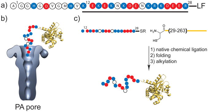
LF binds to PA63 pores via its N-terminal domain, termed LFN, orienting the protein’s N-terminal unstructured, highly charged segment (~29 residues) at the pore entrance. This unstructured segment is believed to enter the pore lumen and interact with the Phe clamp,[10] a structure formed by the Phe427 side chains, thereby blocking ion conductance and initiating N- to C-terminal threading of the polypeptide through the pore (Figure 1).[11] Removal of the first 12 residues of this unstructured segment was found to have little effect on translocation, but truncations of more than 27 residues altered the ability of LFN to block ion conductance and to be translocated through the pore.[11–12]
Figure 1.
Interaction of the N terminus of LF with PA pore. a) The N-terminal 28 amino acid residues of LF, with the highly charged region investigated in this report underlined. b) Illustration of the N-terminal binding domain of Lethal Factor, LFN(1-263), yellow, bound to PA pore. The pore structure was reconstructed from single-pore images obtained by electron microscopy.[18] The blue (basic), red (negative), and white (neutral) circles represent the unstructured N-terminal stretch of LF(12-28), which was not present in the X-ray structure of LF (PDB 1J7N). c) Semisynthesis strategy used to prepare LFN constructs with modifications in the (12-28) amino acid stretch.
In the current study, to probe how structural and electrostatic changes in translocation-competent polypeptides affected translocation, we used native chemical ligation[13] to prepare truncated variants of LFN (residues 12-263 of the native domain), in which residues 12-28 were replaced by synthetic peptides (Figure 1). We also fused each of the LFN variants to the N terminus of the catalytic domain of diphtheria toxin (DTA), to permit the effects of the modifications to be measured on mammalian cells. DTA blocks protein synthesis when introduced into the cytosol of these cells.[14]
Six semisynthetic variants of LFN, SSv1-SSv6, were prepared.[12] Briefly, LFN(12-28)α thioesters were synthesized by manual Boc in-situ neutralization solid phase peptide synthesis[15] and purified by RP-HPLC. N29C-LFN(29-263) was prepared from a His6-SUMO-N29C-LFN(29-263) protein fusion recombinantly expressed in E. coli. Standard ligation conditions were used to couple the LFN(12-28)α thioester and N29C-LFN(29-263), yielding the reaction product N29C-LFN(12-263) (Figure 1C). N29C-LFN(12-263) was alkylated with 2-bromoacetamide to give N29ΨQ-LFN(12-263) (ΨQ = pseudohomoglutamine). LFN-DTA variants were prepared by use of recombinant N29C-LFN(29-263)-DTA[C186S]. Each analogue was characterized by analytical RP-HPLC, high-resolution MS, and circular dichroism (supplementary information). The circular dichroism spectra of all variants were similar to that of LFN(12-263), implying the variants were correctly folded.
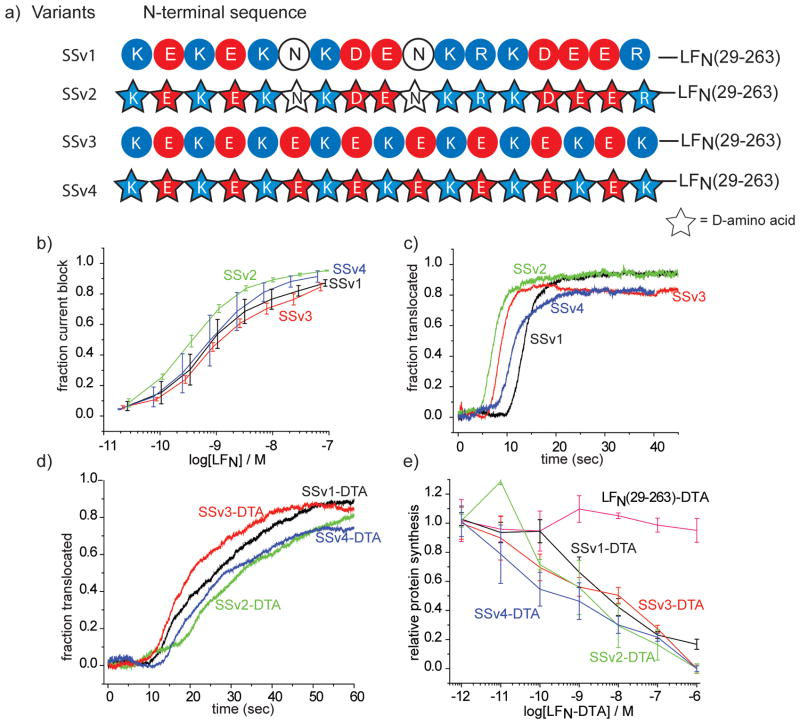
To investigate the possibility that chirality of amino acids could affect translocation, we prepared SSv2, a variant of wild-type LFN in which the residue 12-28 peptide was synthesized from D amino acids (Figure 2). No significant differences were observed between the D and L variants of LFN in ability to inhibit ion conductance through PA pores formed in planar phospholipid bilayers (Figure 2b) or to translocate through those pores in response to a transmembrane pH gradient (cis pH 5.5; trans pH 7.5) (Figure 2c).[16] For all planar phospholipid bilayer experiments the applied potential was Δ Ψ = 20 mV (Δ Ψ = Ψcis − Ψtrans, where Ψtrans ≡ 0). Further, there was no difference between the SSv2-DTA and the wild-type SSv1-DTA fusion proteins in ability to translocate in-vitro or to inhibit protein synthesis in CHO-K1 cells.
Figure 2.
Stereochemical and charge effects on the interaction of of the LFN N-terminus with PA pore. a) Chemical framework of SSv1-SSv4. b) The fraction ion conductance block of PA pore by LFN variants at Δ Ψ = 20 mV (Δ Ψ = Ψcis − Ψtrans, where Ψtrans ≡ 0). For the procedure used to determine the fraction ion conductance block shown in Figure 2 and Figure 3 see the supplementary information. Each blocking experiment was repeated three times. c) Acid triggered translocation of LFN variants through PA pore in response to a pH gradient of ~2 units (cis pH 5.5; trans pH 7.5) at Δ Ψ = 20 mV. d) Translocation of LFN-DTA variants through PA pore in response to a pH gradient of ~2 units (cis pH 5.5; trans pH 7.5) at Δ Ψ = 20 mV. e) Fraction protein synthesis inhibition by LFN-DTA variants in CHO-K1 cells. LFN-DTA variants were incubated at the indicated concentration for 15–17 minutes at 37°C and 5 % CO2. Then, 3H-leucine was added, and after 1 hour the amount of tritium incorporated into cellular protein was measured. The data shown are the average of three experiments. Full experimental details are described in the supplementary information.
The residue 12-28 sequence of native LFN is comprised of 8 basic, 7 acidic, and 2 neutral residues (Figure 1a). When we replaced this segment with a simple sequence of alternating basic and acidic residues (9 Lys and 8 Glu) (Figure 2a), generating variant SSv3, we found no significant change in the ability of the protein to block ion conductance or to be translocated. Also, the corresponding DTA fusion protein, SSv3-DTA, behaved essentially identically to the controls in the translocation assay in bilayers and the cytotoxicity assay in cell culture. Further, SSv4 and SSv4-DTA, having the same alternating Lys/Glu sequence, but synthesized with D amino acids, showed no differences in activity from SSv3 and SSv3-DTA, respectively.
Translocation through PA pores formed in planar bilayers can be driven by applying a transmembrane pH gradient (low pH cis, higher pH trans).[16] This finding, together with the fact that the lumen of the pore is negatively charged and discriminates against the passage of anions, suggested a charge state-dependent Brownian ratchet mechanism.[10,16–17] PA pore is cation selective suggesting the lumen is negatively charged at pH 5.5. According to this model a negative electrostatic barrier within the pore serves to retard the passage of acidic residues of a translocating polypeptide when their side chains are deprotonated. Protonation renders the side chains neutral, allowing the polypeptide to pass the barrier by random thermal motion. Once the residue has passed, deprotonation renders the side chain once again negative, hindering back diffusion across the barrier. A proton gradient across the membrane would therefore be expected to impose directionality upon the thermal motion, driving translocation by virtue of the greater probability of acidic residues being in a protonated state at lower pH values.
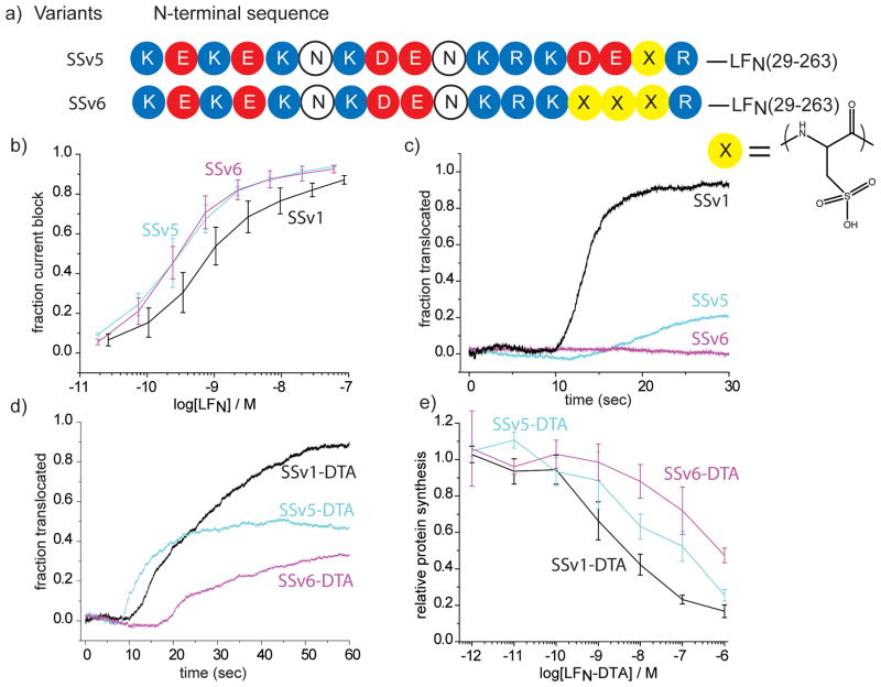
As a test of this hypothesis we prepared variants of LFN(12-263) in which selected acidic residues were replaced with the unnatural amino acid, cysteic acid, which has a negatively charged side chain (pKa -1.9) that protonates only at pH values below the physiological range. In SSv5 we replaced Glu27 alone with cysteic acid, and in SSv6, we replaced three sequential acidic residues: Asp25, Glu26, and Glu27 (Figure 3). Both constructs bound to PA pores and blocked ion conductance as effectively as the wild-type control. Translocation of SSv5 in response to Δ pH was strongly inhibited, however, and with SSv6 no translocation was observed. Like SSv5 and SSv6, the corresponding DTA fusion proteins bound to PA pores in planar bilayers and blocked ion conductance effectively. The LFN-DTA variants showed significant levels of translocation activity in bilayers and of cytotoxicity on cells when combined with PA, but the constructs with a single cysteic acid residue were markedly less active than the wild type constructs, and those with three were even less active. Thus, although non-titratable negative charged residues did not abrogate translocation, they clearly served as a barrier to the process.
Figure 3.
Translocation of LFN variants through PA pore is hindered by cysteic acid. a) Chemical framework of SSv5 and SSv6 (X = cysteic acid). b) The fraction ion conductance block of PA pore by SSv5 and SSv6 at Δ Ψ = 20 mV. c) Translocation of LFN variants through PA pore in response to a pH gradient of ~2 units (cis pH 5.5; trans pH 7.5) at Δ Ψ = 20 mV. d) Acid triggered translocation of LFN-DTA variants through PA pore in response to a pH gradient of ~2 units (cis pH 5.5; trans pH 7.5) at Δ Ψ = 20 mV. e) Fraction protein synthesis inhibition by LFN-DTA variants in CHO-K1 cells. The procedure used was identical to that described in Figure 2e.
Semisynthesis provides the opportunity to test the functional consequences of incorporating chemical structures beyond the standard set of L amino acids into proteins. Our finding that the LFN domain functioned equally well when the segment corresponding to residues 12-28 were built from D amino acids as from L amino acids indicates that the N terminal unstructured segment of LF does not adopt a conformation that interacts with the pore in a stereospecific manner during protein translocation. By extension, we suggest that this property is likely to be characteristic of the entire protein (notwithstanding that the globular portion of LFN binds stereospecifically to sites at the mouth of the pore before it unfolds as a prelude to translocation). Further, if polypeptide segments adopt an α-helical structure during translocation through the β-barrel stem of the pore,[16] a left-handed helix must be accommodated as well as a right-handed one. These concepts are consistent with the notions that the protein-translocation pathway must accommodate all side-chain chemistries of the translocating protein and that interactions with the pore cannot be too strong, lest they arrest the translocation process.
Semisynthesis also allowed us to incorporate a non-natural amino acid, L cysteic acid, as a test of the charge state-dependent Brownian ratchet mechanism proposed earlier.[10, 16] The side chain of cysteic acid would be predicted to be negatively charged under the conditions of our experiment and thus retard translocation. The prediction that replacing an existing acidic residue with cysteic acid would inhibit translocation significantly and that replacing three would be even more inhibitory was fulfilled, supporting the proposed mechanism. Another test, in which negatively charged side chains were introduced by derivatization of introduced Cys residues, also gave results supportive of the mechanism.[17a]
Footnotes
This research was supported by NIAID grants RO1-AI022021 and AI057159. The recombinant proteins employed in the study were prepared in the Biomolecule Production Core of the New England Regional Center of Excellence, supported by NIH grant number AI057159. We thank A. Fischer and A. Barker for helpful suggestions regarding cell cytotoxicity experiments and S. Walker and E. Doud for providing access to their ESI-QTOF MS.
Supporting information for this article is available on the WWW under http://www.angewandte.org or from the author.
References
- 1.Young JA, Collier RJ. Annu Rev Biochem. 2007;76:243. doi: 10.1146/annurev.biochem.75.103004.142728. [DOI] [PubMed] [Google Scholar]
- 2.a) Duesbery NS, Webb CP, Leppla SH, Gordon VM, Klimpel KR, Copeland TD, Ahn NG, Oskarsson MK, Fukasawa K, Paull KD, Vande Woude GF. Science. 1998;280:734. doi: 10.1126/science.280.5364.734. [DOI] [PubMed] [Google Scholar]; b) Vitale G, Pellizzari R, Recchi C, Napolitani G, Mock M, Montecucco C. Biochem Biophys Res Commun. 1998;248:706. doi: 10.1006/bbrc.1998.9040. [DOI] [PubMed] [Google Scholar]
- 3.a) Bradley KA, Mogridge J, Mourez M, Collier RJ, Young JA. Nature. 2001;414:225. doi: 10.1038/n35101999. [DOI] [PubMed] [Google Scholar]; b) Scobie HM, Rainey GJ, Bradley KA, Young JA. Proc Natl Acad Sci U S A. 2003;100:5170. doi: 10.1073/pnas.0431098100. [DOI] [PMC free article] [PubMed] [Google Scholar]
- 4.Molloy SS, Bresnahan PA, Leppla SH, Klimpel KR, Thomas G. J Biol Chem. 1992;267:16396. [PubMed] [Google Scholar]
- 5.Klimpel KR, Molloy SS, Thomas G, Leppla SH. Proc Natl Acad Sci U S A. 1992;89:10277. doi: 10.1073/pnas.89.21.10277. [DOI] [PMC free article] [PubMed] [Google Scholar]
- 6.Milne JC, Furlong D, Hanna PC, Wall JS, Collier RJ. J Biol Chem. 1994;269:20607. [PubMed] [Google Scholar]
- 7.Kintzer AF, Thoren KL, Sterling HJ, Dong KC, Feld GK, Tang, Zhang TT, Williams ER, Berger JM, Krantz BA. J Mol Biol. 2009;392:614. doi: 10.1016/j.jmb.2009.07.037. [DOI] [PMC free article] [PubMed] [Google Scholar]
- 8.Abrami L, Lindsay M, Parton RG, Leppla SH, van der Goot FG. J Cell Biol. 2004;166:645. doi: 10.1083/jcb.200312072. [DOI] [PMC free article] [PubMed] [Google Scholar]
- 9.Leppla SH. Proc Natl Acad Sci U S A. 1982;79:3162. doi: 10.1073/pnas.79.10.3162. [DOI] [PMC free article] [PubMed] [Google Scholar]
- 10.Krantz BA, Melnyk RA, Zhang S, Juris SJ, Lacy DB, Wu Z, Finkelstein A, Collier RJ. Science. 2005;309:777. doi: 10.1126/science.1113380. [DOI] [PMC free article] [PubMed] [Google Scholar]
- 11.Zhang S, Finkelstein A, Collier RJ. Proc Natl Acad Sci U S A. 2004;101:16756. doi: 10.1073/pnas.0405754101. [DOI] [PMC free article] [PubMed] [Google Scholar]
- 12.Pentelute BL, Barker AP, Janowiak BE, Kent SB, Collier RJ. ACS Chem Biol. 2010;5:359. doi: 10.1021/cb100003r. [DOI] [PMC free article] [PubMed] [Google Scholar]
- 13.Dawson PE, Muir TW, Clark-Lewis I, Kent SB. Science. 1994;266:776. doi: 10.1126/science.7973629. [DOI] [PubMed] [Google Scholar]
- 14.a) Sellman BR, Mourez M, Collier RJ. Science. 2001;292:695. doi: 10.1126/science.109563. [DOI] [PubMed] [Google Scholar]; b) Sellman BR, Nassi S, Collier RJ. J Biol Chem. 2001;276:8371. doi: 10.1074/jbc.M008309200. [DOI] [PubMed] [Google Scholar]
- 15.Schnolzer M, Alewood P, Jones A, Alewood D, Kent SB. Int J Pept Protein Res. 1992;40:180. doi: 10.1111/j.1399-3011.1992.tb00291.x. [DOI] [PubMed] [Google Scholar]
- 16.Krantz BA, Finkelstein A, Collier RJ. J Mol Biol. 2006;355:968. doi: 10.1016/j.jmb.2005.11.030. [DOI] [PubMed] [Google Scholar]
- 17.a) Basilio D, Juris SJ, Collier RJ, Finkelstein A. J Gen Physiol. 2009;133:307. doi: 10.1085/jgp.200810170. [DOI] [PMC free article] [PubMed] [Google Scholar]; b) Finkelstein A. Philos Trans R Soc Lond B Biol Sci. 2009;364:209. doi: 10.1098/rstb.2008.0126. [DOI] [PMC free article] [PubMed] [Google Scholar]
- 18.Katayama H, Janowiak BE, Brzozowski M, Juryck J, Falke S, Gogol EP, Collier RJ, Fisher MT. Nat Struct Mol Biol. 2008;15:754. doi: 10.1038/nsmb.1442. [DOI] [PMC free article] [PubMed] [Google Scholar]


