Abstract
Spontaneous spinal epidural haematoma (SSEH) is a rare clinical entity, especially in infants, in whom only a few cases have been reported. In a paediatric emergency setting, SSEH should be considered as part of the differential diagnosis for acute extremity weakness and paraesthesia. Epidural vascular malformations are often suspected in these cases but have rarely been demonstrated. The authors report herein a case of SSEH in a 9-year-old boy arising from an epidural vascular malformation. He initially presented with sudden intense cervicodorsal pain followed by hypotonic lower extremities and progressive motor weakness, with no sensory change. The MRI showed an acute extradural haematoma extending from C7 to T4 with compression of the spinal cord. After submission to decompression surgery, he presented full recovery in 1 month. The histopathological analysis revealed a vascular malformation.
Background
Spontaneous spinal epidural haematomas (SSEH) are a rare clinical entity with an incidence of 0.1 per 100 000 per year.1–4 There is preponderance in older-aged patients, especially those receiving anticoagulant drugs and have bleeding and coagulation defect tendencies. During childhood, they are even more unlikely5–7 with only few cases reported8–19 and their causes are frequently unknown.5 20 The youngest case reported is on a 22-month-old male infant.21 We report a rare case of spontaneous haemorrhage from an epidural vascular malformation in a 9-year-old boy. The haematoma and spinal cord compression was diagnosed by MRI, and the pathology was confirmed by microscopic analysis.
Case presentation
This 9-year-old white male presented with a sudden intense cervicodorsal pain when he rose at dawn. After roughly 30 min, he demonstrated progressive muscular weakness of the lower extremities. There was no history of trauma to the head or spine, nor any bleeding disorder or pertinent family history. He did not have any fever, rhinorrhoea or other upper respiratory tract symptoms.
On arrival at our institution, the pain had subsided but he was non-ambulatory. The neurological examination showed normal upper extremity motor and tone, and normal cranial nerve examination. He demonstrated hypotonic lower extremities, and motor weakness in the lower limbs with no antigravity movement. Deep tendon reflexes were absent in the legs. Sensory evaluation was normal. A complete laboratory work-up, including bleeding time, prothrombin and thromboplastin time, plaque count, serum electrolytes, blood glucose level and calcium, was within normal limits.
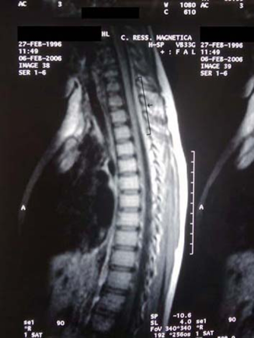
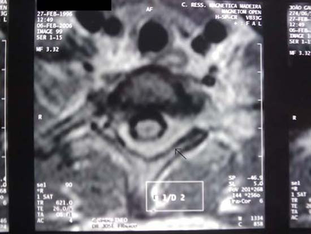
Plain radiographs of the chest were normal. The CT of the spine did not show any evidence of destructive change or fracture of the vertebral body and neural arch. MRI of the spine revealed an extradural mass, compatible with acute haematoma, extending from the C7 to T4 levels, compressing the spinal cord (figures 1 and 2).
Figure 1.

MRI sagittal T1-weighed images: left posterolateral extradural isointense mass spanning C4 to T7 vertebral segments (arrow) and appears inhomogeneous with compression of the spinal cord.
Figure 2.

MRI axial T1-weighed: compression of the spinal cord that is displaced anteriorly and to the right by the mass (arrow).
The patient underwent surgery with C7 to T4 laminectomies and decompression of the extradural haematoma at which time anomalous blood vessels were detected and sent for histopathological examination. Microscopy analysis of the surgical specimen revealed an arteriovenous vascular malformation.
After surgery, the patient began a rehabilitation programme with an uneventful and progressive recovery and was released 26 days after admission with a normal neurological exam.
Investigations
MRI of the spine revealed an extradural mass, compatible with acute haematoma, extending from the C7 to T4 levels, compressing the spinal cord.
Microscopy analysis of the surgical specimen revealed an arteriovenous vascular malformation.
Treatment
The patient underwent surgery with C7 to T4 laminectomies and decompression of the extradural haematoma at which time anomalous blood vessels were detected and sent for histopathological examination.
Outcome and follow-up
After surgery, the patient began a rehabilitation programme with an uneventful recovery and a normal neurological examination within a month.
Discussion
SSEH in children are rare lesions of unclear origin5–7 and in only 10% of the patients is the underlying cause identified.22 A vascular malformation that has been overlooked or obliterated is one possible explanation in idiopathic cases.23
In the case presented, a vascular malformation caused the acute clinical features with no significant medical history, no history of trauma and no warning signs. The presenting symptoms of SSEH are usually a consequence of spinal cord compression and roots compression, resulting in a sudden onset of back and neck pain. In adults, the pain and neurological deficits are characteristically specific and localised, but in children, particularly those under the age of 2 years, the initial symptoms are non-specific. Older children may report back pain, weakness or paraesthesia, whereas infants may have irritability or excessive crying. In some children, there are no significant symptoms to suggest SSEH. Therefore, accurate and early diagnosis may be difficult.8 14 20
The majority of spinal arteriovenous malformations are dural (up to 70%).24 Epidural vascular malformations are rare, particularly in children, and are fed by radicular vessels accompanying the exiting nerve root, more commonly in the lower cervical and thoracic spine, where radicular arteries are more prominent.5 22 23
MRI is the choice of imaging modality and plays an important role in diagnosis and evaluation of SSEH14 25 by simplifying the diagnostic work-up of epidural compression, as it can differentiate soft tissue from vascular and bony pathology22 26–28
Rapid surgical evacuation has been recommended as a treatment of choice for symptomatic SSEH8 12 29. The outcome of patients treated surgically is generally good5 20 23 26. The most significant factors that determine neurological outcome are interval from symptom onset to surgery, and the degree of neurological deficits prior to surgery. The shorter interval from symptom onset to surgery and minor neurological deficit has been reported to result in more favourable outcome.29 The overall good outcome associated with epidural arteriovenous malformations is in distinct contrast to the poorer prognosis associated with dural vascular malformations 26 30 and is likely related to the alteration of spinal cord blood flow rather than to the spinal cord compression.31
Learning points.
-
▶
Spontaneous epidural haematomas should be considered in acute extremity weakness and paraesthesia in children
-
▶
Prompt MRI of the brain and spine is essential for correct diagnosis
-
▶
Immediate surgical decompression offers the best chance for improved outcome
-
▶
Prognosis of epidural vascular malformations is favourable with complete recovery.
Footnotes
Competing interests None.
Patient consent Obtained.
References
- 1.Groen RJ, van Alphen HA. Operative treatment of spontaneous spinal epidural hematomas: a study of the factors determining postoperative outcome. Neurosurgery 1996;39:494–508; discussion 508–9 [DOI] [PubMed] [Google Scholar]
- 2.Holtås S, Heiling M, Lönntoft M. Spontaneous spinal epidural hematoma: findings at MR imaging and clinical correlation. Radiology 1996;199:409–13 [DOI] [PubMed] [Google Scholar]
- 3.Aksay E, Kiyan S, Yuruktumen A, et al. A rare diagnosis in emergency department: spontaneous spinal epidural hematoma. Am J Emerg Med 2008;26:835, e3–5 [DOI] [PubMed] [Google Scholar]
- 4.Riaz S, Jiang H, Fox R, et al. Spontaneous spinal epidural hematoma causing Brown-Sequard syndrome: case report and review of the literature. J Emerg Med 2007;33:241–4 [DOI] [PubMed] [Google Scholar]
- 5.Foo D, Rossier AB. Preoperative neurological status in predicting surgical outcome of spinal epidural hematomas. Surg Neurol 1981;15:389–401 [DOI] [PubMed] [Google Scholar]
- 6.Posnikoff J. Spontaneous spinal epidural hematoma of childhood. J Pediatr 1968;73:178–83 [DOI] [PubMed] [Google Scholar]
- 7.Robertson WC, Jr, Lee YE, Edmonson MB. Spontaneous spinal epidural hematoma in the young. Neurology 1979;29:120–2 [DOI] [PubMed] [Google Scholar]
- 8.Caldarelli M, Di Rocco C, La Marca F. Spontaneous spinal epidural hematoma in toddlers: description of two cases and review of the literature. Surg Neurol 1994;41:325–9 [DOI] [PubMed] [Google Scholar]
- 9.Hehman K, Norrell H. Massive chronic spinal epidural hematoma in a child. Am J Dis Child 1968;116:308–10 [DOI] [PubMed] [Google Scholar]
- 10.JACKSON FE. Spontaneous spinal epidural hematoma coincident with whooping cough. case report. J Neurosurg 1963;20:715–7 [DOI] [PubMed] [Google Scholar]
- 11.Lee JS, Yu CY, Huang KC, et al. Spontaneous spinal epidural hematoma in a 4-month-old infant. Spinal Cord 2007;45:586–90 [DOI] [PubMed] [Google Scholar]
- 12.Liao CC, Lee ST, Hsu WC, et al. Experience in the surgical management of spontaneous spinal epidural hematoma. J Neurosurg 2004;100(1 Suppl Spine):38–45 [DOI] [PubMed] [Google Scholar]
- 13.Licata C, Zoppetti MC, Perini SS, et al. Spontaneous spinal haematomas. Acta Neurochir (Wien) 1988;95:126–30 [DOI] [PubMed] [Google Scholar]
- 14.Pai SB, Maiya PP. Spontaneous spinal epidural hematoma in a toddler–a case report. Childs Nerv Syst 2006;22:526–9 [DOI] [PubMed] [Google Scholar]
- 15.Patel H, Boaz JC, Phillips JP, et al. Spontaneous spinal epidural hematoma in children. Pediatr Neurol 1998;19:302–7 [DOI] [PubMed] [Google Scholar]
- 16.SHENKIN HA, HORN RC, Jr, GRANT FC. Lesions of the spinal epidural space producing cord compression. Arch Surg 1945;51:125–46 [DOI] [PubMed] [Google Scholar]
- 17.Vallée B, Besson G, Gaudin J, et al. Spontaneous spinal epidural hematoma in a 22-month-old girl. J Neurosurg 1982;56:135–8 [DOI] [PubMed] [Google Scholar]
- 18.Lim JJ, Yoon SH, Cho KH, et al. Spontaneous spinal epidural hematoma in an infant: a case report and review of the literature. J Korean Neurosurg Soc 2008;44:84–7 [DOI] [PMC free article] [PubMed] [Google Scholar]
- 19.Lo MD. Spinal cord injury from spontaneous epidural hematoma: report of 2 cases. Pediatr Emerg Care 2010;26:445–7 [DOI] [PubMed] [Google Scholar]
- 20.Beatty RM, Winston KR. Spontaneous cervical epidural hematoma. A consideration of etiology. J Neurosurg 1984;61:143–8 [DOI] [PubMed] [Google Scholar]
- 21.Muhonen MG, Piper JG, Moore SA, et al. Cervical epidural hematoma secondary to an extradural vascular malformation in an infant: case report. Neurosurgery 1995;36:585–7; discussion 587–8 [DOI] [PubMed] [Google Scholar]
- 22.D’Angelo V, Bizzozero L, Talamonti G, et al. Value of magnetic resonance imaging in spontaneous extradural spinal hematoma due to vascular malformation: case report. Surg Neurol 1990;34:343–4 [DOI] [PubMed] [Google Scholar]
- 23.Emery DJ, Cochrane DD. Spontaneous remission of paralysis due to spinal extradural hematoma: case report. Neurosurgery 1988;23:762–4 [DOI] [PubMed] [Google Scholar]
- 24.Lev N, Maimon S, Rappaport ZH, et al. Spinal dural arteriovenous fistulae–a diagnostic challenge. Isr Med Assoc J 2001;3:492–6 [PubMed] [Google Scholar]
- 25.Crisi G, Sorgato P, Colombo A, et al. Gadolinium-DTPA-enhanced MR imaging in the diagnosis of spinal epidural haematoma. Report of a case. Neuroradiology 1990;32:64–6 [DOI] [PubMed] [Google Scholar]
- 26.Avrahami E, Tadmor R, Ram Z, et al. MR demonstration of spontaneous acute epidural hematoma of the thoracic spine. Neuroradiology 1989;31:89–92 [DOI] [PubMed] [Google Scholar]
- 27.Dickman CA, Zabramski JM, Sonntag VK, et al. Myelopathy due to epidural varicose veins of the cervicothoracic junction. Case report. J Neurosurg 1988;69:940–1 [DOI] [PubMed] [Google Scholar]
- 28.Nagel MA, Taff IP, Cantos EL, et al. Spontaneous spinal epidural hematoma in a 7-year-old girl. Diagnostic value of magnetic resonance imaging. Clin Neurol Neurosurg 1989;91:157–60 [DOI] [PubMed] [Google Scholar]
- 29.Lawton MT, Porter RW, Heiserman JE, et al. Surgical management of spinal epidural hematoma: relationship between surgical timing and neurological outcome. J Neurosurg 1995;83:1–7 [DOI] [PubMed] [Google Scholar]
- 30.Coats TJ, King TT. The diagnosis of dural spinal vascular malformations. Br J Neurosurg 1991;5:609–15 [DOI] [PubMed] [Google Scholar]
- 31.Maurice-Williams RS. Spinal dural arteriovenous malformations–a treatable cause of progressive paraparesis in elderly people. Age Ageing 1992;21:412–6 [DOI] [PubMed] [Google Scholar]


