Abstract
Salmonella Cerro prevalence in US dairy cattle has increased significantly during the past decade. Comparison of 237 Salmonella isolates collected from various human and animal sources between 1986 and 2009 using pulsed- field gel electrophoresis, antimicrobial resistance typing, and spvA screening, showed very limited genetic diversity, indicating clonality of this serotype. Improved subtyping methods are clearly needed to analyze the potential emergence of this serotype. Our results thus emphasize the critical importance of population-based pathogen surveillance for the detection and characterization of potentially emerging pathogens, and caution to critically evaluate the adequacy of diagnostic tests for a given study population and diagnostic application.
Keywords: Salmonella Cerro, molecular epidemiology, PFGE, emerging clone
2. INTRODUCTION
In 2007, Salmonella Cerro was one of the most commonly isolated serotypes from healthy lactating dairy cattle in the US, representing a marked increase in prevalence relative to estimates from 1996 and 2002 (Aphis, 2008). In a recent study of Salmonella from dairy cattle in New York serotype Cerro was also the most prevalent serotype and significantly associated with gastrointestinal disease (Cummings et al., 2010). Persistence of Salmonella Cerro in a dairy herd for more than 18 months without clinical disease has also been reported (VanKassel et al., 2007). Furthermore, Salmonella Cerro has been occasionally isolated from healthy humans, clinical human cases and outbreaks among humans such as the 1985 ‘Carne Seca’-associated outbreak in New Mexico (CDC, 1985; Mammina, 2000). Between 1996 and 2006, serotype Cerro represented 0.12% (447/360,948 isolates) of serotyped isolates from human sources in the US (CDC, 2008).
The potential animal and human health concern merits further characterization of this possibly emerging serotype (CDC, 1985). We, thus, selected 237 Salmonella Cerro isolates from sick and healthy cattle, farm environments, humans, and other domestic animals collected over a 20-year period for PFGE analysis, antimicrobial resistance typing and spvA screening.
3. MATERIALS AND METHODS
3.1. Isolate characterization
A total of 141 previously described (Cummings et al., 2010) Salmonella Cerro isolates, collected from cattle and farm environments between 2007 and 2009 (i.e., “recent isolates”, Table), including 115 isolates for which XbaI PFGE patterns have been reported previously and 26 isolates that appeared resistant to one or more antimicrobial drugs on initial testing or that were isolated from clinically sick cattle (Cummings et al., 2010) but for which no PFGE patterns have been reported previously, were compared to a convenience sample of serotype Cerro isolates isolated from clinical human cases (n=3, “human isolates”) and from domestic animals (n=93, “comparison isolates”) collected in the Pacific Northwest between 1986 and 2007. Historical isolates from the Northeastern US were not available for comparison. Comparison isolates originated from cattle (n=87), cats (n=2), dogs (n=1), birds (n=2), and unspecified sources (n=1), and they were collected in the states of Washington (n=91), Utah (n=1), and Nebraska (n=1).
Table.
Overview of study isolates
| Isolate category | Isolate subcategory |
Isolation period | Simpson’s Index of Diversity * | No. isolates tested † | PFGE pattern numbers | No. isolates with PFGE pattern (%) | ||
|---|---|---|---|---|---|---|---|---|
| Region | Host | Attributes | ||||||
| Recent isolates | Northeast | Cattle | Clinical | 2007–2009 | 0.26 (0.12 – 0.40)‡ | 64 | CU-213 | 55 (86%) |
| CU-843 | 1 (2%) | |||||||
| CU-846 | 1 (2%) | |||||||
| CU-848 | 3 (5%) | |||||||
| CU-849 | 1 (2%) | |||||||
| CU-973 | 3 (5%) | |||||||
| Northeast | Cattle | Non-clinical | 2007–2009 | 0.06 (0.00 – 0.18) ‡ | 32 | CU-213 | 31 (97%) | |
| CU-843 | 1 (3%) | |||||||
| Northeast | Farm environment | 2007–2009 | 0.25 (0.08 – 0.42) ‡ | 45 | CU-213 | 39 (87%) | ||
| CU-839 | 1 (2%) | |||||||
| CU-840 | 2 (4%) | |||||||
| CU-843 | 2 (4%) | |||||||
| CU-874 | 1 (2%) | |||||||
| Human isolates | Northeast | Human | Clinical | 2007–2009 | n/a | 3 | CU-213 | 3 (100%) |
| Comparison Isolates | Northwest | Animals | Clinical & Non-clinical | 1986–2007 | 0.43 (0.30 – 0.55) ‡ | 93 | CU-213 | 70 (75%) |
| CU-967 | 5 (5%) | |||||||
| CU-968 | 6 (6%) | |||||||
| CU-969 | 8 (9%) | |||||||
| CU-970 | 1 (1%) | |||||||
| CU-971 | 1 (1%) | |||||||
| CU-972 | 1 (1%) | |||||||
| CU-975 | 1 (1%) | |||||||
Simpson’s Index of Diversity value for all recent isolates combined: 0.21 (0.12 – 0.31);
none of the isolates was resistant to any of the antimicrobial drugs tested, and none of the tested isolates was positive for spvA;
95% Confidence Interval;
3.2. Pulsed field gel electrophoresis (PFGE) pattern analysis
PFGE analysis with restriction enzyme XbaI (Roche Molecular Diagnostics, Pleasanton, CA) was performed according to the CDC PulseNet protocol (Hunter et al., 2005; Ribot et al., 2006). PFGE patterns were analyzed using BioNumerics version 5.1 (Applied Maths, Austin, TX). Similarity analyses were based on Dice coefficients with a maximum space tolerance of 1.5%. Exact 95% binominal confidence intervals (CI) were calculated using SAS version 9.2 (SAS, Cary, NJ). To compare subtype diversity between isolate categories, we calculated Simpson’s Index of Diversity (D) and 95% confidence intervals (Grundemann et al., 2001; Simpson, 1949). A D value of 0 signifies no diversity and a value of 1 signifies complete diversity.
3.3. Antimicrobial resistance typing
Antimicrobial susceptibility testing of all 237 isolates was performed according to the National Antimicrobial Resistance Monitoring System (NARMS) protocol, and results for 141 of the recent isolates have been described previously (Cummings et al., 2010).
3.4. Screening for the presence of spvA
To test for differences in the presence of virulence gene spvA, 41 isolates representing each combination of sample subcategory, PFGE pattern, host species, and initial resistance type, were screened for spvA, using a previously described PCR (Gebreyes et al., 2009). Due to the clearly high level of clonality not all isolates were selected. For each subcategory, a representative isolate was selected randomly (using www.random.org). However, all human isolates were screened to maximize the probability of detecting differences between human and animal isolates. The final isolate set included 23 recent isolates from clinically sick cattle (n=6), clinically healthy cattle (n=12), and environmental isolates (n=5), as well as human isolates (n=3), and 15 comparison isolates from cattle (n=10), birds (n =2), dogs (n=1), and cats (n=2).
4. RESULTS
4.1. PFGE pattern analysis
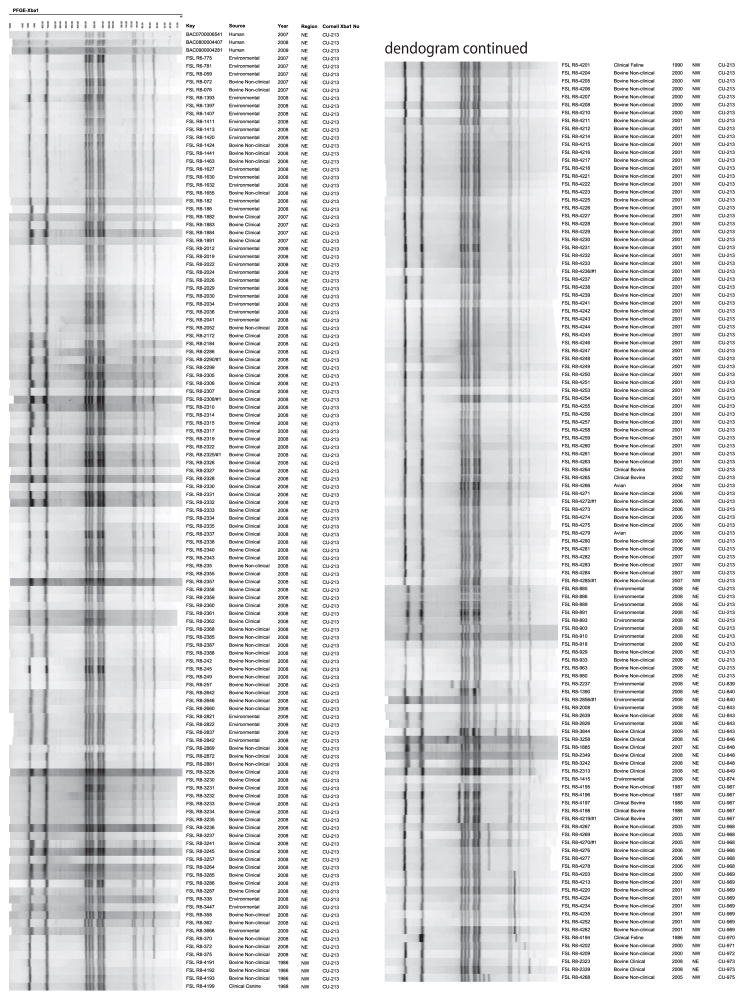
Of the 237 isolates, 198 (84%) shared pattern CU-213 (Table, Figure). The frequency of pattern CU-213 was somewhat higher among the recent isolates (89%, CI: 82 – 93) than among the comparison isolates (75%, CI: 64–83). Pattern CU-213 was detected among 86% (CI: 75 – 93) of isolates from clinically sick cattle, 97% (CI: 84–100) of isolates from clinically healthy cattle, and 87% (CI: 73–95) of environmental isolates (Table). Similarly, all human isolates shared pattern CU-213. Most other PFGE patterns were represented by 1 to 3 isolates, and only pattern CU-843 was detected in more than one isolate subcategory (Table). The comparison isolates with PFGE patterns other than CU-213 represented 7 PFGE patterns and originated from cattle (n=22) and cats (n=1). None of these 7 PFGE patterns were detected among the recent isolates, and the overwhelming majority of PFGE patterns differed from pattern CU-213 by only a single band (Figure).
Figure.
XbaI pulsed-field gel electrophoresis (PFGE) patterns for 237 Salmonella Cerro isolates included in this study. The isolation year, isolate subcategory, PFGE pattern number and geographic area of isolation are indicating adjacent to individual isolates. ‘NE’= isolated in Pacific Northwest; ‘NW’ = isolated in Northeast.
Simpson’s index of diversity values were considerably lower for recent than comparison isolates (Table). Intriguingly, values were similar for isolates from clinically sick cattle and farm environments, but they were considerably lower for isolates from healthy cattle and higher for comparison isolates.
4.2. Antimicrobial resistance typing results
All tested isolates eventually proved susceptible to all antimicrobial drugs. However, on initial analysis, 12 isolates (5%) were resistant to one or more antimicrobial drugs. Yet, PCR – based screening failed to detect resistance genes, and upon retesting using the same protocols and the same glycerol-frozen bacterial cultures, which were stored at −80°C, all 12 isolates were susceptible to all drugs, thus indicating previous false-positive results or, less likely, resistance loss.
4.3. Results of spvA screening
spvA was detected in none of the isolates, even though the gene was readily detected in the positive controls (i.e., Typhimurium isolates FSL S5-800 and FSL- S5-550, see www.pathogentracker.org).
5. DISCUSSION
Our data indicate that Salmonella Cerro strains circulating in the US represent a clonal subtype. The vast majority of cattle isolates, most isolates from other domestic animals, and all human isolates in our study shared a single XbaI PFGE pattern (CU-213), while nearly all other PFGE patterns differed by only one band from this predominant PFGE pattern. Moreover, all isolates were susceptible to all antimicrobial drugs tested, again supporting the hypothesis of one clonal subtype. While further studies are clearly needed, the absence of antimicrobial resistance genes among the 237 tested isolates might indicate that limited antimicrobial selection pressures have so far acted on this serotype, which might be congruent with a predominantly low virulence of this serotype. Our data might further suggest that host or environmental factors, rather than genetic changes, contributed to the recent increase in Salmonella Cerro prevalence. However, genetic changes not detectable by XbaI PFGE or a combination of host and pathogen factors might have also caused the increase in prevalence, and further studies are clearly needed.
Despite Salmonella Cerro being extremely common among dairy cattle in the US, human cases are rarely associated with this serotype. We found though that all three available human Salmonella Cerro isolates had the same XbaI PFGE pattern that was predominant among dairy cattle isolates. It is thus tempting to speculate that at least some Salmonella Cerro strains have some capacity to cause human disease. However, specific virulence characteristics remain to be elucidated. While it is tempting to speculate that the rare occurrence of human Salmonella Cerro cases may be due to reduced virulence of this subtype, scarcity of exposure cannot be excluded as one determining cause. All tested isolates lacked spvA, a generally plasmid-mediated virulence factor found among some strains of certain, predominantly host adapted, serotypes such as Abortusovis, Dublin, or Typhimurium (Chu and Chiu, 2006; Gassama et al., 2006; Guney et al., 1994). Still, absence of this virulence gene is unlikely to explain the rare association of serotype Cerro with human cases.
Importantly, our data also suggest that genetic diversity in contemporary US Salmonella Cerro isolates is low or cannot be adequately captured by standard XbaI PFGE. The development and validation of improved subtyping methods, an integral component of veterinary epidemiology, is clearly needed to allow further studies of this potentially emerging serotype (Belkum et al., 2007).
6. CONCLUSIONS
Overall, our study illustrates the importance of continuous integrated animal and human population-based pathogen surveillance, as this approach may be crucial for the reliable early detection and characterization of emerging pathogens. Moreover, our data provides strong evidence for the necessity to critically evaluate the adequacy of diagnostic tests with regard to the specific study populations and research aims at hand.
Acknowledgments
Support for this project was provided by the National Institute of Allergy and Infectious Diseases, National Institutes of Health, Department of Health and Human Services, under Contract numbers N01-AI-30054 - ZC-006-07 and N01-A1-30055. K.H. was supported by Morris Animal Foundation Fellowship Training Grant D08FE-403
Footnotes
Publisher's Disclaimer: This is a PDF file of an unedited manuscript that has been accepted for publication. As a service to our customers we are providing this early version of the manuscript. The manuscript will undergo copyediting, typesetting, and review of the resulting proof before it is published in its final citable form. Please note that during the production process errors may be discovered which could affect the content, and all legal disclaimers that apply to the journal pertain.
References
- Animal and Plant Health Inspection Service (Aphis), United States Department of Agriculture. Info Sheet. Veterinary Services, Centers for Epidemiology and Animal Health; 2008. Salmonella and Campylobacter on U.S. Dairy Operations, 1996–2007. [Google Scholar]
- Chu C, Chiu CH. Evolution of the virulence plasmids of non-typhoid Salmonella and its association with antimicrobial resistance. Microbes Infect. 2006;8(7):1931–1936. doi: 10.1016/j.micinf.2005.12.026. [DOI] [PubMed] [Google Scholar]
- Centers for Disease Control and Prevention (CDC) Salmonellosis associated with carne seca— New Mexico. MMWR Morb Mortal, Wkly Rep. 1985;34(42):645–646. [PubMed] [Google Scholar]
- Centers for Disease Control and Prevention (CDC) Salmonella Surveillance: Annual Summary, 2006. Atlanta, Georgia: US Department of Health and Human Services; 2008. [Google Scholar]
- Cummings KJ, Warnick LD, Elton M, Rodriguez-Rivera LD, Siler JD, Wright EM, Grohn YT, Wiedmann M. Salmonella enterica Serotype Cerro Among Dairy Cattle in New York: An Emerging Pathogen? Foodborne Pathog Dis. 2010;7(6):659–665. doi: 10.1089/fpd.2009.0462. [DOI] [PMC free article] [PubMed] [Google Scholar]
- Gassama-Sow A, Wane AA, Canu NA, Uzzau S, Kane AA, Rubino S. Characterization of virulence factors in the newly described Salmonella enterica serotype Keurmassar emerging in Senegal (sub-Saharan Africa) Epidemiol Infect. 2006;134(4):741–743. doi: 10.1017/S0950268805005807. [DOI] [PMC free article] [PubMed] [Google Scholar]
- Gebreyes WA, Thakur S, Dorr P, Tadesse DA, Post K, Wolf L. Occurrence of spvA Virulence Gene and Clinical Significance for Multidrug-Resistant Salmonella Strains. J Clin Microbiol. 2009;47(3):777–80. doi: 10.1128/JCM.01660-08. [DOI] [PMC free article] [PubMed] [Google Scholar]
- Grundmann H, Hori S, Tanner G. Determining confidence intervals when measuring genetic diversity and the discriminatory abilities of typing methods for microorganisms. J Clin Microbiol. 2001;39(11):4190–4192. doi: 10.1128/JCM.39.11.4190-4192.2001. [DOI] [PMC free article] [PubMed] [Google Scholar]
- Guiney DG, Fang FC, Krause M, Libby S. Plasmid-mediated virulence genes in non-typhoid Salmonella serovars. FEMS Microbiol Lett. 1994;124(1):1–9. doi: 10.1111/j.1574-6968.1994.tb07253.x. [DOI] [PubMed] [Google Scholar]
- Hunter SB, Vauterin P, Lambert-Fair MA, Van Duyne MS, Kubota K, Graves L, Wrigley D, Barrett T, Ribot E. Establishment of a universal size standard strain for use with the PulseNet standardized pulsed-field gel electrophoresis protocols: converting the national databases to the new size standard. J Clin Microbiol. 2005;43(3):1045–1050. doi: 10.1128/JCM.43.3.1045-1050.2005. [DOI] [PMC free article] [PubMed] [Google Scholar]
- Mammina C, Cannova L, CarfiPavia S, Nastasi A. Endemic presence of Salmonella enterica serotype Cerro in southern Italy. Euro Surveill. 2000;5(7):84–86. doi: 10.2807/esm.05.07.00028-en. [DOI] [PubMed] [Google Scholar]
- Ribot EM, Fair MA, Gautom R, Cameron DN, Hunter SB, Swaminathan B, Barrett TJ. Standardization of pulsed-field gel electrophoresis protocols for the subtyping of Escherichia coli O157:H7, Salmonella, and Shigella for PulseNet. Foodborne Pathog Dis. 2006;3(1):59–67. doi: 10.1089/fpd.2006.3.59. [DOI] [PubMed] [Google Scholar]
- Simpson EH. Measurement of Diversity. Nature. 1949;163:688. [Google Scholar]
- van Belkum A, Tassios PT, Dijkshoorn L, Haeggman S, Cookson B, Fry NK, Fussing V, Geen J, Feil E, Gerner-Smidt P, Brisse S, Struelens M. Guidelines for the validation and application of typing methods for use in bacterial epidemiology. Clin Microbiol Infect. 2007;13(s3):1–46. doi: 10.1111/j.1469-0691.2007.01786.x. [DOI] [PubMed] [Google Scholar]
- van Kessel JS, Karns JS, Wolfgang DR, Hovingh E, Schukken YH. Longitudinal study of a clonal, subclinical outbreak of Salmonella enterica subsp. enterica serovar Cerro in a U.S. dairy herd. Foodborne Pathog, Dis. 2007;4(4):449–461. doi: 10.1089/fpd.2007.0033. [DOI] [PubMed] [Google Scholar]


