

Abstract
The melanocortin MC3 receptor remains the most enigmatic of the melanocortin receptors with regard to its physiological functions. The receptor is expressed both in the CNS and in multiple tissues in the periphery. It appears to be an inhibitory autoreceptor on proopiomelanocortin neurons, yet global deletion of the receptor causes an obesity syndrome. Knockout of the receptor increases adipose mass without a readily measurable increase in food intake or decrease in energy expenditure. And finally, no melanocortin MC3 receptor null humans have been identified and associations between variant alleles of the melanocortin MC3 receptor and disease remain controversial, so the physiological role of the receptor in humans remains to be determined.
Keywords: Melanocortin-3 receptor, melanocortin MC3 receptor, Melanocortin, Obesity, γ-MSH, Proopiomelanocortin
1. Structure and function of the receptor
The melanocortin MC3 receptor belongs to the G-Protein Coupled Receptor family (Gantz et al., 1993; Roselli-Rehfuss et al., 1993). It is positively coupled to adenylyl cyclases through Gs and, upon activation, stimulates cAMP production. A few studies suggest that overexpressed melanocortin MC3 receptor activation can also induce calcium release from intracellular stores (Kim et al., 2002b; Konda et al., 1994; Mountjoy et al., 2001). The mechanism of calcium release is unclear and the role of IP3 generation is controversial (Kim et al., 2002a; Konda et al., 1994; Mountjoy et al., 2001). Based on the discrepancy observed in this signaling cascade when studied in different in-vitro models, it will be important to validate the activation of calcium signaling in melanocortin MC3 receptor neurons in ex-vivo or in-vivo models. Another pathway activated downstream of melanocortin MC3 receptor is the MAPK pathway. Indeed, Chai et al. showed that, in HEK293 cells transfected with the melanocortin MC3 receptor, NDP-αMSH triggers a significant phosphorylation of ERK1/2 (Chai et al., 2007). In addition, they established that melanocortin MC3 receptor-mediated MAPK activation is PI3K dependant and pertussis toxin sensitive (Chai et al., 2007). Interestingly, as with the melanocortin MC4 receptor (Nijenhuis et al., 2001), the melanocortin MC3 receptor was reported to have a constitutive activity (Nijenhuis et al., 2001) but the physiological relevance of this finding is still unclear. Importantly, the melanocortin MC3 receptor is one of the rare G protein-coupled receptors to have a natural inverse agonist, agouti-related protein (Nijenhuis et al., 2001; Ollmann et al., 1997), a protein homologous to agouti (Ollmann et al., 1997). Like most G protein-coupled receptors, following activation, the melanocortin MC3 receptor recruits β-arrestin and internalizes (Breit et al., 2006; Nyan et al., 2008). However, a surprising feature of the melanocortin MC3 receptor was uncovered when Breit et al. showed that both melanocortin MC3 receptor agonists and its natural antagonist agouti-related protein can promote its internalization (Breit et al., 2006). This is very unusual as internalization is thought to be a G protein-couplede receptor signaling “turn off” mechanism, and antagonist-mediated blockade of receptor signaling usually causes a compensatory increase in surface receptor expression rather than receptor internalization. This observation could suggest the existence of a yet unidentified agouti-related protein-mediated signaling pathway.
The natural agonists for the melanocortin receptors are α, β, and γ-melanocyte stimulating hormone, and adrenocorticotropic (ACTH) hormone. They are all proteolytic products of the proopiomelanocortin preprohormone precursor, and all contain the tetrapeptide pharmacophore His-Phe-Arg-Trp. Melanotropins differ in their potency at the five members of the melanocortin receptor family. The melanocortin MC2 receptor is the only melanocortin receptor to be specifically activated by only one of the melanotropins, namely, ACTH. Also, γ-MSH has over 100 fold higher affinity and 45 fold high potency at the melanocortin MC3 receptor than at the other melanocortin receptors. This selectivity is likely to be physiologically important since γ-MSH has been reported to be expressed in the brain (Kawai et al., 1984).
As with most G protein-coupled receptors, the mapping of the melanocortin MC3 receptor’s ligand binding pocket is incomplete. Site directed mutagenesis of amino acids putatively involved in melanocortin MC3 receptor -ligand interaction, based on knowledge acquired from similar studies on melanocortin MC1 receptor and melanocortin MC4 receptor, was performed by Chen et al.(Chen et al., 2006). They showed that alanine substitution of amino acids E131, D154, D158 in TM2 and 3, predicted to form an ionic binding pocket for α-MSH, caused a significant decrease in agonist binding and receptor signaling. Mutagenesis of aromatic amino acids F295 and F296 as well as residue H298, all located in the TM6, also impaired agonist binding and were hypothesized to be part of a hydrophobic binding pocket (Chen et al., 2006). In the same study, the authors also established a requirement for residues D121 and D332 in order to achieve proper expression of the melanocortin MC3 receptor at the plasma membrane; it is however unclear if the lack of receptor at the plasma membrane is due to deficient trafficking, or reduced receptor synthesis or stability. Another interesting finding is the conversion of SHU9119 from antagonist to agonist by mutating the leucine at position 165 in the melanocortin MC3 receptor. This result mimics the previous identification of the same behavior for the corresponding L133 in the TM3 of melanocortin MC4 receptor (Yang et al., 2002) suggesting a role for the described leucine residue in agonist vs. antagonist selectivity for both melanocortin MC3 receptor and melanocortin MC4 receptor.
The development of biologically active melanocortin MC3 receptor specific ligands, both agonist and antagonists, will be instrumental to the elucidation of melanocortin MC3 receptor roles in vivo. To this end, several approaches were used, such as a D-amino acid scan of γ-MSH (Grieco et al., 2000) leading to the discovery of D-Trp8-γ-MSH, a compound reported to be the most selective melanocortin MC3 receptor agonist known today with 250 fold and 300 fold higher potency at the melanocortin MC3 receptor than at the melanocortin MC5 receptor and at the melanocortin MC4 receptor respectively. However, when the same compound was independently tested using a different cAMP assay (Promega P-Glo) the EC50 at the melanocortin MC3 receptor was found to be 0.17 nM, corresponding to a 15 fold selectivity only for melanocortin MC3 receptor compared to melanocortin MC4 receptor (Table 1). These divergent results demonstrate the importance of independent testing of the ligands developed using different methods to validate their potency and specificity. In a separate study, an α-MSH/γ-MSH hybrid (peptide 4) created by Cai et al. showed specific antagonist activity at the melanocortin MC3 receptor with an IC50 of 6 nM (Cai et al., 2005), however, this hybrid is also a potent agonist at melanocortin MC1 receptor and melanocortin MC4 receptor and a partial agonist at melanocortin MC5 receptor. Balse-Srinivasan et al. synthesized a cyclic α-MSH/β-MSH analogue (peptide 9) with potent antagonist properties at the melanocortin MC3 receptor (IC50 = 3 nM), however, this compound is not specific (melanocortin MC5 receptor / melanocortin MC3 receptor = 31) (Balse-Srinivasan et al., 2003). Kavarana et al. synthesized a series of cyclic analogs of α-MSH from which the peptide MK-9 is a potent melanocortin MC3 receptor antagonist with a Ki of 5.9 nM (Kavarana et al., 2002), however this petide is also poorly selective (melanocortin MC4 receptor / melanocortin MC3 receptor = 37) and is a potent agonist at melanocortin MC5 receptor (EC50 = 1.01 nM) (Kavarana et al., 2002). Other studies produced a variety of ligands with activity at the melanocortin MC3 receptor with different affinity, potency and specificity but none of those compounds demonstrated satisfactory selectivity for the melanocortin MC3 receptor over the other melanocortin receptors.
Table 1.
EC50 values for α-MSH and D-Trp-8-γ-MSH at the human melanocortin MC3 receptor and melanocortin MC4 receptor using the pGLO cAMP detection system. Human HEK293 cells were cotransfected with plasmids encoding the human melanocortin MC4 receptor or melanocortin MC3 receptor cDNAs (pCDNA3.1 vector) and with a plasmid encoding an engineered cAMP sensitive luciferase (pGLO sensor™ - 20FcAMP plasmid, Promega) and stable clones were selected for their ability to respond to α-MSH. Cells were seeded in a 384 well plate in 10 µL of culture medium without antibiotics and were incubated by adding 10 µL of the substrate containing media (GloSensor™ cAMP assay, Promega) diluted at 4% in CO2-independent medium (Gibco). The luminescence was recorded before and after injection of a range of concentrations of α-MSH or D-Trp-8-γ-MSH for 15 min to obtain the maximal luminescent responses on a Spectramax M5 (Molecular Devices) plate reader (100 msec integration). Incubations were performed in triplicate and curves and EC50 values were determined using Prism (Graphpad).
| Compound | hMC4-GLO (EC50) | hMC4R-GLO (EC50) |
|---|---|---|
| α-MSH | 2.38×10⁻¹⁰ | 4.5×10⁻¹⁰ |
| D-Trp-8-γ-MSH | 1.7×10⁻¹⁰ | 2.5×10⁻⁹ |
Manipulation of known peptidic ligands of the melanocortin receptors has provided us with a tremendous amount of information in the requirement for receptor binding affinity and selectivity, and additional work will be required to achieve compounds with 100–1000 fold selectivity for melanocortin MC3 receptor. More extensive modification and testing of compounds already available as well as different approaches, like the identification of small molecule ligands or allosteric modulators could prove successful at producing molecules highly specific for the melanocortin MC3 receptor. Such ligands would allow targeted and specific manipulation of melanocortin MC3 receptor signaling in vivo and, hopefully, will lead to a better understanding of the physiological roles of the melanocortin MC3 receptor.
2. Expression of the Receptor
2.1 Central Expression
Both melanocortin MC3 receptor and MC4R are expressed in hypothalamic, midbrain, and brainstem, nuclei, however the similarity in CNS expression ends there (Mountjoy et al., 1994; Roselli-Rehfuss et al., 1993). Studies of the receptor show discrete regions of melanocortin melanocortin MC3 receptor signal independent of other melanocortin receptor subtypes. Therefore, despite the relative redundancy of melanocortin receptor signaling and activation, there are clearly functionally distinct roles for the melanocortin MC3 receptor.
Expression studies of melanocortin melanocortin MC3 receptor mRNA have primarily been focused in the rodent brain. Northern blot hybridization experiments demonstrate the greatest expression of the melanocortin melanocortin MC3 receptor gene is in the hypothalamus (Roselli-Rhefus et al., 1993). In situ hybridization demonstrate approximately 35 different nuclei expressing the receptor, with the highest expression in the arcuate nucleus, ventromedial hypothalamus, medial habenula, ventral tegmental area, and raphe (Roselli-Rhefus et al., 1993). Not surprisingly, melanocortin MC3 receptor mRNA is found primarily in areas of the brain which receive direct innervation from proopiomelanocortin immunoreactive neurons. However, the arcuate nucleus, which contains all of the forebrain proopiomelanocortin expressing neurons, displays moderate levels of melanocortin MC3 receptor mRNA, while the nucleus of the solitary tract (NTS) containing the other central proopiomelanocortin expressing neurons apparently does not express melanocortin MC3 receptor mRNA (Roselli-Rhefus et al., 1993). Expression of melanocortin MC3 receptor in the arcuate nucleus and ventromedial hypothalamus are particularly intriguing with regard to the observations that the receptor appears to inhibit anorexigenic proopiomelanocortin neurons and yet when deleted causes obesity, perhaps via as yet uncharacterized actions in the ventromedial hypothalamus or other nuclei. A mouse line containing GFP under the control of a melanocortin MC3 receptor BAC clone is available, and is a helpful tool for better defining the properties of melanocortin MC3 receptor neurons in the CNS (Fig. 1).
Fig. 1.
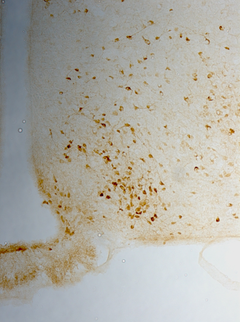
MC3-GFP positive cell bodies in the arcuate nucleus and ventromedial hypothalamic nucleus. A transgenic mouse (MMRRC stock number 00264-UNC) containing the GFP protein under the control of a melanocortin MC3 receptor BAC clone was used for preparation of coronal brain slices. GFP was identified immunohistochemically.
2.2 Development
MC4R receptor binding predominates centrally in various embryonic stages of development, however, a rapid increase in ventromedial and arcuate nuclei expression of melanocortin melanocortin MC3 receptor mRNA postnatally with higher CNS expression evident at postnatal day 27 is described (Kistler-Heer et al., 1998). Interestingly, a transition is shown in melanocortin receptor subtype in regions of the brain including the ventral tegmental area and the dorsoposterior hypothalamus from melanocortin MC4 receptor to melanocortin MC3 receptor -dominant throughout development. This change in receptor subtype suggests a differentiation in regional melanocortin MC3 receptor signaling in order to more acutely regulate specific neuronal populations. Further studies to support this idea show a 3–4 fold increase in melanocortin MC3 receptor mRNA in the ventral tegmental area, habenula, and ventromedial hypothalamus from birth to adulthood in rats (Xia and Wikberg, 1997).
2.3 Peripheral Expression
To add to the complexity of this receptor, expression outside the CNS has been documented, and physiological actions outside the CNS have been demonstrated as well. Northern analysis of poly(A)+ RNA has established the presence of melanocortin MC3 receptor transcripts of the appropriate size in human placenta, and in several human gut tissues including the stomach, duodenum, and pancreas using a combination of RT-PCR and Southern blotting techniques (Gantz et al., 1993). In another study, PCR analysis of human tissues similarly detected melanocortin MC3 receptor cDNA in the heart, while Southern blotting of amplified cDNA detected expression in the testis, ovary, mammary gland, skeletal muscle, and kidney (Chhajlani, 1996). Further studies in rodents have confirmed melanocortin MC3 receptor expression in the kidney and peritoneal macrophages (Getting et al., 2003; Ni et al., 2006b). Melanocortin melanocortin MC3 receptors in these regions may function in modulating natriuresis and immune function, which will be further elaborated later in this review.
3. Genetics
The importance of melanocortin MC3 receptor in human obesity was first suggested by a study that showed a QTL for % body fat in the region of the melanocortin MC3 receptor gene (Lembertas et al., 1997). Further analysis showed that peak LOD scores for body mass index (BMI), fat mass, and subcutaneous fat were localized near the melanocortin MC3 receptor gene (Lembertas et al., 1997). Since then studies have focused on identification of common and rare allelic variants of the melanocortin MC3 receptor that predispose to obesity (Fig. 2).
Fig. 2.

N-terminus to C-terminus FASTA protein sequence of the human melanocortin MC3 receptor with sites of common (green) and rare (red) variants highlighted. Underlined sequences are transmembrane domain regions.
Common variants that may affect melanocortin MC3 receptor expression or function have been found 5’, within, and 3’ of the open reading frame (ORF). 4 common polymorphisms (−201C>G, −239A>G, −762A>T, and −769T>C) have been identified 5’ of the ATG site for melanocortin MC3 receptor (Li et al., 2000). The −239A>G variant falls directly within a GATA binding site, and mutation at this site decreases binding affinity for GATA4 (Schalin-Jantti et al., 2003). In fact, a frequency of 4.5 to 21% has been reported for the −239A>G variant (Obregon et al.; Schalin-Jantti et al., 2003). Despite a known function, this variant has not been shown to be present at a higher rate in obese than in lean controls (Li et al., 2000). In fact, none of the common 5’ variants are present more frequently in obese population than in lean controls.
Within the ORF, two common variants have received the majority of interest. Nucleotide substitutions 17C>A and 241G>A result in missense mutations T6K and V81I, respectively (Obregon et al.; Rutanen et al., 2007; Wong et al., 2002. Minor allele frequency for each of these variants is reported to be 5.6–16% {Schalin-Jantti, 2003 #16). Because they are in linkage disequilibrium, the effects of these variants have been studied in conjunction (Lee et al., 2007; Mencarelli et al., 2008; Rutanen et al., 2007). In a cell culture model, expression of the double mutant has been shown to decrease maximal binding to NDP-MSH by 50% and NDP-stimulated cAMP accumulation by approximately 30% (Feng et al., 2005). Feng et al. report homozygous presentation of both polymorphisms with a prevalence of 15.8 and 1.7% in African American and Caucasian populations, respectively. Together these results suggest that co-presentation of these common variants occurs at a high frequency and may result in a measurable phenotype.
The initial report of the Lys6 Ile81 variants suggested that neither correlated with obesity or glucose tolerance (Wong et al., 2002). However, subsequent studies with larger sample populations have shown that homozygous presentation of the Lys6 Ile81 variant can affect body weight, BMI, fat mass, % body fat, and energy intake (Feng et al., 2005; Lee et al., 2007; Savastano et al., 2009). Additionally, homozygous expression of the Lys6 or Ile81 variants results in decreased HOMA, insulin:glucose ratio, and fasting glucose (Lee et al., 2007). The decreased fasting glucose may result from increased glucose oxidation, previously shown for carriers of the Lys6 and Ile81 alleles (Rutanen et al., 2007). Circulating triglycerides and fasting free fatty acids are lower in homozygous carriers of the Lys6 and Ile81 minor alleles (Lee et al., 2007; Rutanen et al., 2007). Lipid oxidation was lower in carriers of the two minor alleles in both the basal and insulin stimulated state than in individuals that had the Thr6 Val81 genotype (Rutanen et al., 2007). Combined, these results suggest that the common variants T6K and V81I do affect melanocortin MC3 receptor function resulting in measurable phenotypes.
Less well studied common variants include a recently reported ORF variant and a 3’ insertion variant (Boucher et al., 2002; Calton et al., 2009). Calton et al. report an ORF sequence variant R257S that was present in both obese and lean humans and occurred with a prevalence of 0.4%. However, no investigation into possible functions of this variant were reported. The 3’ insertion +2138CAGACC occurs with a minor allele frequency of 17.6% (Obregon et al.). However, the effects of this common variant are not well established (Boucher et al., 2002).
In contrast to the melanocortin MC4 receptor, however, the prevalence of rare melanocortin MC3 receptor variants was not associated with obesity and was found to 0.49% when sequencing 889 obese subjects and 932 lean controls (Calton et al., 2009). A total of 12 rare mutations have been reported in the literature (Calton et al., 2009; Lee et al., 2007; Mencarelli et al., 2008). The most studied of the rare mutations is the I183N mutation, which results in a complete lack of signaling in response to agonist stimulation (Lee et al., 2002; Rached et al., 2004; Tao and Segaloff, 2004). The muted signaling associated with the I183N mutation appears to result from decreased trafficking of the melanocortin MC3 receptor to the cell membrane (Rached et al., 2004; Tao and Segaloff, 2004). Two additional mutations T280S and I335S, first identified in humans, have been found to nearly completely mute melanocortin MC3 receptor activity (Calton et al., 2009; Mencarelli et al., 2008). Similar to the I183N mutation the I335S mutation is shown to eliminate cell surface expression. In fact, I335 of melanocortin MC3 receptor correlates with I301 of melanocortin MC4 receptor, for which the I301T mutation was previously reported to be a loss-of-function mutation (Vaisse et al., 2000). Three additional melanocortin MC3 receptor mutations, first identified in human samples, have been reported to affect receptor activity in a cell culture model. The two robust mutation effects occur with the S69C mutation, which decreases maximal receptor activity to 55% of WT levels, and the F82S mutation which increases the EC50 more than 100 fold and decreases maximal receptor activity more than 50% (Calton et al., 2009). Cells expressing the A70T mutant melanocortin MC3 receptor display a slightly reduced maximal cAMP response to MSH (Lee et al., 2007). Rare mutations for which no obvious signaling defects exist include I87T, A260V, M275T, L297V, A293T, and X361S (Calton et al., 2009; Mencarelli et al., 2008). The X361S mutation abolishes the stop codon leading to the addition of seven extra amino acids to the intracellular C terminus of the receptor (Mencarelli et al., 2008).
Genetic studies have identified mutations in human melanocortin MC1 receptor (Koppula et al., 1997; Valverde et al., 1995), MC2-R (Clark et al., 1993; Tsigos et al., 1993), melanocortin MC4 receptor (Farooqi et al., 2000; Vaisse et al., 2000), and proopiomelanocortin (Krude et al., 1998) that lead to distincy syndromes. Yet, despite the significant obesity syndrome seen on deletion of the melanocortin MC3 receptor in the mouse, an obesity syndrome associated with loss of melanocortin MC3 receptor expression in humans has not yet been clearly demonstrated.
4. Melanocortin MC3 receptor as an autoreceptor
Melanocortin MC3 receptor is expressed widely within the CNS with abundant expression in the proopiomelanocortin and NPY Neurons of the arcuate nucleus (Bagnol et al., 1999; Mounien et al., 2005). In fact, melanocortin MC3 receptor is expressed in a rostral caudal gradient in both proopiomelanocortin (43%→13%) and AgRP/NPY (55→28%) neurons. An auto-inhibitory functional role of melanocortin MC3 receptor on proopiomelanocortin neurons was first suggested when it was shown that bath application of the melanocortin MC3 receptor agonist, D-trp8-γ-MSH (7 nM), increased IPSC frequency on proopiomelanocortin neurons (Cowley et al., 2001). This was in direct opposition to the inhibitory effect of NPY (100 nM) on IPSC frequency in proopiomelanocortin neurons (Cowley et al., 2001). Subsequently, in vivo effects of D-trp8-γ-MSH have supported a role for melanocortin MC3 receptor in the dampening of proopiomelanocortin neuronal activity. Peripheral administration of D-trp8-γ-MSH has been shown to cause a dose responsive increase in food intake that peaked at 5 µg/animal and was absent in the melanocortin MC3 receptor −/− mouse (Marks et al., 2006). Using the melanocortin MC4 receptor −/− mouse, reductions in food intake at higher doses of D-trp8-γ-MSH were shown to result from non-specific activity at the MC4R (Marks et al., 2006). Lending further credence to an auto-inhibitory role of melanocortin MC3 receptor on proopiomelanocortin neurons, 66 h melanocortin MC3 receptor stimulation by ICV infusion of D-trp8-γ-MSH decreases proopiomelanocortin mRNA expression (Lee et al., 2008). While melanocortin MC4 receptor −/− mice appear relatively insensitive to the food intake and body weight effect of illness induced cachexia, melanocortin MC3 receptor −/− mice are hypersensitive to these same cachexia models (Marks et al., 2003). The opposite effects of melanocortin MC3 receptor and melanocortin MC4 receptor ablation in cachexia models provides further evidence for a role of melanocortin MC3 receptor as a brake on proopiomelanocortin neuron activity and subsequently melanocortin MC4 receptor stimulation.
5. Physiology of the melanocortin MC3 receptor
Our understanding of the physiology of the melanocortin MC3 receptor has lagged behind that of the other centrally expressed receptor, melanocortin MC4 receptor. Much of what we do know about the physiological function of this receptor has come from studies using the melanocortin MC3 receptor −/− mouse and the comparatively melanocortin MC3 receptor specific agonist γ-MSH and its stable analogues.
5.1 Energy homeostasis
The melanocortin MC3 receptor −/− mouse has a unique phenotype characterized by an increase in adiposity on a standard chow diet in the absence of a notable difference in body weight, total food intake or energy expenditure (Butler et al., 2000; Chen et al., 2000). The increase in adiposity in these animals is exacerbated by feeding a high-fat diet (Butler et al., 2000; Ellacott et al., 2007; Sutton et al., 2006; Trevaskis et al., 2007) suggesting an alteration nutrient partitioning. Despite the increase in adiposity in the melanocortin MC3 receptor −/− mouse these animals are relatively protected from the development of metabolic syndrome, compared with other mouse models with comparative levels of adiposity, due to a reduced inflammatory response to obesity (Ellacott et al., 2007; Trevaskis et al., 2007). The obesity phenotype in the melanocortin MC3 receptor −/− mouse occurs by a mechanism distinct from the obesity phenotype in the melanocortin MC4 receptor deficient animal as combined melanocortin MC3 receptor / melanocortin MC4 receptor deletions have an additive effect on adiposity (Chen et al., 2000). Recent studies have proposed that elements of the phenotype in the melanocortin MC3 receptor −/− mouse may be caused by an alteration in circadian rhythm in this model (for review see (Begriche et al., 2009)), which will be discussed in a later section of this review.
Despite the increase in adiposity in the melanocortin MC3 receptor −/− mouse, a striking phenotype in these animals is an increased susceptibility to weight loss in experimental models of cachexia (Marks et al., 2003; Marks et al., 2001). This is in sharp contrast to the melanocortin MC4 receptor −/− mouse which is protected from weight loss in numerous cachexia paradigms relative to wild-type animals (Cheung et al., 2005; Marks et al., 2003; Marks et al., 2001; Scarlett et al.). The differential response to cachexia in these two models of central melanocortin receptor deficiency, which both show increased adiposity, is likely to be connected at least in part to the differences in lean body mass phenotype. In melanocortin MC4 receptor −/− animals obesity is associated with increased lean body mass (Huszar et al., 1997) while the increased adiposity in the melanocortin MC3 receptor −/− mouse does not correlate with an increase in lean mass (Butler et al., 2000; Chen et al., 2000). Furthermore, the enhanced cachexia seen in the melanocortin MC3 receptor −/− animals may support the hypothesis that the melanocortin MC3 receptor functions, at least in part, as an autoinhitory receptor on hypothalamic proopiomelanocortin neurons.
There are a limited number of pharmacological studies examining the role of the melanocortin MC3 receptor in the regulation of energy homeostasis. The melanocortin MC3 receptor agonist D-Trp8-γ-MSH, a stabilized γ-MSH analogue which has significant selectivity for the melanocortin MC3 receptor over the melanocortin MC4 receptor (Grieco et al., 2000), has been used to examine alterations in feeding behavior in response to pharmacological modulation of the melanocortin MC3 receptor. Intracerebroventricular (i.c.v.) administration of d-trp8-γ-MSH in rats (Lee et al., 2008) and peripheral administration of the same compound in mice (Marks et al., 2006) stimulates food intake. In these studies intake was measured in freely-feeding animals following chronic administration via an osmotic mini-pump (Lee et al., 2008) or following acute administration prior to the normal nocturnal intake period or in satiated animals (Marks et al., 2006). Some early studies using γ-MSH as opposed to d-trp8-γ-MSH failed to see any effect of i.c.v. administration of this peptide on food intake rodents in a fast-induced refeeding paradigm (Abbott et al., 2000; Kask et al., 2000). The differences in outcome between these studies are likely due to be related to the increased stability of D-Trp8-γ-MSH compared with γ-MSH and the different feeding paradigms used. Currently, due to a lack of pharmacological antagonists with the ability to cleanly differentiate between melanocortin MC3 receptor and melanocortin MC4 receptor much of what we know about the effect of loss of melanocortin MC3 receptor signaling in the regulation of energy homeostasis comes from studies in animals with genetic deficiency of these receptors, as described above.
5.2 Natriuresis
The cardiovascular and natriuretic effects of γ-MSH in rodents have been documented since 1985 (Callahan et al., 1985; Lymangrover et al., 1985). While there is some debate over whether the cardiovascular effects of exogenously administered γ-MSH are mediated via the melanocortin MC3 receptor (Gruber et al., 2009; Mioni et al., 2003; Ni et al., 2006b), the importance of the melanocortin MC3 receptor in mediating natriuresis is established. Gamma-MSH plays a critical role in reflex natriuresis after unilateral nephrectomy (Lin et al., 1987; Ni et al., 1998). In addition to circulating γ-MSH being elevated after unilateral nephrectomy (Lin et al., 1987; Ni et al., 1998), both circulating γ-MSH and kidney melanocortin MC3 receptor mRNA levels are also increased in rodents following ingestion of a high-salt diet (Chandramohan et al., 2009; Mayan et al., 1996; Ni et al., 2006a) implicating melanocortin MC3 receptor signaling in mediating the natriuretic response in two distinct paradigms. The results of these studies are reinforced by studies demonstrating that the genetic disruption of γ-MSH signaling in either the melanocortin MC3 receptor deficient mouse or pro-hormone convertase 2 (PC2) deficient mouse (which is unable to process proopiomelanocortin to γ-MSH) results in salt-sensitive hypertension (Ni et al., 2003). In the case of the PC2 deficient animal, this salt-sensitive hypertension can be overcome by infusion of exogenous NDP-γ-MSH, a stable γ-MSH analogue (Ni et al., 2003). A detailed review of the cardiovascular and renal actions of γ-MSH can be found elsewhere (Humphreys, 2007).
5.3 Immune function
Peptides of the melanocortin family have been shown to exert anti-inflammatory effects in vivo and in vitro (for review see (Catania et al., 2004)). Many of the anti-inflammatory effects of melanocortin peptides are believed to be mediated via the melanocortin MC1 receptor, which is expressed on cell types involved in mediating the inflammatory response including neutrophils, monocytes, dendritic cells and B-lymphocytes (for review see (Catania, 2007)). Melanocortin MC3 receptor mRNA has also been detected in macrophages (Getting et al., 1999a; Getting et al., 1999b). melanocortin MC3 receptors on macrophages have been proposed to mediate some of the anti-inflammatory effects of γ-MSH. In a model of gouty arthritis, D-Trp8-γ-MSH dose dependently reduces interleukin-1 and chemokine CXCL1 release from primary peritoneal macrophages induced by monosodium urate crystals in a melanocortin MC3 receptor dependent mechanism (Getting et al., 2006a; Getting and Perretti, 2001). The anti-inflammatory effects of D-Trp8-γ-MSH in this paradigm are intact in macrophages obtained from the recessive yellow e/e mouse which has defective melanocortin MC1 receptor signaling, further supporting the contribution of the melanocortin MC3 receptor in mediating this effect (Getting et al., 2006b). The same group also report melanocortin MC3 receptor dependent anti-inflammatory efficacy of D-Trp8-γ-MSH in models of vascular inflammation (Leoni et al., 2008), and lung inflammation (Getting et al., 2008).
5.4 Circadian Rhythm
Rodents maintain a clear circadian rhythm of food intake, with the majority of energy intake taking place during the night. Animals with defective clock genes exhibit defects in this rhythm, with increased food intake during the day and increased susceptibility to diet-induced obesity (Turek et al., 2005). Behavioral and endocrine rhythms are entrained to photic cues by virtue of the retinal hypothalamic tract, however diurnal energy intake patterns also impact on circadian oscillators. For example, restricting food intake to limited time periods can uncouple peripheral clocks from the central circadian oscillator in the suprachiasmatic nucleus (Damiola et al., 2000). One assay of this coordinated effect of photic and metabolic cues is food anticipatory locomotor activity (FAA). Briefly, when animals are food restricted to approximately 50–60% of their normal intake during a 4 hr window of time during the day, they will eventually exhibit increased locomotor activity 1–2 hours prior to the presentation of food. Butler and colleagues have discovered that melanocortin MC3 receptor −/− mice exhibit a striking reduction in FAA (Sutton et al., 2008). The pattern of expression of cellular clock genes in peripheral tissues, such as Bmal1 and Rev-erbα, can also be shifted with this restricted daytime food presentation. However again, the melanocortin MC3 receptor −/− shows a defect in the entrainment of peripheral clock genes in this food restriction paradigm (Sutton et al.). While perturbations in circadian rhythms are associated with obesity (Fonken et al.), circadian rhythms of food intake and locomotor activity are largely intact in the melanocortin MC3 receptor −/−. Additional data will be required to determine if the defective entrainment to restricted food presentation can elucidate a mechanism for the obesity syndrome and metabolic defects in the melanocortin MC3 receptor −/− mouse.
6. Conclusions
Many potential functions of the melanocortin MC3 receptor have now been elucidated, primarily using the melanocortin MC3 receptor −/− mouse model. These include effects on lean and adipose mass, natriuresis, immune function, susceptibility to aspects of metabolic function, and entrainment to restricted food presentation. In the case of the melanocortin MC4 receptor, we understand basics of many of the regulatory inputs, neural circuits and effector pathways that cause the obesity syndrome in mice and humans with defective melanocortin MC4 receptor signaling. However, in the case of the melanocortin MC3 receptor there remain more questions than answers. What are the effector pathways mediating these physiological responses to melanocortin MC3 receptor blockade? For example, what are the endocrine or autonomic mechanisms by which melanocortin MC3 receptor blockade causes obesity and reduced metabolic syndrome? What are the behavioral pathways mediating the defective entrainment to reduced food presentation? Are there specific inputs to melanocortin MC3 receptor neurons in the CNS, such as γ-MSH? What is the source of the physiological ligand for peripheral melanocortin MC3 receptor? And, what are the respective contributions of central and peripheral melanocortin MC3 receptor to the physiological functions identified thus far? Future research in the field is needed to address these problems.
Acknowledgements
This work was supported by NIH grant DK078850 (RDC).
Footnotes
Publisher's Disclaimer: This is a PDF file of an unedited manuscript that has been accepted for publication. As a service to our customers we are providing this early version of the manuscript. The manuscript will undergo copyediting, typesetting, and review of the resulting proof before it is published in its final citable form. Please note that during the production process errors may be discovered which could affect the content, and all legal disclaimers that apply to the journal pertain.
Bibliography
- Abbott CR, Rossi M, Kim M, AlAhmed SH, Taylor GM, Ghatei MA, Smith DM, Bloom SR. Investigation of the melanocyte stimulating hormones on food intake. Lack Of evidence to support a role for the melanocortin-3-receptor. Brain Res. 2000;869:203–210. doi: 10.1016/s0006-8993(00)02386-6. [DOI] [PubMed] [Google Scholar]
- Bagnol D, Lu XY, Kaelin CB, Day HE, Ollmann M, Gantz I, Akil H, Barsh GS, Watson SJ. The anatomy of an endogenous antagonist: relationship between agouti- related protein and proopiomelanocortin in brain. J Neurosci. 1999;19:RC26. doi: 10.1523/JNEUROSCI.19-18-j0004.1999. [DOI] [PMC free article] [PubMed] [Google Scholar]
- Balse-Srinivasan P, Grieco P, Cai M, Trivedi D, Hruby VJ. Structure-activity relationships of novel cyclic alpha-MSH/beta-MSH hybrid analogues that lead to potent and selective ligands for the human melanocortin MC3 receptor and human MC5R. J Med Chem. 2003;46:3728–3733. doi: 10.1021/jm030111j. [DOI] [PubMed] [Google Scholar]
- Begriche K, Sutton GM, Fang J, Butler AA. The role of melanocortin neuronal pathways in circadian biology: a new homeostatic output involving melanocortin-3 receptors? Obes Rev. 2009;10 Suppl 2:14–24. doi: 10.1111/j.1467-789X.2009.00662.x. [DOI] [PMC free article] [PubMed] [Google Scholar]
- Boucher N, Lanouette CM, Larose M, Perusse L, Bouchard C, Chagnon YC. A +2138InsCAGACC polymorphism of the melanocortin receptor 3 gene is associated in human with fat level and partitioning in interaction with body corpulence. Mol Med. 2002;8:158–165. [PMC free article] [PubMed] [Google Scholar]
- Breit A, Wolff K, Kalwa H, Jarry H, Buch T, Gudermann T. The natural inverse agonist agouti-related protein induces arrestin-mediated endocytosis of melanocortin-3 and -4 receptors. J Biol Chem. 2006;281:37447–37456. doi: 10.1074/jbc.M605982200. [DOI] [PubMed] [Google Scholar]
- Butler AA, Kesterson RA, Khong K, Cullen MJ, Pelleymounter MA, Dekoning J, Baetscher M, Cone RD. A unique metabolic syndrome causes obesity in the melanocortin-3 receptor-deficient mouse. Endocrinology. 2000;141:3518–3521. doi: 10.1210/endo.141.9.7791. [DOI] [PubMed] [Google Scholar]
- Cai M, Mayorov AV, Cabello C, Stankova M, Trivedi D, Hruby VJ. Novel 3D pharmacophore of alpha-MSH/gamma-MSH hybrids leads to selective human MC1R and melanocortin MC3 receptor analogues. J Med Chem. 2005;48:1839–1848. doi: 10.1021/jm049579s. [DOI] [PubMed] [Google Scholar]
- Callahan MF, Kirby RF, Wolff DW, Strandhoy JW, Lymangrover JR, Johnson AK, Gruber KA. Sympathetic nervous system mediation of acute cardiovascular actions of gamma 2-melanocyte-stimulating hormone. Hypertension. 1985;7:1145–1150. doi: 10.1161/01.hyp.7.3_pt_2.i145. [DOI] [PubMed] [Google Scholar]
- Calton MA, Ersoy BA, Zhang S, Kane JP, Malloy MJ, Pullinger CR, Bromberg Y, Pennacchio LA, Dent R, McPherson R, Ahituv N, Vaisse C. Association of functionally significant Melanocortin-4 but not Melanocortin-3 receptor mutations with severe adult obesity in a large North American case-control study. Hum Mol Genet. 2009;18:1140–1147. doi: 10.1093/hmg/ddn431. [DOI] [PMC free article] [PubMed] [Google Scholar]
- Catania A. The melanocortin system in leukocyte biology. J Leukoc Biol. 2007;81:383–392. doi: 10.1189/jlb.0706426. [DOI] [PubMed] [Google Scholar]
- Catania A, Gatti S, Colombo G, Lipton JM. Targeting melanocortin receptors as a novel strategy to control inflammation. Pharmacol Rev. 2004;56:1–29. doi: 10.1124/pr.56.1.1. [DOI] [PubMed] [Google Scholar]
- Chai B, Li JY, Zhang W, Ammori JB, Mulholland MW. Melanocortin-3 receptor activates MAP kinase via PI3 kinase. Regul Pept. 2007;139:115–121. doi: 10.1016/j.regpep.2006.11.003. [DOI] [PubMed] [Google Scholar]
- Chandramohan G, Durham N, Sinha S, Norris K, Vaziri ND. Role of gamma melanocyte-stimulating hormone-renal melanocortin 3 receptor system in blood pressure regulation in salt-resistant and salt-sensitive rats. Metabolism. 2009;58:1424–1429. doi: 10.1016/j.metabol.2009.04.022. [DOI] [PubMed] [Google Scholar]
- Chen AS, Marsh DJ, Trumbauer ME, Frazier EG, Guan XM, Yu H, Rosenblum CI, Vongs A, Feng Y, Cao L, Metzger JM, Strack AM, Camacho RE, Mellin TN, Nunes CN, Min W, Fisher J, Gopal-Truter S, MacIntyre DE, Chen HY, Van der Ploeg LH. Inactivation of the mouse melanocortin-3 receptor results in increased fat mass and reduced lean body mass. Nat Genet. 2000;26:97–102. doi: 10.1038/79254. [DOI] [PubMed] [Google Scholar]
- Chen M, Aprahamian CJ, Celik A, Georgeson KE, Garvey WT, Harmon CM, Yang Y. Molecular characterization of human melanocortin-3 receptor ligand-receptor interaction. Biochemistry. 2006;45:1128–1137. doi: 10.1021/bi0521792. [DOI] [PubMed] [Google Scholar]
- Cheung W, Yu PX, Little BM, Cone RD, Marks DL, Mak RH. Role of leptin and melanocortin signaling in uremia-associated cachexia. J Clin Invest. 2005;115:1659–1665. doi: 10.1172/JCI22521. [DOI] [PMC free article] [PubMed] [Google Scholar]
- Chhajlani V. Distribution of cDNA for melanocortin receptor subtypes in human tissues. Biochem. Mol. Biol. Int. 1996;38:73–80. [PubMed] [Google Scholar]
- Clark AJ, McLoughlin L, Grossman A. Familial glucocorticoid deficiency associated with point mutation in the adrenocorticotropin receptor. Lancet. 1993;341:461–462. doi: 10.1016/0140-6736(93)90208-x. [DOI] [PubMed] [Google Scholar]
- Cowley MA, Smart JL, Rubinstein M, Cerdan MG, Diano S, Horvath TL, Cone RD, Low MJ. Leptin activates anorexigenic proopiomelanocortin neurons through a neural network in the arcuate nucleus. Nature. 2001;411:480–484. doi: 10.1038/35078085. [DOI] [PubMed] [Google Scholar]
- Damiola F, Le Minh N, Preitner N, Kornmann B, Fleury-Olela F, Schibler U. Restricted feeding uncouples circadian oscillators in peripheral tissues from the central pacemaker in the suprachiasmatic nucleus. Genes Dev. 2000;14:2950–2961. doi: 10.1101/gad.183500. [DOI] [PMC free article] [PubMed] [Google Scholar]
- Ellacott KL, Murphy JG, Marks DL, Cone RD. Obesity-induced inflammation in white adipose tissue is attenuated by loss of melanocortin-3 receptor signaling. Endocrinology. 2007;148:6186–6194. doi: 10.1210/en.2007-0699. [DOI] [PubMed] [Google Scholar]
- Farooqi IS, Yeo GS, Keogh JM, Aminian S, Jebb SA, Butler G, Cheetham T, O'Rahilly S. Dominant and recessive inheritance of morbid obesity associated with melanocortin 4 receptor deficiency. J Clin Invest. 2000;106:271–279. doi: 10.1172/JCI9397. [DOI] [PMC free article] [PubMed] [Google Scholar]
- Feng N, Young SF, Aguilera G, Puricelli E, Adler-Wailes DC, Sebring NG, Yanovski JA. Co-occurrence of two partially inactivating polymorphisms of melanocortin MC3 receptor is associated with pediatric-onset obesity. Diabetes. 2005;54:2663–2667. doi: 10.2337/diabetes.54.9.2663. [DOI] [PMC free article] [PubMed] [Google Scholar]
- Fonken LK, Workman JL, Walton JC, Weil ZM, Morris JS, Haim A, Nelson RJ. Light at night increases body mass by shifting the time of food intake. Proc Natl Acad Sci U S A. 107:18644–18669. doi: 10.1073/pnas.1008734107. [DOI] [PMC free article] [PubMed] [Google Scholar]
- Gantz I, Konda Y, Tashiro T, Shimoto Y, Miwa H, Munzert G, Watson SJ, DelValle J, Yamada T. Molecular Cloning of a Novel Melanocortin Receptor. J. Biol. Chem. 1993;268:8246–8250. [PubMed] [Google Scholar]
- Getting SJ, Christian HC, Lam CW, Gavins FN, Flower RJ, Schioth HB, Perretti M. Redundancy of a functional melanocortin 1 receptor in the anti-inflammatory actions of melanocortin peptides: studies in the recessive yellow (e/e) mouse suggest an important role for melanocortin 3 receptor. J Immunol. 2003;170:3323–3330. doi: 10.4049/jimmunol.170.6.3323. [DOI] [PubMed] [Google Scholar]
- Getting SJ, Flower RJ, Perretti M. Agonism at melanocortin receptor type 3 on macrophages inhibits neutrophil influx. Inflamm Res. 1999a;48 Suppl 2:S140–S141. doi: 10.1007/s000110050557. [DOI] [PubMed] [Google Scholar]
- Getting SJ, Gibbs L, Clark AJ, Flower RJ, Perretti M. proopiomelanocortin gene-derived peptides activate melanocortin type 3 receptor on murine macrophages, suppress cytokine release, and inhibit neutrophil migration in acute experimental inflammation. J Immunol. 1999b;162:7446–7453. [PubMed] [Google Scholar]
- Getting SJ, Lam CW, Chen AS, Grieco P, Perretti M. Melanocortin 3 receptors control crystal-induced inflammation. FASEB J. 2006a;20:2234–2241. doi: 10.1096/fj.06-6339com. [DOI] [PubMed] [Google Scholar]
- Getting SJ, Lam CW, Leoni G, Gavins FN, Grieco P, Perretti M. [D-Trp8]-gamma-melanocyte-stimulating hormone exhibits anti-inflammatory efficacy in mice bearing a nonfunctional MC1R (recessive yellow e/e mouse) Mol Pharmacol. 2006b;70:1850–1855. doi: 10.1124/mol.106.028878. [DOI] [PubMed] [Google Scholar]
- Getting SJ, Perretti M. Arthritic diseases: melanocortin type 3 receptor agonists as potential therapeutics. Curr Opin Investig Drugs. 2001;2:1064–1069. [PubMed] [Google Scholar]
- Getting SJ, Riffo-Vasquez Y, Pitchford S, Kaneva M, Grieco P, Page CP, Perretti M, Spina D. A role for melanocortin MC3 receptor in modulating lung inflammation. Pulm Pharmacol Ther. 2008;21:866–873. doi: 10.1016/j.pupt.2008.09.004. [DOI] [PubMed] [Google Scholar]
- Grieco P, Balse PM, Weinberg D, MacNeil T, Hruby VJ. D-Amino acid scan of gamma-melanocyte-stimulating hormone: importance of Trp(8) on human MC3 receptor selectivity. J Med Chem. 2000;43:4998–5002. doi: 10.1021/jm000211e. [DOI] [PubMed] [Google Scholar]
- Gruber KA, Fan W, Akerberg H, Larhammar D, Chee MJ, Colmers WF, Cone RD. Neuropeptide Y and gamma-melanocyte stimulating hormone (gamma-MSH) share a common pressor mechanism of action. Endocrine. 2009 doi: 10.1007/s12020-008-9141-3. [DOI] [PMC free article] [PubMed] [Google Scholar]
- Humphreys MH. Cardiovascular and renal actions of melanocyte-stimulating hormone actions. Current Opinion in Nephrology and Hypertension. 2007;16:32–38. doi: 10.1097/MNH.0b013e3280117fb5. [DOI] [PubMed] [Google Scholar]
- Huszar D, Lynch CA, Fairchild-Huntress V, Dunmore JH, Fang Q, Berkemeier LR, Gu W, Kesterson RA, Boston BA, Cone RD, Smith FJ, Campfield LA, Burn P, Lee F. Targeted disruption of the melanocortin-4 receptor results in obesity in mice. Cell. 1997;88:131–141. doi: 10.1016/s0092-8674(00)81865-6. [DOI] [PubMed] [Google Scholar]
- Kask A, Rago L, Wikberg JE, Schioth HB. Differential effects of melanocortin peptides on ingestive behaviour in rats: evidence against the involvement of MC(3) receptor in the regulation of food intake. Neurosci Lett. 2000;283:1–4. doi: 10.1016/s0304-3940(00)00837-5. [DOI] [PubMed] [Google Scholar]
- Kavarana MJ, Trivedi D, Cai M, Ying J, Hammer M, Cabello C, Grieco P, Han G, Hruby VJ. Novel cyclic templates of alpha-MSH give highly selective and potent antagonists/agonists for human melanocortin-3/4 receptors. J Med Chem. 2002;45:2644–2650. doi: 10.1021/jm020021z. [DOI] [PubMed] [Google Scholar]
- Kawai Y, Inagaki S, Shiosaka S, Shibasaki T, Ling N, Tohyama M, Shiotani Y. The distribution and projection of gamma-melanocyte-stimulating hormone in the rat brain: an immunohistochemical analysis. Brain Res. 1984;297:21–32. doi: 10.1016/0006-8993(84)90539-0. [DOI] [PubMed] [Google Scholar]
- Kim CS, Lee SH, Kim RY, Kim BJ, Li SZ, Lee IH, Lee EJ, Lim SK, Bae YS, Lee W, Baik JH. Identification of domains directing specificity of coupling to G-proteins for the melanocortin MC3 and MC4 receptors. J Biol Chem. 2002a;277:31310–31317. doi: 10.1074/jbc.M112085200. [DOI] [PubMed] [Google Scholar]
- Kim MS, Rossi M, Abbott CR, AlAhmed SH, Smith DM, Bloom SR. Sustained orexigenic effect of Agouti related protein may be not mediated by the melanocortin 4 receptor. Peptides. 2002b;23:1069–1076. doi: 10.1016/s0196-9781(02)00039-6. [DOI] [PubMed] [Google Scholar]
- Kistler-Heer V, Lauber ME, Lichtensteiger W. Different developmental patterns of melanocortin MC3 and MC4 receptor mRNA: predominance of MC4 in fetal rat nervous system. J. Neuroendocrinol. 1998;10:133–146. doi: 10.1046/j.1365-2826.1998.00180.x. [DOI] [PubMed] [Google Scholar]
- Konda Y, Gantz I, DelValle J, Shimoto Y, Miwa H, Yamada T. Interaction of dual intracellular signaling pathways activated by the melanocortin-3 receptor. Journal of Biological Chemistry. 1994;269:13162–13166. [PubMed] [Google Scholar]
- Koppula SV, Robbins LS, Lu D, Baack E, White CR, Jr, Swanson NA, Cone RD. Identification of common polymorphisms in the coding sequence of the human MSH receptor (MCIR) with possible biological effects. Hum Mutat. 1997;9:30–36. doi: 10.1002/(SICI)1098-1004(1997)9:1<30::AID-HUMU5>3.0.CO;2-T. [DOI] [PubMed] [Google Scholar]
- Krude H, Biebermann H, Luck W, Horn R, Brabant G, Gruters A. Severe early-onset obesity, adrenal insufficiency and red hair pigmentation caused by proopiomelanocortin mutations in humans. Nature Genetics. 1998;19:155–157. doi: 10.1038/509. [DOI] [PubMed] [Google Scholar]
- Lee M, Kim A, Conwell IM, Hruby V, Mayorov A, Cai M, Wardlaw SL. Effects of selective modulation of the central melanocortin-3-receptor on food intake and hypothalamic proopiomelanocortin expression. Peptides. 2008;29:440–447. doi: 10.1016/j.peptides.2007.11.005. [DOI] [PMC free article] [PubMed] [Google Scholar]
- Lee YS, Poh LK, Kek BL, Loke KY. The role of melanocortin 3 receptor gene in childhood obesity. Diabetes. 2007;56:2622–2630. doi: 10.2337/db07-0225. [DOI] [PubMed] [Google Scholar]
- Lee YS, Poh LK, Loke KY. A novel melanocortin 3 receptor gene (melanocortin MC3 receptor) mutation associated with severe obesity. J Clin Endocrinol Metab. 2002;87:1423–1426. doi: 10.1210/jcem.87.3.8461. [DOI] [PubMed] [Google Scholar]
- Lembertas AV, Perusse L, Chagnon YC, Fisler JS, Warden CH, Purcell-Huynh DA, Dionne FT, Gagnon J, Nadeau A, Lusis AJ, Bouchard C. Identification of an obesity quantitative trait locus on mouse chromosome 2 and evidence of linkage to body fat and insulin on the human homologous region 20q. J. Clin Invest. 1997;100:1240–1247. doi: 10.1172/JCI119637. [DOI] [PMC free article] [PubMed] [Google Scholar]
- Leoni G, Patel HB, Sampaio AL, Gavins FN, Murray JF, Grieco P, Getting SJ, Perretti M. Inflamed phenotype of the mesenteric microcirculation of melanocortin type 3 receptor-null mice after ischemia-reperfusion. FASEB J. 2008;22:4228–4238. doi: 10.1096/fj.08-113886. [DOI] [PMC free article] [PubMed] [Google Scholar]
- Li WD, Joo EJ, Furlong EB, Galvin M, Abel K, Bell CJ, Price RA. Melanocortin 3 receptor (melanocortin MC3 receptor) gene variants in extremely obese women. Int J Obes Relat Metab Disord. 2000;24:206–210. doi: 10.1038/sj.ijo.0801114. [DOI] [PubMed] [Google Scholar]
- Lin S-Y, Wiedemann E, Deschepper CF, Alper RH, Humpreys MH. Prevention of reflex natiuresis after acute unilateral nephrectomy by neonatal administration of MSG. Am. Physiol. Soc. 1987:F276–F282. doi: 10.1152/ajprenal.1987.252.2.F276. [DOI] [PubMed] [Google Scholar]
- Lymangrover JR, Buckalew VM, Harris J, Klein MC, Gruber KA. Gamma-2 MSH is natriuretc in the rat. Endocrinol. 1985;116:1227–1229. doi: 10.1210/endo-116-3-1227. [DOI] [PubMed] [Google Scholar]
- Marks DL, Butler AA, Turner R, Brookhart G, Cone RD. Differential role of melanocortin receptor subtypes in cachexia. Endocrinology. 2003;144 doi: 10.1210/en.2002-221099. in press. [DOI] [PubMed] [Google Scholar]
- Marks DL, Hruby V, Brookhart G, Cone RD. The regulation of food intake by selective stimulation of the type 3 melanocortin receptor (melanocortin MC3 receptor) Peptides. 2006;27:259–264. doi: 10.1016/j.peptides.2005.01.025. [DOI] [PMC free article] [PubMed] [Google Scholar]
- Marks DL, Ling N, Cone RD. Role of the central melanocortin system in cachexia. Cancer Res. 2001;61:1432–1438. [PubMed] [Google Scholar]
- Mayan H, Ling KT, Lee EY, Wiedemann E, Kalinyak JE, Humphreys MH. Dietary sodium intake modulates pituitary proopiomelanocortin mRNA abundance. Hypertension. 1996;28:244–249. doi: 10.1161/01.hyp.28.2.244. [DOI] [PubMed] [Google Scholar]
- Mencarelli M, Walker GE, Maestrini S, Alberti L, Verti B, Brunani A, Petroni ML, Tagliaferri M, Liuzzi A, Di Blasio AM. Sporadic mutations in melanocortin receptor 3 in morbid obese individuals. Eur J Hum Genet. 2008;16:581–586. doi: 10.1038/sj.ejhg.5202005. [DOI] [PubMed] [Google Scholar]
- Mioni C, Giuliani D, Cainazzo MM, Leone S, Bazzani C, Grieco P, Novellino E, Tomasi A, Bertolini A, Guarini S. Further evidence that melanocortins prevent myocardial reperfusion injury by activating melanocortin MC3 receptors. Eur J Pharmacol. 2003;477:227–234. doi: 10.1016/s0014-2999(03)02184-8. [DOI] [PubMed] [Google Scholar]
- Mounien L, Bizet P, Boutelet I, Vaudry H, Jegou S. Expression of melanocortin MC3 and MC4 receptor mRNAs by neuropeptide Y neurons in the rat arcuate nucleus. Neuroendocrinology. 2005;82:164–170. doi: 10.1159/000091737. [DOI] [PubMed] [Google Scholar]
- Mountjoy KG, Kong PL, Taylor JA, Willard DH, Wilkison WO. Melanocortin receptor-mediated mobilization of intracellular free calcium in HEK293 cells. Physiol Genomics. 2001;5:11–19. doi: 10.1152/physiolgenomics.2001.5.1.11. [DOI] [PubMed] [Google Scholar]
- Mountjoy KG, Mortrud MT, Low MJ, Simerly RB, Cone RD. Localization of the melanocortin-4 receptor (MC4-R) in neuroendocrine and autonomic control circuits in the brain. Mol. Endo. 1994;8:1298–1308. doi: 10.1210/mend.8.10.7854347. [DOI] [PubMed] [Google Scholar]
- Ni XP, Bhargava A, Pearce D, Humphreys MH. Modulation by dietary sodium intake of melanocortin 3 receptor mRNA and protein abundance in the rat kidney. Am J Physiol Regul Integr Comp Physiol. 2006a;290:R560–R567. doi: 10.1152/ajpregu.00279.2005. [DOI] [PubMed] [Google Scholar]
- Ni XP, Butler AA, Cone RD, Humphreys MH. Central receptors mediating the cardiovascular actions of melanocyte stimulating hormones. J Hypertens. 2006b;24:2239–2246. doi: 10.1097/01.hjh.0000249702.49854.fa. [DOI] [PubMed] [Google Scholar]
- Ni XP, Kesterson RA, Sharma SD, Hruby VJ, Cone RD, Wiedemann E, Humphreys MH. Prevention of reflex natriuresis after acute unilateral nephrectomy by melanocortin receptor antagonists. Am J Physiol. 1998;274:R931–R938. doi: 10.1152/ajpregu.1998.274.4.R931. [DOI] [PubMed] [Google Scholar]
- Ni XP, Pearce D, Butler AA, Cone RD, Humphreys MH. Genetic disruption of gamma-melanocyte-stimulating hormone signaling leads to salt-sensitive hypertension in the mouse. J Clin Invest. 2003;111:1251–1258. doi: 10.1172/JCI16993. [DOI] [PMC free article] [PubMed] [Google Scholar]
- Nijenhuis WA, Oosterom J, Adan RA. AgRP(83-132) acts as an inverse agonist on the human-melanocortin-4 receptor. Mol Endocrinol. 2001;15:164–171. doi: 10.1210/mend.15.1.0578. [DOI] [PubMed] [Google Scholar]
- Nyan DC, Anbazhagan R, Hughes-Darden CA, Wachira SJ. Endosomal colocalization of melanocortin-3 receptor and beta-arrestins in CAD cells with altered modification of AKT/PKB. Neuropeptides. 2008;42:355–366. doi: 10.1016/j.npep.2007.12.007. [DOI] [PubMed] [Google Scholar]
- Obregon AM, Amador P, Valladares M, Weisstaub G, Burrows R, Santos JL. Melanocortin-3 receptor gene variants: association with childhood obesity and eating behavior in Chilean families. Nutrition. 2010;26:760–765. doi: 10.1016/j.nut.2009.07.005. [DOI] [PubMed] [Google Scholar]
- Ollmann MM, Wilson BD, Yang Y-K, Kerns JA, Chen Y, Gantz I, Barsh GS. Antagonism of central melanocortin receptors in vitro and in vivo by agouti-related protein. Science. 1997;278:135–137. doi: 10.1126/science.278.5335.135. [DOI] [PubMed] [Google Scholar]
- Rached M, Buronfosse A, Begeot M, Penhoat A. Inactivation and intracellular retention of the human I183N mutated melanocortin 3 receptor associated with obesity. Biochim Biophys Acta. 2004;1689:229–234. doi: 10.1016/j.bbadis.2004.03.009. [DOI] [PubMed] [Google Scholar]
- Roselli-Rehfuss L, Mountjoy KG, Robbins LS, Mortrud MT, Low MJ, Tatro JB, Entwistle ML, Simerly RB, Cone RD. Identification of a receptor for gamma melanotropin and other proopiomelanocortin peptides in the hypothalamus and limbic system. Proc Natl Acad Sci U S A. 1993;90:8856–8860. doi: 10.1073/pnas.90.19.8856. [DOI] [PMC free article] [PubMed] [Google Scholar]
- Roselli-Rhefus L, Mountjoy KG, Robbins LS, Mortrud MT, Low MJ, Tatro JB, Entwistle ML, Simerly RB, Cone RC. Identification of a receptor for gamma melanotropin and other proopiomelanocortin peptides in the hypothalamus and limbic system. Proc.Natl.Acad.Sci.USA. 1993;90:8856–8860. doi: 10.1073/pnas.90.19.8856. [DOI] [PMC free article] [PubMed] [Google Scholar]
- Rutanen J, Pihlajamaki J, Vanttinen M, Salmenniemi U, Ruotsalainen E, Kuulasmaa T, Kainulainen S, Laakso M. Single nucleotide polymorphisms of the melanocortin-3 receptor gene are associated with substrate oxidation and first-phase insulin secretion in offspring of type 2 diabetic subjects. J Clin Endocrinol Metab. 2007;92:1112–1117. doi: 10.1210/jc.2006-1201. [DOI] [PubMed] [Google Scholar]
- Savastano DM, Tanofsky-Kraff M, Han JC, Ning C, Sorg RA, Roza CA, Wolkoff LE, Anandalingam K, Jefferson-George KS, Figueroa RE, Sanford EL, Brady S, Kozlosky M, Schoeller DA, Yanovski JA. Energy intake and energy expenditure among children with polymorphisms of the melanocortin-3 receptor. Am J Clin Nutr. 2009;90:912–920. doi: 10.3945/ajcn.2009.27537. [DOI] [PMC free article] [PubMed] [Google Scholar]
- Scarlett JM, Bowe DD, Zhu X, Batra AK, Grant WF, Marks DL. Genetic and pharmacologic blockade of central melanocortin signaling attenuates cardiac cachexia in rodent models of heart failure. J Endocrinol. 206:121–130. doi: 10.1677/JOE-09-0397. [DOI] [PMC free article] [PubMed] [Google Scholar]
- Schalin-Jantti C, Valli-Jaakola K, Oksanen L, Martelin E, Laitinen K, Krusius T, Mustajoki P, Heikinheimo M, Kontula K. Melanocortin-3-receptor gene variants in morbid obesity. Int J Obes Relat Metab Disord. 2003;27:70–74. doi: 10.1038/sj.ijo.0802184. [DOI] [PubMed] [Google Scholar]
- Sutton GM, Begriche K, Kumar KG, Gimble JM, Perez-Tilve D, Nogueiras R, McMillan RP, Hulver MW, Tschop MH, Butler AA. Central nervous system melanocortin-3 receptors are required for synchronizing metabolism during entrainment to restricted feeding during the light cycle. FASEB J. 24:862–872. doi: 10.1096/fj.09-142000. [DOI] [PMC free article] [PubMed] [Google Scholar]
- Sutton GM, Perez-Tilve D, Nogueiras R, Fang J, Kim JK, Cone RD, Gimble JM, Tschop MH, Butler AA. The melanocortin-3 receptor is required for entrainment to meal intake. J Neurosci. 2008;28:12946–12955. doi: 10.1523/JNEUROSCI.3615-08.2008. [DOI] [PMC free article] [PubMed] [Google Scholar]
- Sutton GM, Trevaskis JL, Hulver MW, McMillan RP, Markward NJ, Babin MJ, Meyer EA, Butler AA. Diet-genotype interactions in the development of the obese, insulin-resistant phenotype of C57BL/6J mice lacking melanocortin-3 or -4 receptors. Endocrinology. 2006;147:2183–2196. doi: 10.1210/en.2005-1209. [DOI] [PMC free article] [PubMed] [Google Scholar]
- Tao YX, Segaloff DL. Functional characterization of melanocortin-3 receptor variants identify a loss-of-function mutation involving an amino acid critical for G protein-coupled receptor activation. J Clin Endocrinol Metab. 2004;89:3936–3942. doi: 10.1210/jc.2004-0367. [DOI] [PubMed] [Google Scholar]
- Trevaskis JL, Gawronska-Kozak B, Sutton GM, McNeil M, Stephens JM, Smith SR, Butler AA. Role of adiponectin and inflammation in insulin resistance of Mc3r and Mc4r knockout mice. Obesity (Silver Spring) 2007;15:2664–2672. doi: 10.1038/oby.2007.318. [DOI] [PMC free article] [PubMed] [Google Scholar]
- Tsigos C, Arai K, Hung W, Chrousos GP. Hereditary isolated glucocorticoid deficiency is associated with abnormalities of the adrenocorticotropin receptor gene. J. Clin. Invest. 1993;92:2458–2461. doi: 10.1172/JCI116853. [DOI] [PMC free article] [PubMed] [Google Scholar]
- Turek FW, Joshu C, Kohsaka A, Lin E, Ivanova G, McDearmon E, Laposky A, Losee-Olson S, Easton A, Jensen DR, Eckel RH, Takahashi JS, Bass J. Obesity and metabolic syndrome in circadian Clock mutant mice. Science. 2005;308:1043–1045. doi: 10.1126/science.1108750. [DOI] [PMC free article] [PubMed] [Google Scholar]
- Vaisse C, Clement K, Durand E, Hercberg S, Guy-Grand B, Froguel P. Melanocortin-4 receptor mutations are a frequent and heterogenous cause of morbid obesity. J. Clin. Invest. 2000 doi: 10.1172/JCI9238. [DOI] [PMC free article] [PubMed] [Google Scholar]
- Valverde P, Healy E, J I, JL R, Thody AJ. Variants of the melanocyte-stimulating hormone receptor gene are associated with red hair and fair skin in humans. Nature Genetics. 1995;11:328–330. doi: 10.1038/ng1195-328. [DOI] [PubMed] [Google Scholar]
- Wong J, Love DR, Kyle C, Daniels A, White M, Stewart AW, Schnell AH, Elston RC, Holdaway IM, Mountjoy KG. Melanocortin-3 receptor gene variants in a Maori kindred with obesity and early onset type 2 diabetes. Diabetes Res Clin Pract. 2002;58:61–71. doi: 10.1016/s0168-8227(02)00126-2. [DOI] [PubMed] [Google Scholar]
- Xia Y, Wikberg JES. Postnatal expression of melanocortin-3 receptor in rat diencephalon and mesencephalon. Neuropharmacology. 1997;36:217–224. doi: 10.1016/s0028-3908(96)00151-7. [DOI] [PubMed] [Google Scholar]
- Yang Y, Chen M, Lai Y, Gantz I, Georgeson KE, Harmon CM. Molecular determinants of human melanocortin-4 receptor responsible for antagonist SHU9119 selective activity. J Biol Chem. 2002;277:20328–20335. doi: 10.1074/jbc.M201343200. [DOI] [PubMed] [Google Scholar]