Abstract
Background
The aim of this study was to answer the following two questions: 1) Do the radiological parameters of dysplasia have significant correlations between themselves or with the parameters of the proximal femoral deformity and vice versa? 2) Do the physical parameters have a significant correlation with the radiological parameters of hip dysplasia and proximal femoral deformity?
Methods
Four hundred and twenty eight consecutive patients with no clinical evidence of hip osteoarthritis and who underwent pelvic radiography in the supine position for hip contusion or a routine health check were analyzed for the relationships between the center-edge (CE) angle, acetabular depth, acetabular angle, the head-neck ratio and the neck-shaft angle as well as the relationships of the above-mentioned variables with age, gender, body height and the body mass index.
Results
The CE angle, acetabular depth and acetabular angle showed a strong correlation with each other. The neck-shaft angle and the head-neck ratio showed no correlation with each other or with the CE angle, acetabular depth and acetabular angle. Age was positively associated with the CE angle, and inversely associated with the acetabular depth or acetabular angle. Male gender was significantly associated with the increased neck-shaft angle, and inversely associated with the head-neck ratio.
Conclusions
The radiological parameters of hip dysplasia are all strongly, if not perfectly, inter-correlated. Age was associated with the radiological parameters of hip dysplasia whereas gender was associated with the radiological parameters of a proximal femoral deformity.
Keywords: Hip, Osteoarthritis, Radiology, Parameters, Correlation
Osteoarthritis (OA) of the hip is one of the main causes of disability in the elderly. OA has several predisposing factors including mechanical factors.1) Aberrant shapes of the acetabulum or the proximal femur have been identified as risk factors for the development of OA in the hip joint.2,3) The mechanism leading to the onset of premature OA is related to increased joint stress and pressure under these conditions.4) Apart from severe acetabular dysplasia and overt disease of the femoral head, some minor acetabular dysplasia or subtle variations in the proximal femoral morphology might compromise the joint biomechanics, which can lead to OA.2,5) However, the correlation between one radiological parameter and another, as well as the correlations between physical parameters, such as age, gender, height and body mass index (BMI), has not been studied extensively in normal hip joints. This study was conducted to answer the following two questions: 1) Do the radiological parameters of dysplasia (center-edge [CE] angle, acetabular depth [AD], and acetabular angle [AA]) have significant correlations with each other or with the parameters of the proximal femoral deformity and vice versa? 2) Do the physical parameters (age, BMI, height, and gender) have significant correlation with the radiological parameters of hip dysplasia and proximal femoral deformity?
METHODS
Patients
The study group was comprised of 428 consecutive patients without clinical evidence of hip OA and who underwent anteroposterior (AP) pelvic radiography in the supine position for a hip contusion (170, 40%) or for a routine health check (258, 60%) at our hospital. The group comprise the same pool of patients who were investigated in the authors' previous study.6) Those patients with chronic hip pain (≥ 1 month), symptomatic hip OA, any inflammatory arthritis, prior hip pathology or had previously undergone surgery on their lower extremities were excluded. The study protocol was approved by the Institutional Review Board at our institution. The films with incorrect patient positioning (misalignment of the sacrum-pubic symphysis vertical axis ≥ 1.5 cm) were also excluded. All patients had their height and body weight measured, and their BMI were calculated.
Radiographic Studies
The AP radiographic views of the pelvis were obtained and standardized for the position of the beam and radiographic penetration. The patients were placed in the supine position with their hips both extended and rotated internally by 15°. The distance between the X-ray tube and cassette was 100 cm. The central radiographic ray was perpendicular to the cassette, and was centered 5 cm superior to the symphysis pubis. Each radiograph was defined as adequate if there was visualization of the entire joint space between the femoral head and acetabulum. The radiographic OA observed on the X-rays was graded using the Kellgren and Lawrence severity scale.7) Patients with more than grade 2 were excluded. All the radiographs were digitized using picture archiving communication system (PACS) software (PiViewStar®, Infinit Technology, Seoul, Korea) in our hospital, and all distances and angles were measured using the calipers and the goniometer provided by the software. The software enables straight-line measurements with an accuracy of 0.01 mm and 0.01°. The CE angle, angle between the line joining the center of the femoral head to the lateral margin of the acetabular roof and the line perpendicular to the line joining the centers of the femoral heads were also measured using the goniometer provided by the software. The acetabular depth, which is greatest perpendicular distance from the acetabular roof to a line joining the lateral margin of the acetabular roof and the upper corner of the symphysis pubis, was also measured. The AA was formed by the angle between a line connecting the left and right sides of the pelvic teardrop and a line joining the lateral edge of the acetabular roof and the inferior tip of the pelvic tear drop. The head-to-neck ratio was determined by dividing the maximal head diameter by the minimum parallel neck diameter. The neck-shaft angle was the angle formed by the mid-cervical axis and the mid-shaft axis. All measurements were carried out on the right hips of the patients. All radiographs were read by a single observer chosen from three competitors based on reproducibility. The observer was "blinded" to the patients' information.
Reproducibility
The intraobserver reproducibility was assessed by a randomly chosen subset of 50 radiographs that were read one month apart. The levels of agreement were qualified using kappa statistics. The reproducibility of the continuous variables was assessed using the method reported by Bland and Altman.8) The reproducibility of the radiological parameters was good, and the kappa values were all > 0.7.
Statistical Analysis
SPSS ver. 11 (SPSS Inc., Chicago, IL, USA) was used for statistical analysis. The Pearson's correlation coefficient was calculated to determine the linear relationships between the radiological parameters (CE angle, acetabular depth, head-neck ratio, neck-shaft angle, and pelvic width) and the physical parameters (age, BMI, height and gender). Multiple regression analysis was also performed to determine the association between the physical parameters and the radiological parameters. p-values < 0.05 were considered significant.
RESULTS
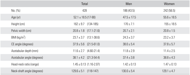
Four hundred twenty-eight patients (186 males and 242 females) were evaluated. Their ages ranged from 17 to 92 years (mean age, 52.1; SD, 18.5 years). The mean height and BMI was 162 ± 9.7 cm and 23.7 ± 3.7 kg/m2, respectively. There were 352, 67, and 9 hips with a Kellgren-Lawrence grade of 0, 1, and 2, respectively. The mean CE angle and AD was 37.9 ± 5.6° and 11.6 ± 2.7 mm, respectively. The mean AA was 38.1 ± 4.2°. The mean head-neck ratio and neck-shaft angle was 1.45 ± 0.13 and 129.6 ± 5.1, respectively (Table 1).
Table 1.
Characteristics of the Study Patients
Values are presented as mean ± SD (range) unless otherwise indicated.
BMI: body mass index, CE: center-edge.
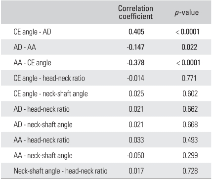
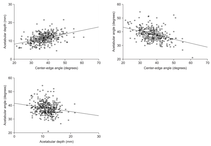
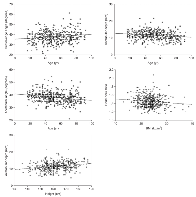
From the results of correlation analysis, the CE angle correlated with the AD (r = 0.405, p < 0.0001), whereas while was inversely correlated with the AA (r = -0.378, p < 0.0001). The AA was inversely correlated with the AD (r = -0.147, p = 0.022). None of the three landmarks of hip dysplasia correlated significantly with the head-neck ratio or the neck-shaft angle. There was no significant correlation between the neck-shaft angle and head-neck ratio (Table 2, Fig. 1). Age was correlated with the CE angle (r = 0.152, p = 0.002), whereas it was inversely correlated with the AD (r = -0.175, p < 0.0001) and AA (r = -0.227, p < 0.0001). The BMI was inversely correlated with the head-neck ratio (r = -0.121, p = 0.012), whereas the height was correlated positively with the AD (r = 0.235, p < 0.0001) and inversely with the head-neck ratio (r = -0.121, p = 0.0012). Male gender was significantly related to the neck-shaft angle (r = 0.122, p = 0.012) but inversely related to the AA (r = -0.138, p = 0.004) and head-neck ratio (r = -0.182, p < 0.0001) (Table 3, Fig. 2).
Table 2.
Correlation between Radiological Parameters
CE: center-edge, AD: acetabular depth, AA: acetabular angle.
Fig. 1.
The correlations between the radiological parameters of hip dysplasia.
Table 3.
Correlation of the Radiological Parameters with Age, BMI, Height, and Gender
BMI: body mass index, CE: center-edge.
*Male: 1, Female: 0.
Fig. 2.
The correlation between age and the radiological parameters of hip dysplasia.
Multiple linear regression analysis was performed to determine the association between a physical parameter and radiological parameters with the influence of other physical variables excluded. Age was associated significantly with the CE angle (p = 0.0009), but inversely with the AD (p = 0.005) and the AA (p = 0.014). Male gender was associated significantly with the increased neck-shaft angle (p = 0.036) but inversely with the head-neck ratio (p = 0.015) (Table 4).
Table 4.
Results of the Multiple Linear Regression Analysis
BMI: body mass index, CE: center-edge.
*Male: 1, Female: 0
DISCUSSION
Hip dysplasia is defined as a CE angle < 25°, an AD < 9 mm, or an AA > 42°. These three radiological parameters have been used to predict the development of OA.9,10) The concept of the CE angle was developed by Wiberg as a measurement of the degree of acetabular development, and the degree of dislocation of the femoral head in children.11) Fredensborg11) reported that the CE angle increased slowly until the age of 15 and was maintained at a constant value with no differences between genders. Murphy et al.1) reported that OA ultimately developed in the hip with a CE angle < 15°. The AD was proposed to compensate for the inaccuracy of the CE angle, which can be caused by the formation of a bony spur. The difficulty in localizing the center of the femoral head had been pointed out as another drawback of using the CE angle when the subluxation of the femoral head was present.12) The AA was devised to assess the degree of a patient's hip dysplasia without considering the position of the femoral head.12) Han et al.13) examined 591 Korean hip radiographs and found that the CE angle and aceatabular angle are more useful parameters for a diagnosis of acetabular dysplasia because of the insignificant age- or gender-dependent variations. From these results, the correlation between the CE angle and AD was the strongest, followed by that between the CE angle and AA, and lastly that between the AD and AA. This means that the CE angle predicts the other two parameters, and suggests that the CE angle, despite its limitations, is a very useful tool for defining hip dysplasia.
The Pearson correlation coefficients were obtained to examine the correlation between the physical parameters and radiological parameters of hip dysplasia. BMI and height as well as age and gender correlated with several radiological parameters. However, when multiple linear regression analysis was performed to exclude the influence of other physical parameters, only age was significantly associated with the radiological parameters of hip dysplasia. These results revealed a small, but statistical significant age-related increase in the CE angle and decrease in the AD and AA. The meaning of this is up to conjecture, but the increase in the CE angle and decrease in the AA might be the result of the age-related build-up of bony support on the lateral edge of the acetabulum. The age-related decrease in the AD is more difficult to interpret because it showed an opposite tendency to the other two variables.
A decreased head-neck ratio and an extremely increased/ decreased neck-shaft angle have also been reported to be associated with developing OA.3,14) The Pearson correlation showed no correlation of these parameters with each other or with the CE angle, AD or AA. On the other hand, these parameters correlated positively or inversely with the physical parameters. When multiple linear regression analysis was performed, only gender was left as a significantly associated factor to the head-neck ratio or neck-shaft angle, even though the differences were not large between men and women.
In conclusion, the radiological parameters of hip dysplasia are strongly, if not perfectly, inter-correlated. Age and gender have a significant associa tion with the radiological parameters of hip dysplasia or proximal femoral deformity.
ACKNOWLEDGEMENTS
This study was supported by the Korea Research Foundation Grant funded by the Korean Government (KRF-2006-311-E00359).
Footnotes
No potential conflict of interest relevant to this article was reported.
References
- 1.Murphy SB, Ganz R, Muller ME. The prognosis in untreated dysplasia of the hip: a study of radiographic factors that predict the outcome. J Bone Joint Surg Am. 1995;77(7):985–989. doi: 10.2106/00004623-199507000-00002. [DOI] [PubMed] [Google Scholar]
- 2.Reijman M, Hazes JM, Pols HA, Koes BW, Bierma-Zeinstra SM. Acetabular dysplasia predicts incident osteoarthritis of the hip: the Rotterdam study. Arthritis Rheum. 2005;52(3):787–793. doi: 10.1002/art.20886. [DOI] [PubMed] [Google Scholar]
- 3.Doherty M, Courtney P, Doherty S, et al. Nonspherical femoral head shape (pistol grip deformity), neck shaft angle, and risk of hip osteoarthritis: a case-control study. Arthritis Rheum. 2008;58(10):3172–3182. doi: 10.1002/art.23939. [DOI] [PubMed] [Google Scholar]
- 4.Jessel RH, Zurakowski D, Zilkens C, Burstein D, Gray ML, Kim YJ. Radiographic and patient factors associated with pre-radiographic osteoarthritis in hip dysplasia. J Bone Joint Surg Am. 2009;91(5):1120–1129. doi: 10.2106/JBJS.G.00144. [DOI] [PubMed] [Google Scholar]
- 5.Daysal GA, Goker B, Gonen E, et al. The relationship between hip joint space width, center edge angle and acetabular depth. Osteoarthritis Cartilage. 2007;15(12):1446–1451. doi: 10.1016/j.joca.2007.05.016. [DOI] [PubMed] [Google Scholar]
- 6.Im GI, Kim JY. Radiological joint space width in the clinically normal hips of a Korean population. Osteoarthritis Cartilage. 2010;18(1):61–64. doi: 10.1016/j.joca.2009.08.001. [DOI] [PubMed] [Google Scholar]
- 7.Kellgren JH, Lawrence JS. Radiological assessment of osteoarthrosis. Ann Rheum Dis. 1957;16(4):494–502. doi: 10.1136/ard.16.4.494. [DOI] [PMC free article] [PubMed] [Google Scholar]
- 8.Bland JM, Altman DG. Statistical methods for assessing agreement between two methods of clinical measurement. Lancet. 1986;1(8476):307–310. [PubMed] [Google Scholar]
- 9.Wiberg G. Studies on dysplastic acetabula and congenital subluxation of the hip joint with special reference to the complication of osteo-arthritis. Acta Chir Scand. 1939;83(Suppl 58):1–135. [Google Scholar]
- 10.Murray RO. The aetiology of primary osteoarthritis of the hip. Br J Radiol. 1965;38(455):810–824. doi: 10.1259/0007-1285-38-455-810. [DOI] [PubMed] [Google Scholar]
- 11.Fredensborg N. The CE angle of normal hips. Acta Orthop Scand. 1976;47(4):403–405. doi: 10.3109/17453677608988709. [DOI] [PubMed] [Google Scholar]
- 12.Sharp IK. Acetabular dysplasia: the acetabular angle. J Bone Joint Surg Br. 1961;43(2):268–272. [Google Scholar]
- 13.Han CD, Yoo JH, Lee WS, Choe WS. Radiographic parameters of acetabulum for dysplasia in Korean adults. Yonsei Med J. 1998;39(5):404–408. doi: 10.3349/ymj.1998.39.5.404. [DOI] [PubMed] [Google Scholar]
- 14.Goker B, Sancak A, Arac M, Shott S, Block JA. The radiographic joint space width in clinically normal hips: effects of age, gender and physical parameters. Osteoarthritis Cartilage. 2003;11(5):328–334. doi: 10.1016/s1063-4584(03)00023-2. [DOI] [PubMed] [Google Scholar]


