Abstract
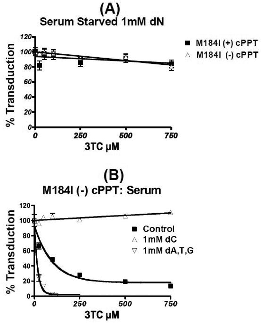
We recently reported that the M184I 3TC resistant mutation reduces RT binding affinity to dNTP substrates. First, the HIV-1 M184I mutant vector displays reduced transduction efficiency compared to wild type (WT) RT vector, which could be rescued by both elevating the cellular dNTP concentration and incorporating WT RT molecules into the M184I vector particles. Second, the central polypurine tract (cPPT) mutation and M184I mutation additively reduced the vector transduction to almost undetectable levels, particularly in nondividing cells. Third, the M184I (−) cPPT vector became significantly more sensitive to 3TC than the M184I (+) cPPT vector, but not to AZT or Nevirapine in the dividing cells. Finally, this 3TC sensitizing effect of the cPPT inactivation of the M184I vector was reversed by elevating dCTP level, but not by other three dNTPs. These data support a mechanistic interaction between cPPT and M184I RT with respect to viral replication and sensitivity to 3TC.
Keywords: HIV-1, cPPT, RT inhibitor sensitivity
Introduction
The ability of lentiviruses to infect terminally differentiated/nondividing macrophages is unique among retroviruses (Lewis, Hensel, and Emerman, 1992; Lewis and Emerman, 1994; Weinberg et al., 1991) and also important for their utility as gene delivery tools for nondividing cell types such as neurons (Naldini et al., 1996). The infection of macrophages by Human Immunodeficiency Virus type 1 (HIV-1) produces a landmark viral phenotype in the early phase of HIV-1 pathogenesis (Crowe, Zhu, and Muller, 2003; Verani, Gras, and Pancino, 2005). Various viral elements have been identified that enable lentiviruses to infect nondividing cells, such as Vpr, the central polypurine tract (cPPT)-DNA flap, and reverse transcriptase (RT) (as reviewed by Yamashita and Emerman) (De Rijck et al., 2007; Yamashita and Emerman, 2006). We have also previously reported that lentiviral RTs are able to aid in the infection of non-diving cells by efficiently synthesize proviral DNA in limited dNTP pools, such as those found in macrophage, due to their unique high affinity to dNTP substrates (Diamond et al., 2004).
The cPPT sequence, which is found among lentiviruses (Charneau, Alizon, and Clavel, 1992), lies at the exact center of the ~9.6 kb long HIV-1 RNA genome within the coding region of the integrase gene. A key function of the cPPT is to generate an additional RNA primer to initiate second (+) strand viral DNA synthesis (Charneau, Alizon, and Clavel, 1992). The cPPT-central termination sequence (CTS) has also been proposed to create a flap at the end of (+) strand DNA synthesis, which is recognized by unknown host/viral factors (Arhel et al., 2007). This flap is believed to enhance the nuclear import of the pre-integration complex (PIC) in nondividing cells (Zennou et al., 2000). However, since the flap does not have to be generated at the exact middle in order to be recognized by host or viral factors (De Rijck, Van Maele, and Debyser, 2005), the reason why the cPPT lies at the center of the genome still remains unclear. Furthermore, the role of the cPPT in nuclear import also remains controversial, since it has been shown that HIV-1 lacking the cPPT still efficiently replicates in macrophages and CD4+ T cells (Dvorin et al., 2002; Marsden and Zack, 2007).
We have recently reported that the addition of the cPPT can compensate for the delayed proviral DNA synthesis in a 3-vector system under the control of cytomeglovirus promoter. The HIV-1 vectors harbored the RT mutants, V148I and Q151N, which are defective in binding to dNTPs and thus fail to synthesize proviral DNA in cells with low dNTP concentrations, such as macrophages (Skasko and Kim, 2008). This finding suggested that the cPPT is necessary for HIV-1 to accelerate (+) DNA synthesis by creating an additional priming site, which can shorten the length of (+) DNA synthesis replicated by one primer from 9.6 kb to ~5 kb. Thus, it would be reasonable to assume that the cPPT becomes more critical for HIV-1 replication in macrophages where the viral replication kinetics are slow due to limited dNTP pools (Diamond et al., 2004), and during the delayed replication of HIV-1 vector containing partially defective RT mutations such as Q151N and V148I (Diamond et al., 2004; Jamburuthugoda, Chugh, and Kim, 2006; Jamburuthugoda et al., 2008).
The HIV-1 M184I RT mutation is clinically important due to its 3TC resistance, which can be generated through the deamination of cytosine at this residue by APOBEC3G (Mulder, Harari, and Simon, 2008). This RT mutation appears consistently in 3TC therapy, and ultimately results in a M184V mutation (Frost et al., 2000). The transient nature of the M184I mutation is likely due to its limited replicational fitness, compared to the M184V mutation (Domaoal et al., 2008; Frost et al., 2000). In fact several biochemical defects in the M184I mutant RT have been reported (Gao et al., 2008; Jamburuthugoda et al., 2008; Sarafianos et al., 1999). The β-branched side chain of isoleucine in M184I reduces the binding affinity of RT for dNTP substrates and raises its Kd approximately 50-fold (from 1 µM of WT RT Kd to 56 µM) (Jamburuthugoda et al., 2008). Thus M184I RT exhibits a severely decreased activity at low dNTP concentrations (Jamburuthugoda et al., 2008), whereas M184V RT, which displays WT levels of dNTP binding affinity (Domaoal et al., 2008), remains enzymatically active at both the low and high dNTP concentrations found in macrophages and activated CD4+ T cells, respectively (Aquaro et al., 2005; Diamond et al., 2004). Structural studies also demonstrated that the M184I mutation alters the template-primer interactions of RT, which explains its reduced processivity (Gao et al., 2008).
In this report, we investigated the mechanistic interplay between host dNTP pools, HIV-1 RT dNTP binding affinity and cPPT with respect to viral sensitivity and resistance to RT inhibitors. We have identified a specific compensatory relationship between the viral resistance of M184I HIV-1 RT and its dependence on the cPPT and cellular dNTP concentrations.
Materials and Procedures
Cells and pseudotyped vector production
MRC5 primary human fetal lung fibroblasts were a kind gift from Dr. Toru Takimoto (University of Rochester). The D3HIV-GFP virus expresses eGFP and all the HIV-1 proteins except for Nef and Env. The virus was pseudotyped with vesicular stomatitis virus glycoprotein (VSV-G) as described prior to this work (Jamburuthugoda et al., 2008). The central polypurine tract was mutated in the pD3HIV-GFP plasmid by PCR-based site-directed mutagenesis, through the use of a quick-change kit (Stratagene). PCR was performed using the following primers to mutate the cPPT: F primer – 5’ GTATTCATCCACAA ATTTTAAAGCGCAAACCCTCCTATTGGGGGGTACAGTC 3’; R primer – 5’ CACTGTACCCCCCAATACCACCTTTGCGCTTAAAATTGTGGATGAATAC 3’. The M184I mutation was generated in a similar fashion as previously described (Diamond et al., 2004). 293FTs were transfected using 1mg/ml of PEI (Polysciences) with 25 µg of HIV-1 vector DNA and 5 µg of VSV-G vector DNA. The 293FTs were washed 24 hours post transfection and supernatant was collect at 48 and 72 hours after transfection. The collected supernatant was spun in an ultra centrifuge (Beckman) with a SW-28 rotor at 22k for 2 hours. The virus-containing pellets from each spin were collected, treated with DNase, and stored at −80°C. The resulting pseudotyped vectors were normalized by p24 levels, which were determined through an enzyme-linked immunosorbent assay system (PerkinElmer).
Cell transduction
HLFs were treated with 10µg/ml of Polybrene (Sigma) prior to transduction with vector that was normalized to equal p24 levels (2.55 × 10⁵ pg). HLFs were washed with DPBS 24 hours after transduction. 48 hours post transduction, cells were washed again with DPBS and visualized by fluorescence microscopy (Zeiss). The 48 hour time-point was chosen because it is sufficient for the completion of reverse transcription of WT RT vector. Also it is unlikely that the delayed reverse transcription of the M184I mutant might eventually catch up to a WT level of the reverse transcription at later time points, because the preintegration (PIC) of HIV has a half-life of 1 day (Pierson et al., 2002), Following imaging, HLFs were trypsinized and monitored for GFP expression by fluorescent activated cell sorting using the FL-1 channel of a FACS Caliber (Becton Dickinson). The percent transduction was determined using FlowJo software (version 8.8.5, Tree Star, Inc).
2LTR Circle and late product Q-PCR
HLFs were serum-starved for 2 days before transduction with the WT (+) or (−) cPPT vectors 48 hours after the transduction, genomic extracts were prepared using SV genomic extraction kits (Wizard). The genomic extracts were then assayed for 2 LTR circles or LTR-Gag DNA by real time PCR using the following primers for the LTR regions of 2LTR Circles: 5’LTR region – 5’ GTGCCCGTCTG TTGTGTGACT-3’ and 3’ LTR region – 5’ CTTGTCTTCTTTGGGAGTGAATT AGC-3’, and the probe 5’-6-carboxylfluorsecein-TCCACACTGACTAAAAGGGTCTGAGGGATCTCT-carboxytetramethylrhodamine-3’ (IDT) or LTR-Gag region: 5’LTR region – 5’ CTAGGGAACCCACTG-‘3 and Gag – 5’ CAGACCCTTTTAGTCAGTGTGGAA-3’ and the probe 5’-6-carboxylfluorsecein- TCTCTAGCAGTGGCGCC-carboxytetramethylrhodamine-3’ (IDT) For the late product PCR, DNase digestions (Turbo DNase, Ambion) were conducted to correct for vector contamination in late product Q-PCR. For the PCR background control, the cells were pretreated with 2µM Nevirapine and 1µM AZT (NIH AIDS reagent program). Digestions were preformed on vector prior to transduction and on the transduced cells prior to lyses. The amplification and quantitation were carried out as previously described (Jamburuthugoda, Chugh, and Kim, 2006).
dN Effect Assay and Serum Starvation
To elevate the dNTP concentration, HLFs were treated with 1) 1.0mM of all dNs; (Sigma) 2 hours prior to the transduction of virus or 2) 1.0mM dC, or 3) 1.0mM all dNs without dC (dA, dT, and dG), 2 hours after 3TC treatment, and then 24 hours after transduction. To lower the cellular dNTP concentration, cells were cultured in DMEM supplemented with 0.2% FBS 48 hours prior to and throughout vector transduction.
Altering the ratio of WT and M184I RT packaged in a virion
To produce vectors with varying concentrations of packaged RT, 293FT cells were transfected with the following amounts of WT (−) cPPT to M184I (−) cPPT vector DNA: 0:25, 2.5:22.5, 12.5:12.5, 22.5:2.5, and 25µg: 0µg. Each transfection included 5 µg of VSV-G vector DNA.
Generation of IC50 values for 3TC Nevirapine and AZT in HLFs
HLFs cultured in DMEM supplemented with 10% FBS were pre-treated 2 hours before transduction and 24 hours after transduction (Moravek Biochemicals) in the following concentrations (0, 25, 100, 250, 500 and 750 µM/0, 0.25, 0.5, 1, 2, 4, and 6 µM) with 3TC, (0, 2.5, 5, 10, 30, 50, 75, 100, 150, 200 nM/0, 75, 100, 150, 200, 300, and 400 nM) of Nevirapine, or (0.32, 0.64, 1.28, 4, 8, 16µM) of AZT (NIH AIDS reagent program). The HLFs were transduced with equal p24 levels (2.77 × 10⁵ pg). HLFs expressing eGFP were quantitated at 48 hours post transduction through FACS. IC50 values were calculated using a best-fit curve (Prism).
Results
The effect of the cPPT and cellular dNTP concentrations on HIV-1 replication
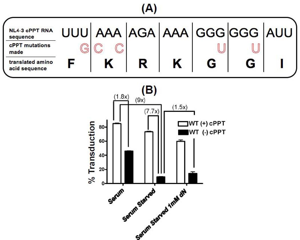
First, we investigated the effects of the cPPT mutation and the cellular dNTP concentrations on the transduction efficiency of a HIV-1 vector in primary human lung fibroblasts (HLF). HLFs have been used as an ideal model system for both dividing and nondividing cells because their cell cycles can be easily manipulated (Jamburuthugoda et al., 2008; Skasko and Kim, 2008). In addition, the cellular dNTP concentrations of HLFs can be modulated by both deoxynucleoside (dN) treatment and serum starvation (Jamburuthugoda, Chugh, and Kim, 2006). Dividing HLFs cultured in 10% serum have an intracellular dNTP concentration of approximately 150–300 nM (Jamburuthugoda, Chugh, and Kim, 2006), which is higher than the dNTP concentration found in human macrophages (~50 nM), but lower than activated CD4+ T cells (2 – 4 µM) (Charneau, Alizon, and Clavel, 1992; Diamond et al., 2004). Treating dividing and nondividing HLFs with deoxynucleosides (dNs) elevates their cellular dNTP concentration to 50 µM and ~1µM, respectively (Jamburuthugoda et al., 2008). In this study, we employed a VSV-G pseudotyped D3HIV-GFP vector which encodes a ~9.6kb long HIV-1 NL4-3 based RNA genome similar to the length of an infectious HIV genome, and expresses all HIV-1 proteins except for Env and Nef, which was replaced with GFP. The cPPT sequence of this vector was inactivated by mutating a series of purine bases to pyrimidine bases without altering the amino acid sequence of the integrase gene (Figure 1A), as described previously (Charneau, Alizon, and Clavel, 1992).
Figure 1. Effect of cPPT mutation and cellular dNTP level on transduction efficiency of HIV-1 vector.
(A) Nucleotide sequences of wild type and mutant NL4-3 cPPTs used in this study. The translated amino acid sequence of the cPPT is a part of HIV-1 integrase. (B) Effect of the cPPT mutation on the transduction of the WT RT vector in dividing (serum) and nondividing primary human lung fibroblasts (HLFs) with (serum starved with 1mM dN) and without (serum starved) the elevation of cellular dNTP levels. Primary HLFs were plated, 2.5×10⁵, in media containing 10% serum and then in media containing 0.2% serum for 48 hours in the presence and absence of 1mM dNs. HLFs cultured under these conditions were transduced with 2.77×10⁵ pg of WT RT HIV-1 vector with (+) or without (−) the cPPT. The transduced cells were analyzed by FACS for GFP expression at 48 hours post transduction and the transduction efficiency was plotted. The experiments presented in this figure were performed at least in triplicate.
First, we transduced HLFs with an equivalent p24 level (2.77×10⁵ pg) of the (+) and (−) cPPT WT RT HIV-1 vectors and cultured them in three separate media conditions; 10% serum that allows for cellular division, 0.2% serum (nondividing), and also 0.2% serum supplemented with 1mM of dNs. GFP expression was determined through FACS analysis at 48 hours post transduction. As shown in Figure 1B, the cPPT mutation had only a 1.8-fold decrease in the WT RT vector transduction in dividing HLFs, while a 7.7-fold decrease was observed by the cPPT inactivation in nondividing HLFs. Combining cPPT inactivation and nondividing culture conditions led to a 9-fold decrease of the WT RT vector transduction. This large decrease in transduction with nondividing HLFs could be due to loss of the cPPT functions in both nuclear import and the replication enhancement during (+) strand DNA synthesis. However, as seen in Figure 1B, the treatment with dNs only slightly (1.5×) compensated for the 9-fold transduction decrease seen in the nondividing HLFs. This minimal compensation by the dN treatment supports the idea that a major factor for the cPPT inactivation, which induced 9-fold decrease of the WT RT vector transduction in nondividing cells, is the loss of the nuclear import function of cPPT which cannot be compensated by the elevation of cellular dNTP concentrations. In addition, the minimal effect of the dN treatment is predictable because WT RT alone is sufficient for the efficient proviral DNA synthesis even at very low concentrations found in nondividing cells without the dNTP level elevation.
Roles of the cPPT and cellular dNTP pools in a HIV-1 vector harboring the 3TC resistant M184I RT mutant
Next, we investigated the effects of the cPPT mutation and cellular dNTP concentrations on the transduction of the HIV-1 vector bearing the M184I RT variant. M184I RT displays reduced polymerase activity and is clinically relevant due to its 3TC resistance (Jamburuthugoda et al., 2008; Li et al., 2009). This mutant appears transiently but consistently during 3TC treatment and ultimately results in M184V (Frost et al., 2000). Importantly, we have previously reported that the M184I RT displays a significantly reduced dNTP binding affinity when compared to the wild type (WT) RT. However, the reduced activity of M184I RT was rescued by increasing the dNTP concentration (Jamburuthugoda et al., 2008).
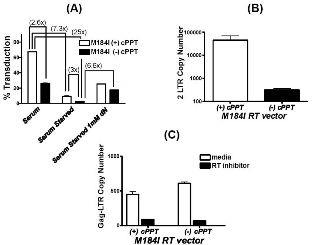
First, we characterized the transduction of the M184I HIV-1 with respect to cell division, cellular dNTP concentration and inactivation of the cPPT primer. HLFs were cultured in media with 1) 10% serum, 2) 0.2% serum, and 3) 0.2% serum with 1mM dNs. They were transduced with M184I (+) and (−) cPPT vectors with the same p24 level (2.77×10⁵ pg) used for the WT RT vectors in Figure 1B. The transduction efficiencies of each vector were determined by FACS analysis for GFP expression at 48 hours post transduction. As shown in Figure 2A, the M184I mutant vector showed ~70% transduction efficiency in dividing HLFs, which is only slightly reduced when compared to WT (~80%, Figure 1B). The cPPT mutation in the M184I vector induced a 2.6-fold transduction decrease in dividing cells, and this reduction is also very similar to the reduction observed with the WT vector (Figure 1B). In nondividing HLFs, the transduction of the M184I vector decreased 7.3-fold (to 8%), compared to the dividing cells (Figure 2A), and this is contrasted to the WT RT vector which displayed a similar transduction efficiency in both dividing (~80%) and nondividing HLFs (~70%) (Figure 1B). In nondividing cells, the cPPT mutation in the M184I vector caused a 3-fold further decrease in transduction (Figure 2A), while the WT RT vector showed a 7.7-fold decrease (Figure 1B). Basically, the M184I (−) cPPT vector almost failed to transduce nondividing HLF (<2%), supporting that the cPPT inactivation and M184I RT mutation additively reduced the vector transduction in nondividing HLFs. However, when the cellular dNTP pools were elevated by the dN treatment (Figure 2A), both (+) and (−) cPPT M184I vector significantly gained transduction efficiency, supporting that the dNTP concentration increase can at least partially compensate for both M184I and cPPT mutations. Therefore, it is apparent that the elevation of the cellular dNTP could significantly compensate the replicational delay induced by the M184I mutation and low dNTP concentration of the nondividing cells, leading to a larger increase (6.6×) of the M184I vector transduction efficiency in the nondividing cells treated with dNs, compared to the WT RT vector (1.5×, Figure 1B).
Figure 2. Effect of cPPT mutation on the transduction efficiency and proviral DNA synthesis of HIV-1 vector harboring M184I mutant RTs.
(A) Transduction efficiency of (+) and (−) cPPT M184I vectors in HLFs cultured in 10% serum, 0.2% serum and 0.2% serum with 1mM dNs. HLFs were transduced with an equal p24 level, 2.77×10⁵ pg of (+) and (−) cPPT M184I RT HIV-1 vectors, and the transduction efficiency was determined as described in Figure 1B. (B) Quantitative 2LTR circle PCR assay of the (+) and (−) cPPT M184I vector in the serum-starved HLFs. Genomic DNA of serum-starved HLFs transduced with the M184I virus was prepared at 48 hours post transduction and used for the quantitative 2LTR PCR to monitor the kinetics of the proviral DNA synthesis and normalized with genomic cellular DNA as previously described (Jamburuthugoda, Chugh, and Kim, 2006; Skasko and Kim, 2008). (C) The LTR/gag late reverse transcription product synthesized from the 3’ LTR was measured for the (+) and (−) M184I vector in the serum-starved HLF at 48 hours post transduction as described in Materials and Procedures. The treatment with 2µM Nevirapine and 1µM AZT, which block viral reverse transcription, was employed for assessing the PCR background control. The experiments presented in this figure were performed at least in triplicate.
Next, we investigated if the cPPT inactivation, which further reduced the transduction efficiency of the M184I vector in nondividing HLFs, directly impacted proviral DNA synthesis kinetics. For this, we conducted 2LTR Q-PCR assay; 2-LTR circles are a result of the end joining of proviral DNA in the nucleus post completion of reverse transcription and nuclear import, and have been used for a marker for completed proviral DNA synthesis and nuclear import (Butler, Johnson, and Bushman, 2002; Van Maele et al., 2003). We also simultaneously examined the copy number of LTR-gag late product which is replicated from 3’ PPT (Sun et al., 1997). As shown in Figure 2B and 2C, a drastic decrease in the 2LTR copy number in the M184I (−) cPPT vector was observed, compared to the M184I (+) cPPT vector in nondividing HLFs, while the copy number of the late product synthesized from the 3’ PPT was not affected by the cPPT deletion. Thus, the reduction of the 2LTR circle copy number was likely due to the deletion of the additional PPT primer and flap, which affects the (+) strand synthesis kinetics and nuclear import, respectively.
Rescue of the M184I (−) cPPT mutation by WT RT molecules
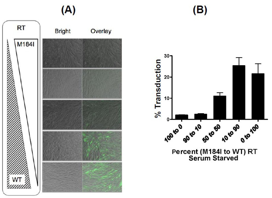
Next, we tested if the reduced transduction of the M184I RT (−) cPPT vector in nondividing HLFs could be rescued by introducing WT RT molecules into the virion of the M184I vector. It was predicted that co-packaging of the WT HIV-1 RT together with the M184I RT within the same capsid could rescue the biochemical defects induced by the M184I RT mutation. We prepared hybrid HIV-1 vectors, which have been previously described (Julias et al., 2001), harboring both WT and M184I RT molecules in different ratios within the same virion. Briefly, these hybrid vectors were prepared by transfecting 293FT cells with varying ratios of WT to M184I RT packaging plasmids, and used to transduce serum-starved HLFs. The expression and packaging of these two RT constructs are equally efficient as demonstrated by the almost equal p24 levels of the vector particles produced from the different mixtures of the two RT plasmids (Data not shown). The transduction efficiency of each vector was determined through FACS analysis and fluorescence microscopy as presented in Figure 3A and 3B. Indeed, the M184I vectors with higher ratios of co-packaged WT RT protein, exhibited an increased number of GFP expressing cells (Figure 3A), and thus a higher transduction efficiency (Figure 3B). These data confirm that the cause of the decreased transduction efficiency of the M184I vector in nondividing cells was due to delayed reverse transcription resulting from the decreased polymerase kinetic activity of M184I RT, especially at low cellular dNTP pools found in nondividing cells.
Figure 3. Compensation of the transduction defect of the M184I (−) cPPT HIV-1 vector by the incorporation of WT HIV-1 RT.
(A) (−) cPPT HIV-1 vectors containing different percentages of WT RT to M184I RT in a single viral particle (100% M184I, 90% M184I to 10% WT, 50% M184I to 50% WT, 10% M184I to 90% WT, 100% WT) were prepared. The serum-starved HLFs were transduced with an equal p24 amount of the constructed hybrid vectors. The pictures of the transduced cells in bright field and GFP were taken at 48 hours post transduction, and the overlaid pictures were constructed. At the same time, the transduction efficiency was determined by FACS analysis for the GFP-expressing cells (B). The experiments presented in this figure were performed at least in triplicate.
Effect of the cPPT mutation on the sensitivity of HIV-1 M184I vector to 3TC and other RT inhibitors
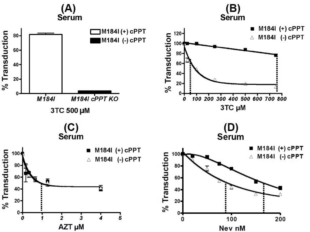
Next, we tested if the replication delay induced by the cPPT mutation could affect the sensitivity of the HIV-1 vector to RT inhibitors, which directly interrupt proviral synthesis. To test this we investigated the effect of the cPPT mutation on the sensitivity of the M184I vector to 3TC. M184I RT has been previously shown to have a high resistance to 3TC due to the steric hindrance between the isoleucine of RT and 3TC (Sarafianos et al., 1999). Dividing HLFs were pre-treated with varying concentrations of 3TC, and these cells were transduced with an equal p24 level of the (+) and (−) cPPT M184I vectors. As shown in Figure 4A, HLFs treated with 500µM 3TC, which is ~300 times higher than the previously published IC50 values of the WT RT HIV-1 (Yoshimura et al., 1999), still maintained efficient transduction levels with the M184I (+) cPPT vector, confirming the high 3TC resistance of the M184I RT. In contrast, a large transduction decrease was clearly observed at 500 µM of 3TC in the M184I (−) cPPT vector, suggesting that the cPPT inactivation counteracts the strong 3TC resistance of the M184I RT mutation. Next, we examined a 3TC dose dependence of with the M184I RT (+) and (−) cPPT vectors. As seen in Figure 4B, the M184I (+) cPPT vector exhibited a high resistance to 3TC, and only a minimal decrease in transduction was seen with this vector even at 750 µM 3TC. In contrast, the M184I (−) cPPT vector displayed a significant decrease in its resistance to 3TC. Since M184I (+) cPPT vector displays a high 3TC resistance, we were only able to determine the IC25 value, the drug concentration yielding 25% inhibition (Figure 4B). Indeed, we found that cPPT inactivation causes a 30-fold increase in viral sensitivity to 3TC.
Figure 4. Effect of the cPPT mutation on 3TC sensitivity of HIV-1 M184I vector.
(A) The transduction efficiency of the M184I (+) and (−) cPPT HIV-1 vectors in dividing HLFs in the presence of 50µM 3TC. (B) 3TC dose dependence of the M184I (+) and (−) cPPT vectors. (C) AZT dose dependence of the M184I (+) and (−) cPPT vectors. (D) Nevirapine dose dependence on the M184I (+) and (−) cPPT vectors. HLFs were transduced with an equal p24 level of the (+) and (−) cPPT vector in dividing HLFs pretreated with varying concentrations of 3TC, AZT or Nevirapine. The transduction efficiencies at each of the concentrations in (B), (C) and (D) were normalized with that of the drug free control (100%). The IC25 and IC50values were marked with a dotted line. The experiments presented in this figure were performed at least in triplicates.
Next, we tested if the same drug sensitization in the M184I (−) cPPT vector could be observed with AZT or Nevirapine. Dividing HLFs were pretreated with various concentrations of AZT (Figure 4C) or Nevirapine (NEV, Fig. 4D), and were transduced with the M184I (+) and (−) cPPT vectors. The IC50 values with each drug treatment were determined and summarized in Table 1. Indeed, both the M184I (+) and (−) cPPT vectors showed very similar IC50 values for AZT and NEV. This suggests that the sensitivity of the M184I (−) cPPT vector is exclusive to 3TC.
Table 1.
IC50 values of WT and M184I (+) and (−) cPPT HIV-1 vectors to 3TC, AZT and NEV in dividing HLFs.
| WT RT Vectors |
IC50 values | ||
|---|---|---|---|
| 3TC | AZT | NEV | |
| (+) cPPT | 2.4µM ± 4.98 | 0.8µM ± 12.63 | 169nM ± 4.26 |
| (−) cPPT | 2.4µM ± 2.88 | 0.8µM ± 9.463 | 93nM ± 4.628 |
| M184I RT Vectors |
IC50 values | ||
| 3TC | AZT | NEV | |
| (+) cPPT | >750µM ± 2.09 | 0.8µM ± 12.03 | 166nM ± 4.13 |
| (−) cPPT | 10µM ± 6.821 | 0.9µM ± 7.16 | 93nM ± 5.875 |
Effect of specific cellular dNTP pool changes on the 3TC sensitization property of the M184I (−) cPPT vector
Next, we investigated possible mechanistic explanations of the 3TC sensitization effect of the cPPT inactivation in M184I vector. First, we measured the 3TC sensitivity of the M184I (+) and (−) vectors in the HLFs with elevated pools of all four dNTPs. As shown in Figure 5A, both M184I (+) and (−) cPPT vectors displayed strong 3TC resistance, and thus the 3TC sensitization activity of the cPPT mutation could be effectively eliminated by the 1mM dN treatment (Figure 5A). Next, we tested if the loss of the 3TC sensitization effect is nucleotide-specific because it is possible that 3TC may only compete with dC, not the other three dNs. For this test, we treated HLFs with 1mM dC alone or a mixture of dG, dT and dA, and measured the 3TC sensitivity of the M184I (−) cPPT. As seen in Figure 5B, upon dC treatment, the M184I (−) cPPT vector gained a strong resistance to 3TC. In contrast, when increasing the remaining other three dNTPs, the M184I (−) cPPT vector remained sensitive to 3TC, demonstrating that a high concentration of only cellular dCTP abolishes the 3TC sensitivity of the M184I vector gained by the cPPT inactivation.
Figure 5. Effect of dN treatment on M184I (−) HIV-1 vector sensitivity to 3TC.
(A) 3TC dose dependence of the M184I (+) and (−) cPPT vectors in HLFs pretreated with 1mM dN. (B) The transduction of the M184I (−) cPPT vectors in dividing HLFs in the presence and absence of 1mM dC or a mixture of 1mM dA, dT, and dG. HLFs were transduced with an equal p24 level of the (−) cPPT M184I RT vectors in dividing HLFs pretreated with varying concentrations of 3TC. The transduction efficiency at each of the concentrations was normalized with that of the drug free control (100%). The experiments presented in this figure were performed at least in triplicate.
Finally, we examined if the WT (−) cPPT vector would also display an increased sensitivity to 3TC, AZT or Nevirapine treatment. As shown in Table 1, the cPPT mutation did not increase the sensitivity of WT RT to 3TC. In addition, the IC50 values of AZT and Nevirapine for the WT (+) and (−) cPPT vectors remained the same. These data demonstrate that the drug sensitization effect of cPPT inactivation is exclusive to 3TC and the M184I (−) cPPT vector.
Discussion
Proviral DNA synthesis of retroviruses is a very complex process requiring both host and viral factors for its efficient completion, particularly in nondividing cells (Yamashita and Emerman, 2006). In this study we further characterized these elements, identified interactive relationships between them, and assessed their contributions to viral RT inhibitor sensitivity. Predictably, elements that can affect the DNA polymerization rate of RT, such as dNTP concentration and the binding affinity of RT to dNTPs, were able to alter proviral DNA synthesis kinetics. We have previously reported that the RTs of lentiviruses uniquely synthesize DNA efficiently at the low dNTP concentrations found in nondividing cells, such as macrophages (Diamond et al., 2004; Jamburuthugoda et al., 2008). This is a unique biochemical feature and can be contrasted to the RTs of oncoretroviruses that fail to synthesize DNA efficiently at low dNTP concentrations and infect only dividing cells. These observations suggested that HIV-1 and other lentiviral RTs, may have evolved a unique ability to utilize substrates that fall within a broad concentration range varying between its two main target cell types, macrophage and activated CD4+ T cells.
We have also recently identified the cPPT as a viral element that contributes toward efficient proviral DNA synthesis of HIV-1, by dually initiating the (+) strand synthesis and thus shortening the genome distance per primer required to compete the (+) strand HIV-1 DNA synthesis (Skasko and Kim, 2008). This efficient proviral DNA synthesis was found to be particularly important in cell types bearing low dNTP concentrations. We were also able to demonstrate that the cPPT could compensate for the delayed proviral DNA synthesis induced by dNTP defective binding RT mutants such as Q151N and V184I (Skasko and Kim, 2008).
Here we investigate the cPPT and the cellular dNTP concentrations with respect to the transduction of the M1841 vector and its sensitivity to 3TC. Our data demonstrates that the cPPT inactivation and the M184I RT mutation additively reduce the vector transduction efficiency and proviral DNA synthesis kinetics. This reduced transduction could become further pronounced by eliminating cellular division, which both lowers cellular dNTP concentration and requires the nuclear import function of cPPT. This reduction is partially rescued by elevating both cellular dNTP concentrations and viral DNA polymerization capacity by incorporating WT RT molecules to the M184I vector. It can be presumed that by increasing intracellular dNTP pools, the M184I vector is rescued from its dNTP binding defect. However, this rescue seen with dN treatment is unique to dividing cells. This is likely due to the additional requirement for the nuclear import function of the cPPT flap in nondividing cells, which cannot be compensated by an increased rate of proviral DNA synthesis.
Importantly, the M184I mutant vector lost its 3TC resistance upon the cPPT inactivation. Since this effect was observed in dividing cells, this sensitization could not be due to the loss of the nuclear import function of the cPPT. However, this drug sensitization effect seen with the M184I (−) cPPT vector was observed only with 3TC, not AZT or Nevirapine. In addition, the WT (−) cPPT vector did not display an increased sensitivity to any of the drugs used in this study. Therefore, we believe the sensitivity to 3TC observed is exclusive to the M184I (−) cPPT vector. One possible cause for this specificity could be the use of high concentrations of 3TC during the drug titration curves of the M184I vector. These high concentrations of 3TC may result in a decrease in the cellular concentrations of dCTP and/or all dNTPs, by forcing a majority of cellular nucleoside phosphorylation machinery to 3TC. The resulting limited dNTP pool would not affect the proviral DNA synthesis of WT RT. WT RT is able to synthesize proviral DNA efficiently even at very low dNTP concentrations due to its high binding affinity to dNTPs, but the limited dNTP pools induced by the treatment with high 3TC concentrations would affect the M184I RT due to its reduced dNTP binding affinity. We tested this possibility by increasing the cellular dC concentrations after HLFs were treated with 3TC. Indeed, we observed that only the elevation of dCTP, not the other three NTPs, abolished the sensitivity of the M184I (−) cPPT vector to 3TC. Thus this data supports the idea that, instead of the direct replication inhibition induced by the 3TCTP incorporation, the cellular dCTP limitation induced by the high concentration of 3TC may be a cause of the replication restriction of the M184I vector especially when the replication kinetic role of cPPT is inactivated by the cPPT mutation. Importantly, it is also possible that the pretreatment with dC may also reduce the phosphorylation of 3TC to 3TCTP which is an active drug form of 3TC. But this possibility was excluded because the limitation of the 3TCTP synthesis should result in the increase of the 3TC resistance, and instead we observed the decrease of the 3TC resistance.
One would expect that the M184I (−) cPPT vector would become more sensitive to AZT because, like 3TC, AZT is a nucleoside analog that also requires phosphorylation by cellular machinery (Sommadossi, 1998). However, the cPPT inactivation did not affect the sensitivity of the M184I (−) cPPT vector to AZT. Importantly, unlike 3TC, the low concentrations of AZT in this study (<6 µM) were sufficient to inhibit the replication of the M184I vector because the M184I RT is still sensitive to 3TCTP like WT RT, and the treatment with the low AZT concentration should not affect the cellular dNTP (dTTP) pools (Sommadossi, 1998). Indeed, it has been previously shown that 5µM of AZT only inhibits 50% of thymidine phosphorylation (Sommadossi, 1998; Susan-Resiga et al., 2007). Interestingly, a slight decrease of the NEV sensitivity was induced by the cPPT inactivation in the M184I vector. While it is possible that the delayed replication by the cPPT inactivation may be further delayed by the reduction of the enzymatically capable free M184I RTs induced by NEV binding, this effect appears to be minimal.
Finally, we also observed that the WT (−) cPPT did not display any sensitivity to the drugs used in this study. There could be two possible explanations for this. First, the low concentrations of AZT and 3TC used in the drug titration curves with WT vector were not sufficient to affect the dNTP synthesis machinery. Second, unlike M184I RT, WT HIV-1 RT is highly functional even at low dNTP concentrations. Therefore, if the dNTP pool were to decrease, it would have little effect on the polymerization capability of WT RT due to its high binding affinity for dNTPs.
In this study, we did not conduct experiments with the M184V RT vector, because, unlike M184I RT, the pre-steady state kinetic analysis demonstrated that M184V RT has a WT RT level of the dNTP binding affinity (Domaoal et al., 2008). This biochemical data also explains why the M184I RT further mutates to M184V during the 3TC treatment. Therefore, considering M184V RT in terms of its dNTP binding affinity, we postulate that cPPT removal should not significantly affect the 3TC resistance of the M184V vector even though the treatment with high concentrations of 3TC may restrict the cellular dNTP pools.
Here we present a comprehensive study that inter-connects various host and viral factors that HIV-1 employs to achieve efficient proviral DNA synthesis. By altering these factors such as RT activity and cellular dNTP concentrations, we were able to detect the specific interplay of the cPPT sequence with the 3TC resistance of the M184I HIV-1 vector.
Acknowledgements
This research was supported by AI049781 (B.K.), NIH T-32AI049815SKVCH) and R25 GM064133 (WD).
Footnotes
Publisher's Disclaimer: This is a PDF file of an unedited manuscript that has been accepted for publication. As a service to our customers we are providing this early version of the manuscript. The manuscript will undergo copyediting, typesetting, and review of the resulting proof before it is published in its final citable form. Please note that during the production process errors may be discovered which could affect the content, and all legal disclaimers that apply to the journal pertain.
References
- Aquaro S, Svicher V, Ceccherini-Silberstein F, Cenci A, Marcuccilli F, Giannella S, Marcon L, Caliò R, Balzarini J, Perno CF. Limited development and progression of resistance of HIV-1 to the nucleoside analogue reverse transcriptase inhibitor lamivudine in human primary macrophages. J Antimicrob Chemother. 2005;55(6):872–878. doi: 10.1093/jac/dki104. [DOI] [PubMed] [Google Scholar]
- Arhel NJ, Souquere-Besse S, Munier S, Souque P, Guadagnini S, Rutherford S, Prévost MC, Allen TD, Charneau P. HIV-1 DNA Flap formation promotes uncoating of the pre-integration complex at the nuclear pore. EMBO J. 2007;26(12):3025–3037. doi: 10.1038/sj.emboj.7601740. [DOI] [PMC free article] [PubMed] [Google Scholar]
- Butler SL, Johnson EP, Bushman FD. Human immunodeficiency virus cDNA metabolism: notable stability of two-long terminal repeat circles. Journal of Virology. 2002;76(8):3739–3747. doi: 10.1128/JVI.76.8.3739-3747.2002. [DOI] [PMC free article] [PubMed] [Google Scholar]
- Charneau P, Alizon M, Clavel F. A second origin of DNA plus-strand synthesis is required for optimal human immunodeficiency virus replication. Journal of Virology. 1992;66(5):2814–2820. doi: 10.1128/jvi.66.5.2814-2820.1992. [DOI] [PMC free article] [PubMed] [Google Scholar]
- Crowe S, Zhu T, Muller WA. The contribution of monocyte infection and trafficking to viral persistence, and maintenance of the viral reservoir in HIV infection. J Leukoc Biol. 2003;74(5):635–641. doi: 10.1189/jlb.0503204. [DOI] [PubMed] [Google Scholar]
- De Rijck J, Van Maele B, Debyser Z. Positional effects of the central DNA flap in HIV-1-derived lentiviral vectors. Biochem Biophys Res Commun. 2005;328(4):987–994. doi: 10.1016/j.bbrc.2005.01.052. [DOI] [PubMed] [Google Scholar]
- De Rijck J, Vandekerckhove L, Christ F, Debyser Z. Lentiviral nuclear import: a complex interplay between virus and host. Bioessays. 2007;29(5):441–451. doi: 10.1002/bies.20561. [DOI] [PubMed] [Google Scholar]
- Diamond TL, Roshal M, Jamburuthugoda VK, Reynolds HM, Merriam AR, Lee KY, Balakrishnan M, Bambara RA, Planelles V, Dewhurst S, Kim B. Macrophage tropism of HIV-1 depends on efficient cellular dNTP utilization by reverse transcriptase. J Biol Chem. 2004;279(49):51545–51553. doi: 10.1074/jbc.M408573200. [DOI] [PMC free article] [PubMed] [Google Scholar]
- Domaoal RA, McMahon M, Thio CL, Bailey CM, Tirado-Rives J, Obikhod A, Detorio M, Rapp KL, Siliciano RF, Schinazi RF, Anderson KS. Pre-steady-state kinetic studies establish entecavir 5'-triphosphate as a substrate for HIV-1 reverse transcriptase. J Biol Chem. 2008;283(9):5452–5459. doi: 10.1074/jbc.M707834200. [DOI] [PMC free article] [PubMed] [Google Scholar]
- Dvorin JD, Bell P, Maul GG, Yamashita M, Emerman M, Malim MH. Reassessment of the roles of integrase and the central DNA flap in human immunodeficiency virus type 1 nuclear import. Journal of Virology. 2002;76(23):12087–12096. doi: 10.1128/JVI.76.23.12087-12096.2002. [DOI] [PMC free article] [PubMed] [Google Scholar]
- Frost SD, Nijhuis M, Schuurman R, Boucher CA, Brown AJ. Evolution of lamivudine resistance in human immunodeficiency virus type 1-infected individuals: the relative roles of drift and selection. Journal of Virology. 2000;74(14):6262–6268. doi: 10.1128/jvi.74.14.6262-6268.2000. [DOI] [PMC free article] [PubMed] [Google Scholar]
- Gao L, Hanson M, Balakrishnan M, Boyer P, Roques B, Hughes S, Kim B, Bambara R. Apparent defects in processive DNA synthesis, strand transfer, and primer elongation of M184 mutants of HIV-1 reverse transcriptase derive solely from a dNTP utilization defect. Journal of Biological Chemistry. 2008:21. doi: 10.1074/jbc.M710148200. [DOI] [PMC free article] [PubMed] [Google Scholar]
- Jamburuthugoda VK, Chugh P, Kim B. Modification of human immunodeficiency virus type 1 reverse transcriptase to target cells with elevated cellular dNTP concentrations. J Biol Chem. 2006;281(19):13388–13395. doi: 10.1074/jbc.M600291200. [DOI] [PubMed] [Google Scholar]
- Jamburuthugoda VK, Santos-Velazquez JM, Skasko M, Operario DJ, Purohit V, Chugh P, Szymanski EA, Wedekind JE, Bambara RA, Kim B. Reduced dNTP Binding Affinity of 3TC-resistant M184I HIV-1 Reverse Transcriptase Variants Responsible for Viral Infection Failure in Macrophage. Journal of Biological Chemistry. 2008;283(14):9206. doi: 10.1074/jbc.M710149200. [DOI] [PMC free article] [PubMed] [Google Scholar]
- Julias JG, Ferris AL, Boyer PL, Hughes SH. Replication of phenotypically mixed human immunodeficiency virus type 1 virions containing catalytically active and catalytically inactive reverse transcriptase. Journal of Virology. 2001;75(14):6537–6546. doi: 10.1128/JVI.75.14.6537-6546.2001. [DOI] [PMC free article] [PubMed] [Google Scholar]
- Lewis P, Hensel M, Emerman M. Human immunodeficiency virus infection of cells arrested in the cell cycle. EMBO J. 1992;11(8):3053–3058. doi: 10.1002/j.1460-2075.1992.tb05376.x. [DOI] [PMC free article] [PubMed] [Google Scholar]
- Lewis PF, Emerman M. Passage through mitosis is required for oncoretroviruses but not for the human immunodeficiency virus. Journal of Virology. 1994;68(1):510–516. doi: 10.1128/jvi.68.1.510-516.1994. [DOI] [PMC free article] [PubMed] [Google Scholar]
- Li J, Li L, Li HP, Zhuang DM, Liu SY, Liu YJ, Bao ZY, Wang Z, Li JY. Competitive capacity of HIV-1 strains carrying M184I or Y181I drug-resistant mutations. Chin Med J. 2009;122(9):1081–1086. [PubMed] [Google Scholar]
- Marsden MD, Zack JA. Human immunodeficiency virus bearing a disrupted central DNA flap is pathogenic in vivo. Journal of Virology. 2007;81(11):6146–6150. doi: 10.1128/JVI.00203-07. [DOI] [PMC free article] [PubMed] [Google Scholar]
- Mulder LC, Harari A, Simon V. Cytidine deamination induced HIV-1 drug resistance. P Natl Acad Sci Usa. 2008;105(14):5501–5506. doi: 10.1073/pnas.0710190105. [DOI] [PMC free article] [PubMed] [Google Scholar]
- Naldini L, Blömer U, Gallay P, Ory D, Mulligan R, Gage FH, Verma IM, Trono D. In vivo gene delivery and stable transduction of nondividing cells by a lentiviral vector. Science. 1996;272(5259):263–267. doi: 10.1126/science.272.5259.263. [DOI] [PubMed] [Google Scholar]
- Pierson TC, Zhou Y, Kieffer TL, Ruff CT, Buck C, Siliciano RF. Molecular characterization of preintegration latency in human immunodeficiency virus type 1 infection. Journal of Virology. 2002;76(17):8518–8531. doi: 10.1128/JVI.76.17.8518-8531.2002. [DOI] [PMC free article] [PubMed] [Google Scholar]
- Sarafianos SG, Das K, Clark AD, Ding J, Boyer PL, Hughes SH, Arnold E. Lamivudine (3TC) resistance in HIV-1 reverse transcriptase involves steric hindrance with beta-branched amino acids. P Natl Acad Sci Usa. 1999;96(18):10027–10032. doi: 10.1073/pnas.96.18.10027. [DOI] [PMC free article] [PubMed] [Google Scholar]
- Skasko M, Kim B. Compensatory role of human immunodeficiency virus central polypurine tract sequence in kinetically disrupted reverse transcription. Journal of Virology. 2008;82(15):7716–7720. doi: 10.1128/JVI.00120-08. [DOI] [PMC free article] [PubMed] [Google Scholar]
- Sommadossi JP. Cellular nucleoside pharmacokinetics and pharmacology: a potentially important determinant of antiretroviral efficacy. AIDS. 1998;12 Suppl 3:S1–S8. [PubMed] [Google Scholar]
- Sun Y, Pinchuk LM, Agy MB, Clark EA. Nuclear import of HIV-1 DNA in resting CD4+ T cells requires a cyclosporin A-sensitive pathway. J Immunol. 1997;158(1):512–517. [PubMed] [Google Scholar]
- Susan-Resiga D, Bentley AT, Lynx MD, LaClair DD, McKee EE. Zidovudine inhibits thymidine phosphorylation in the isolated perfused rat heart. Antimicrob Agents Chemother. 2007;51(4):1142–1149. doi: 10.1128/AAC.01227-06. [DOI] [PMC free article] [PubMed] [Google Scholar]
- Van Maele B, De Rijck J, De Clercq E, Debyser Z. Impact of the central polypurine tract on the kinetics of human immunodeficiency virus type 1 vector transduction. Journal of Virology. 2003;77(8):4685–4694. doi: 10.1128/JVI.77.8.4685-4694.2003. [DOI] [PMC free article] [PubMed] [Google Scholar]
- Verani A, Gras G, Pancino G. Macrophages and HIV-1: dangerous liaisons. Mol Immunol. 2005;42(2):195–212. doi: 10.1016/j.molimm.2004.06.020. [DOI] [PubMed] [Google Scholar]
- Weinberg JB, Matthews TJ, Cullen BR, Malim MH. Productive human immunodeficiency virus type 1 (HIV-1) infection of nonproliferating human monocytes. J Exp Med. 1991;174(6):1477–1482. doi: 10.1084/jem.174.6.1477. [DOI] [PMC free article] [PubMed] [Google Scholar]
- Yamashita M, Emerman M. Retroviral infection of non-dividing cells: old and new perspectives. Virology. 2006;344(1):88–93. doi: 10.1016/j.virol.2005.09.012. [DOI] [PubMed] [Google Scholar]
- Yoshimura K, Feldman R, Kodama E, Kavlick MF, Qiu YL, Zemlicka J, Mitsuya H. In vitro induction of human immunodeficiency virus type 1 variants resistant to phosphoralaninate prodrugs of Z-methylenecyclopropane nucleoside analogues. Antimicrob Agents Chemother. 1999;43(10):2479–2483. doi: 10.1128/aac.43.10.2479. [DOI] [PMC free article] [PubMed] [Google Scholar]
- Zennou V, Petit C, Guetard D, Nerhbass U, Montagnier L, Charneau P. HIV-1 genome nuclear import is mediated by a central DNA flap. Cell. 2000;101(2):173–185. doi: 10.1016/S0092-8674(00)80828-4. [DOI] [PubMed] [Google Scholar]


