
Fig. 3.
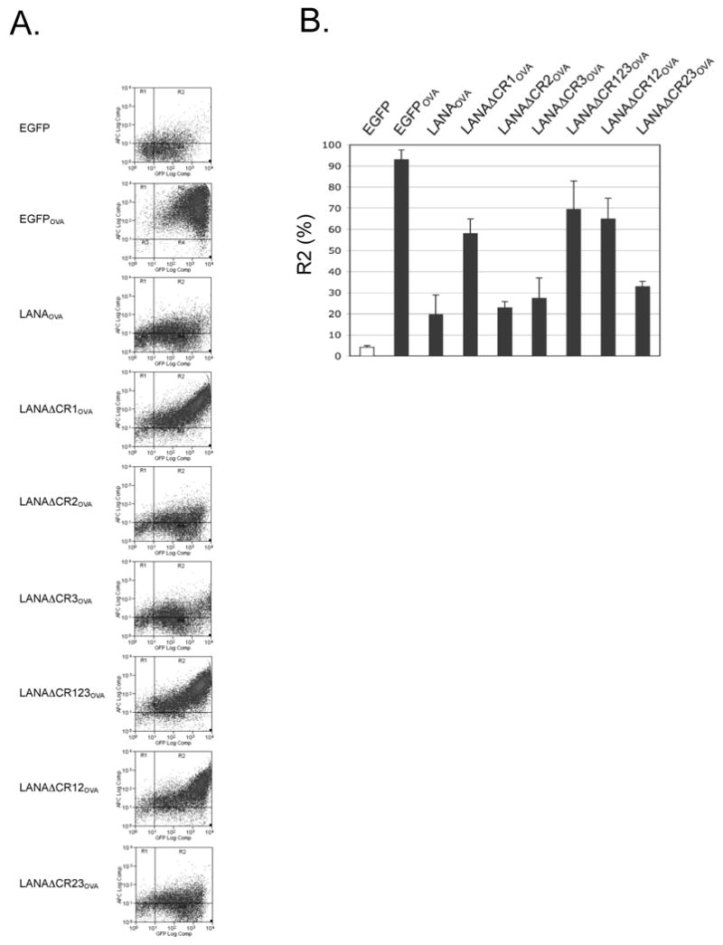
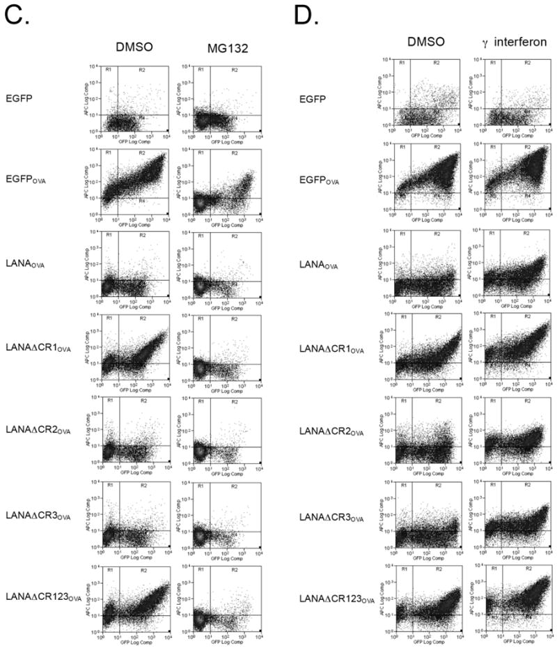
The CR1 region of LANA1 inhibits MHC I peptide antigen presentation in cis. (A) Representative results from flow cytometry analysis for SIINFEKL presentation on 293KbC2 cells expressing EGFP-LANA1OVA constructs (Fig. 2A). Cells were stained with APC anti-mouse MHC class I Kb-SIINFEKL (25-D1.16) 24 h after transfection. Samples were gated for EGFP positivity to ensure construct expression and then the percentage SIINFEKL presenting cells were determined by APC positivity. All experiments were repeated at least three times using either APC or Phycoerythrin (PE) 25-D1.16 (eBioscience) for detection and show similar results. (B) Quantitation of SIINFEKL presentation from three independent experiments (mean±S.D.). P=0.0001 (EGFP and EGFPOVA), 0.0023 (EGFPOVA and LANAOVA), 0.0049 (LANAOVA and LANAΔCR1OVA), 0.0135 (LANAOVA and LANAΔCR123OVA), 0.0311 (LANAOVA and LANAΔCR12OVA). P values between different groups, were obtained by two-tailed Student's t test using Prism software (GraphPad). (C, D) The effect of different pretreatment conditions on antigen presentation. Cells were pretreated with MG132 (proteasome inhibitor) (C), interferon gamma (immunoproteasome inducer) (D) and then SIINFEKL peptide presentation was determined by flow cytometry analysis.