Abstract
As digital slides need a lot of storage space, lack of a singular method to acquire and store these large, two-dimensional images has been a major stumbling block in the universal acceptance of this technology. The DICOMS Standard Committee Working Group 26 has put in a tremendous effort to standardize storage methods so that they are more in line with currently available PACS in most hospitals for storage of radiology images. A recent press release (Supplement 145) of these standards was hailed by one and all involved in the field of digital pathology as it will make it easier for hospitals to integrate digital pathology into their already established systems without adding too much overhead costs. Besides, it will enable different vendors developing the scanners to upgrade their products to storage systems that are common across all systems.
Keywords: Digital pathology, z-planes, DICOM
INTRODUCTION
Recently, new standardization was finalized for storage and retrieval of digital images which can enhance interplay between digital pathology systems and tools and other systems such as PACS systems used for Radiology and other medical imaging modalities. Digital Imaging and Communications in Medicine (DICOM), synonymous with ISO (International Organization for Standardization) standard 12052, is the global standard for medical imaging and is used in all electronic medical record systems that include imaging as part of the patient record.[1] Its continuing evolution is managed by a collaboration of 40 professional societies, government agencies, trade associations, and imaging equipment manufacturers, under the Medical Imaging and Technology Alliance (MITA). DICOM Standards Committee Working Group 26 is a group of people dedicated to the advancement of digital pathology. This group, along with the College of American Pathologist's Diagnostic Intelligence and Health Information Technology (DIHIT) Committee, developed standards for making digital pathology more universal.[2,3] This effort has helped move the field of digital pathology closer toward having a true, enterprise-wide Picture Archiving and Communication System (PACS) that can be used by all specialties in health care systems. Currently, most hospitals use a PACS to manage and store radiology images.[4] Until now, PACS software and the DICOM core standard did not accommodate pathology whole slide images (WSI).[5,6] The technical specifications approved for use by the Working group 26 will make it possible for clinical laboratories and pathology groups to store digital pathology images in a form that is compatible with the same DICOM archive systems currently being used by hospitals and other providers to distribute, view and store radiology images. The standard will enable the electronic display, sharing, storage, and management of static and WSI, or even larger images, used in pathology. It will also provide health care professionals with flexibility, such as panning and zooming, when interacting with the image.
Although the DICOM standards have tried to standardize all of the steps involved in digitalizing slides (including image creation, acquisition, processing, analyzing, distribution, visualization, and data management), the most important impact has been on image storage, which actually has been one of the main stumbling blocks for universal acceptance of this technology. Standardization of all digital imaging aspects will also help develop open system scanners that readily store images into a commercially available PACS system using one DICOM-standard messaging. As of now, DICOM standards did not provide for singular methods to acquire and store large two-dimensional images, nor did they incorporate any way to handle stored images or multiple images of the same slide at various resolutions. Not only is storage important, but so too is retrieval so that one can easily interact with the images. These deficiencies have now been mostly resolved and standardized.
IMAGE STORAGE
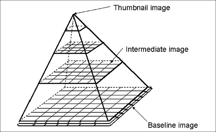
The whole slide images (WSI) made by digitizing glass slides at diagnostic resolutions are very large. Typically, an H & E slide needs 4-20 GB of storage space, but some WSI may take up even larger space. Currently, the DICOM standard does not have provision for storage of these large two-dimensional images, nor does it incorporate a way to handle images so that they can be retrieved easily. The new recommendations suggest that the basic approach to storing large multi-resolution multi-planar WSI is to map subregions from each layer into a DICOM series. This is referred to as a tiled organization in which images are stored in squares or rectangular tiles, which are then stored in two-dimensional arrays. In simple terms, it is like cutting a large picture into smaller squares or rectangular fragments and then storing them together in a box. Multiple resolutions are stored in the same way. The highest resolution has the most data and will form the broadest row. Each row of less resolution will stack up on top of rows containing more resolution, forming a sort of pyramid. Each layer can then be retrieved and put together, to either form part of an image or the entire picture. This makes it relatively easy to randomly access any subregion of the image without loading large amounts of data. The images are stored at several precomputed resolutions, facilitating rapid zooming. Thus, the typical organization of a WSI for pathology will resemble a pyramid of image data. The highest resolutions captured by an image scanner will sit at the base of the pyramid, while the apex corresponds to the zoom or low power image. When multiple z-plane (vertical level) images are present, each plane will be stored separately on one level [Figure 1]. This approach required relatively minor conceptual changes in the DICOM standard and will make implementation by vendors with existing DICOM-compatible products easier, helping to foster adoption of this standard.
Figure 1.

The highest resolutions captured by an image scanner will sit at the base of the pyramid, while the apex corresponds to the zoom or low power image. When multiple z-plane (vertical level) images are present, each plane will be stored separately on one level (This figure has been modified and redrawn using Supplement 145 as reference)
IMAGE COMPRESSION
Previously WSI data was compressed using either JPEG or JPEG2000 modalities. As JPEG2000 compressions yield higher compression and fewer image artifacts, it has advantages over other available systems for image compression standard and coding systems. However, other compression methods can also be used, including Moving Picture Experts Group (MPEG) or even private compression schemes. The pixel data can be monochrome, color (RGB or YCB, CR) or palette color.
WSI FRAME OF REFERENCE
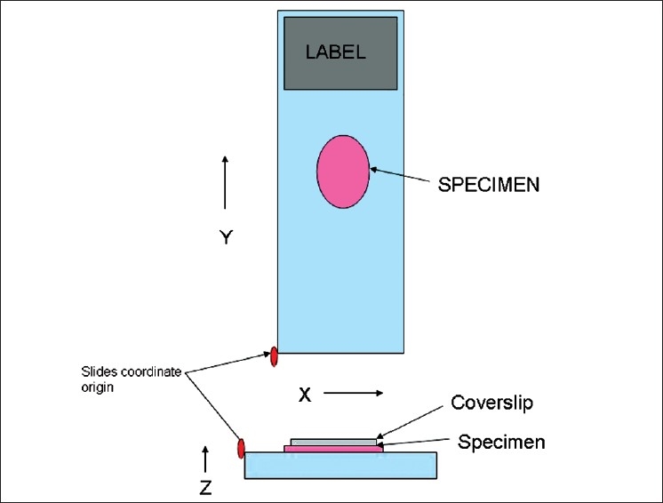
In order that all digital slides be processed and handled similarly, the DICOM standards specify a particular corner of the slide as a normal reference origin and a right-handed (X, Y, Z) coordinate system for position from that origin. The top left corner of the total imaging area is used as the point of reference as shown in Figure 2.
Figure 2.

The top left corner of the total imaging area is used as the point of reference and a right-handed (X, Y, Z) coordinate system is followed for position from that origin (This figure has been modified and redrawn using Supplement 145 as reference)
Z-PLANES
For WSI z-planes are imaginary focal planes described at a certain nominal physical height (in microns) of the image above a reference level, which is the glass slide in WSI. It provides information for spatial positioning of imaginary planes and all tiles from one z-plane should be stored at the same level within the pyramid model. It can thus help to match tiles form one z-plane when an image is being retrieved.
ANNOTATIONS
As annotations are created at a later time from the image acquisition, DICOM standards state that they will be recorded in a separate series (as a DICOM series is limited to objects of single modality, produced by single equipment, in a single procedure step).
WORKFLOW MANAGEMENT
Techniques that were in place for Radiology have been found adequate and useful for pathology and automated slide scanning modalities. These include the modalities wordlist (MWL) and modalities performance procedure setup (MPPS). These allow a scanner to query for slides by slide barcode. The scanner scans the label along with the slide and information from both these regions is sent together to the laboratory information system. On retrieval, all patient information comes along with the slide image. Both gross specimen photography and automated slide scanning modalities were incorporated into the MWL and MPPS services.
CONCLUSION
In addition, Supplement 145 added 56 new data elements to the DICOM Standard in 14 new or revised modules. Also included are seven new or revised DICOM structured data templates and 18 new defined value sets. Moreover, 80 new coded terminology concepts were added to SNOMED, and 36 to DICOM.
This achievement has been hailed by those involved in the world of digital pathology. It will certainly make it easy for hospitals to integrate digital pathology into their already established radiology information systems. There is no need to reinvent the wheel. Secondly, digital pathology and other medical imaging vendors such as those who make PACS systems will now be in a position to enhance their products to support this new standard, enabling interoperability between systems for digital pathology images.
Footnotes
Available FREE in open access from: http://www.jpathinformatics.org/text.asp?2011/2/1/23/80719
REFERENCES
- 1.Supplement 145: Whole Slide Microscopic Image IOD and SOP Classes. Digital Imaging and Communications in Medicine (DICOM) 2010 Sep [Google Scholar]
- 2.Le Bozec C, Henin D, Fabiani B, Schrader T, Garcia-Rojo M, Beckwith B. Refining DICOM for pathology--progress from the IHE and DICOM pathology working groups. Stud Health Technol Inform. 2007;129:434–8. [PubMed] [Google Scholar]
- 3.Daniel C, García Rojo M, Bourquard K, Henin D, Schrader T, Della Mea V, et al. Standards to support information systems integration in anatomic pathology. Arch Pathol Lab Med. 2009;133:1841–9. doi: 10.5858/133.11.1841. [DOI] [PubMed] [Google Scholar]
- 4.Kahn CE, Jr, Carrino JA, Flynn MJ, Peck DJ, Horii SC. DICOM and radiology: Past, present, and future. J Am Coll Radiol. 2007;4:652–7. doi: 10.1016/j.jacr.2007.06.004. [DOI] [PubMed] [Google Scholar]
- 5.Tuominen VJ, Isola J. Linking whole-slide microscope images with DICOM by using JPEG2000 interactive protocol. J Digit Imaging. 2010;23:454–62. doi: 10.1007/s10278-009-9200-1. [DOI] [PMC free article] [PubMed] [Google Scholar]
- 6.Morelli S, Grigioni M, Giovagnoli MR, Balzano S, Giansanti D. Picture archiving and communication systems in digital cytology. Ann Ist Super Sanita. 2010;46:130–7. doi: 10.4415/ANN_10_02_05. [DOI] [PubMed] [Google Scholar]


