Abstract
The ability of Francisella tularensis to replicate in macrophages is critical for its pathogenesis, therefore intracellular growth assays are important tools for assessing virulence. We show that two lysis solutions commonly used in these assays, deionized water and deoxycholate in PBS, lead to highly inaccurate measurements of intracellular bacterial survival.
Keywords: Francisella tularensis, intracellular growth, lysis
Report
Francisella tularensis, one of the most deadly respiratory pathogens in existence, is considered a potential biological weapon because of its ease of aerosolization and its high degree of infectivity in and lethality to humans. F. tularensis replicates within multiple cell types, mostly macrophages (Hall et al., 2008), and this ability is critical for F. tularensis virulence (for a review, see Meibom and Charbit, 2010). Therefore, intracellular growth assays are important tools for studying F. tularensis pathogenesis. To measure the number of intracellular bacteria, a common method is to lyse the infected host cells and plate the lysates on agar for colony-forming unit (CFU) determination. In the literature on F. tularensis, a wide array of solutions have been used for cell lysis including ultrapure water (dH2O) to disrupt host cells by osmotic pressure and detergents such as sodium deoxycholate (DOC), sodium dodecyl sulfate (SDS), or saponin. However, some of these lytic solutions may lead to an inaccurate measurement of intracellular bacterial numbers. When comparing data from three different studies, in which similar conditions were used (same macrophage-like cell line, J774A.1, similar multiplicity of infection (moi)), but different lysis solutions, the number of intracellular bacteria per cell at time zero of infection was drastically different: 0.005 bacteria/cell using dH2O (Qin et al., 2008), 0.2 using 0.1% DOC (Sammons-Jackson et al., 2008), or 5 using 0.05% SDS (Mohapatra et al., 2008) (Table 1). Here we address whether commonly used lysis solutions are similarly suitable to measure the number of F. tularensis intracellular bacteria.
Table 1.
Measurement of the number of intracellular F. tularensis bacteria per cell, at time zero of infection of J774A.1 macrophage-like cell line, in three different studies.
| Lysis solution | moi | Bacteria/cell (t = 0) | Reference |
|---|---|---|---|
| 0.1% DOC | 100 | 0.2 | Sammons-Jackson et al., 2008 |
| 0.05% SDS | 50 | 5 | Mohapatra et al., 2008 |
| dH2O | 50 | 0.005 | Qin et al., 2008 |
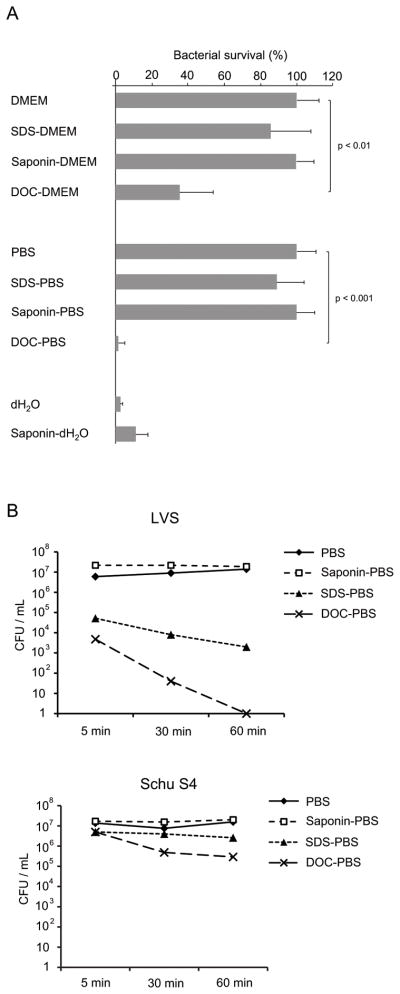
A suitable lysis solution should not affect bacterial viability. The sensitivity of F. tularensis Live Vaccine Strain (LVS) to the most commonly used lysis solutions was assessed. LVS bacteria were grown in Mueller-Hinton broth to mid-logarithmic phase, pelleted, and incubated for 5 min in three different detergents: 0.1% DOC, 0.1% SDS, or 1% saponin (same concentration throughout the study), prepared in Dulbecco’s modified Eagle medium (DMEM, Invitrogen) or in PBS. Saponin had no effect on LVS viability (Fig. 1A). By contrast, DOC killed LVS organisms: 36 ± 18 % of bacteria survived after incubation in DOC-DMEM, and only 2 ± 3% survived in DOC-PBS (Fig. 1A). This decrease is statistically significant relative to diluent controls DMEM and PBS (t-test) (Fig. 1). LVS sensitivity to two other commonly used lysis solutions, dH2O and dH2O supplemented with 1% saponin, was also assessed. The survival rate was 3 ± 1% in dH2O, and 11 ± 7% in dH2O-saponin, demonstrating that LVS organisms were sensitive to hypotonic shock (Fig. 1A). After a longer exposure (60 min), LVS was almost eradicated in DOC-PBS, but was not affected by saponin in PBS (Fig. 1B). LVS sensitivity to SDS in PBS exhibited some variability: after a 5-min exposure, the number of CFU was sometimes the same as in PBS control (Fig. 1A), sometimes drastically reduced by 2 logs (Fig. 1B). Therefore we recommend to avoid the use of SDS as a lysis reagent. To determine whether the virulent F. tularensis Schu S4 strain presented the same sensitivity pattern, Schu S4 bacteria were exposed to PBS, DOC-PBS, SDS-PBS and saponin-PBS, for 5, 30 and 60 min. Like LVS, Schu S4 bacteria were sensitive to SDS (1 log reduction in CFU after 60 min) and DOC (2-log reduction in CFU after 60 min), but not to saponin (Fig. 1B). It is noteworthy that Schu S4 bacteria were more resistant to SDS and DOC than LVS bacteria (Fig. 1B), as Schu S4 strain is more virulent than LVS.
Figure 1.
Francisella tularensis LVS and Schu S4 sensitivity to various lysis solutions. (A) Mid-log LVS bacteria were exposed for 5 min to 0.1% SDS, 1% saponin, or 0.1% DOC prepared in DMEM or in PBS, or to dH2O, supplemented with (or without) 1% saponin. CFUs were determined before and after exposure. Data are presented as percent survival (mean ± SD of three independent experiments). Unpaired Student’s t-test was used for statistical analysis. A p-value < 0.05 was considered significant. (B) Mid-log LVS and Schu S4 bacteria were exposed to 0.1% SDS, 1% saponin, or 0.1% DOC prepared in PBS. CFUs were determined after 5, 30 and 60 min.
To know whether LVS sensitivity to lysis solution could affect the quantitation of intracellular bacterial number, J774A.1 macrophage-like cells were seeded overnight in 24-well plates (2 × 10⁵ cells/well) in DMEM supplemented with 10% fetal bovine serum, and infected for 2 h with mid-log LVS at an moi of 200 (bacteria to cell). Cells were then washed three times with PBS, treated with gentamicin (100 μg/mL) for 1 h to kill extracellular bacteria, and incubated for 5 min in various lysis solutions: DOC-DMEM, SDS-PBS, saponin-PBS, DOC-PBS, dH2O or saponin-dH2O. Serial dilutions of lysate were plated onto cysteine heart agar supplemented with 1% hemoglobin, and CFU were counted. To determine the number of intracellular bacteria per cell, the number of macrophages per well was measured after trypsin treatment. The highest number of intracellular bacteria per cell was obtained when using DOC-DMEM (1.8 bacteria/cell), while numbers using DOC-DMEM, SDS-PBS, saponin-PBS, or saponin-dH2O were slightly but significantly lower (between 0.4 and 0.7 bacteria/cell) (Table 2). The number of viable bacterial cells was drastically reduced when using DOC-PBS (0.01 bacteria/cell), or dH2O (0.07 bacteria/cell) (Table 2). Therefore, LVS sensitivity to DOC-PBS and dH2O (Fig. 1) affected LVS survival. However while LVS was also sensitive to DOC-DMEM, SDS-PBS and saponin-dH2O (Fig. 1), these lysis solutions did not affect the quantitation of intracellular bacteria, compared to saponin-PBS to which LVS is resistant (Table 2). Perhaps the LVS strain was less sensitive when localized in macrophage monolayers rather than as mid-log phase bacteria growing in broth. Whether this was due to a different bacterial physiological state, or to a protective consequence of the complex environment of the macrophage lysate remains unknown. Importantly, the use of DOC-PBS and dH2O led to a 1- to 2-log underestimation of the number of viable intracellular bacteria/cell, and these lysis solutions should be avoided.
Table 2.
Number of intracellular LVS bacteria per J774A.1 macrophage, determined by different methods : by lysis with DOC-DMEM, SDS-PBS, saponin-PBS, DOC-PBS, dH2O or dH2O-saponin, or by confocal microscopy.
| DOC-DMEM | SDS-PBS | saponin-PBS | DOC-PBS | dH2O | saponin dH2O | Confocal microscopy | |
|---|---|---|---|---|---|---|---|
| Bacteria/cell | 1.8 ± 0.2a | 0.7 ± 0.1b | 0.5 ± 0.1b | 0.01 ± 0.002b | 0.07 ± 0.01b | 0.4 ± 0.1b | 1.75 ± 0.02 |
Mean ± SD, two independent experiments.
Statistically different relative to confocal microscopy and DOC-DMEM results (t-test, p < 0.01)
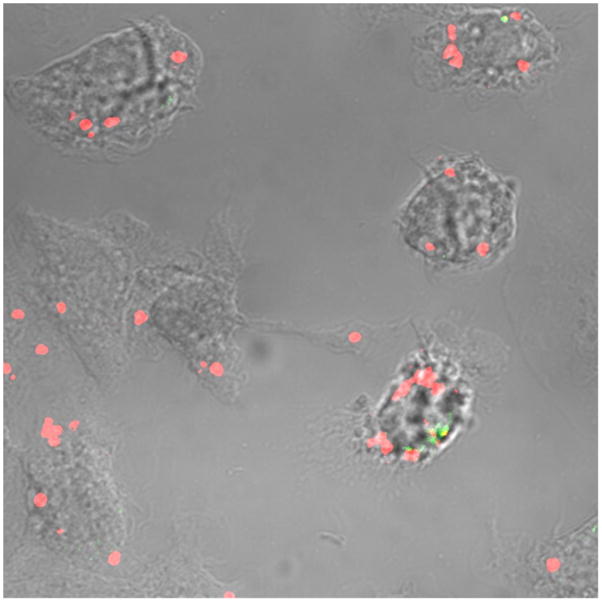
Confocal microscopy was used to determine whether our data were representative of the actual number of intracellular bacteria. J774A.1 cells were infected as described above, fixed in 2% paraformaldehyde, and incubated with rabbit anti-LVS antibodies (1:500, Lampire Biological Laboratories (Pipersville, PA)), and donkey anti-rabbit IgG conjugated to AlexaFluor-488 (Invitrogen) as a secondary antibody. Cells were then permeabilized in 0.1% Triton X-100, incubated with rabbit anti-LVS as primary antibody and donkey anti-rabbit IgG conjugated to AlexaFluor-568 (Invitrogen) as a secondary antibody. Samples were observed by confocal microscopy (Carl Zeiss LSM). This sequential staining procedure allowed the differentiation of extracellular bacteria, stained with AlexaFluor-488 (green), from intracellular bacteria, stained with AlexaFluor-568 (red) (Fig. 2). In a total of 500 macrophages, the average number of intracellular bacteria per cell was 1.75 ± 0.02 (Table 2). Therefore, intracellular bacterial quantification with DOC-DMEM lysis was statistically similar to the number determined by confocal microscopy, while measures with SDS-PBS, saponin-PBS and saponin-dH2O were slightly but significantly lower (Table 2).
Figure 2.

Immunostaining of LVS intracellular organisms in J774A.1 macrophage-like cells, visualized by confocal microscopy. Red bacteria are intracellular, green bacteria are extracellular.
In summary, we show here that dH2O and DOC in PBS, commonly used as lysis buffers in F. tularensis intracellular viability assays, should be avoided, as these reagents strongly affect LVS bacteria viability, resulting in a 1- to 2-log underestimation of the actual number of intracellular bacteria. This finding could explain the small number of bacteria per cell reported by Qin et al. when using dH2O (Table 1). DOC in DMEM, SDS and saponin are suitable for F. tularensis intracellular growth assays, the first giving the most accurate data relative to confocal microscopy observations. However, lysis in DOC in DMEM, SDS in PBS and saponin in dH2O should be carefully performed, as these lysis buffers show some toxicity toward the LVS strain of F. tularensis. DOC and SDS in PBS should also be avoided in F. tularensis Schu S4 intracellular viability assays, as this strain is sensitive to these lysis solutions.
Acknowledgments
We are grateful to Colette Cywes-Bentley (Brigham and Women’s Hospital, Boston) for technical assistance. This study was supported by New England Center of Excellence in Biodefense and Emerging Infectious Disease, grant AI057159 from the National Institute of Allergy and Infectious Diseases.
Abbrevations used
- CFU
colony-forming unit
- dH2O
ultrapure water
- SDS
sodium dodecyl sulfate
- DOC
sodium deoxycholate
- DMEM
Dulbecco’s modified Eagle medium
- PBS
phosphate buffer saline
Footnotes
Publisher's Disclaimer: This is a PDF file of an unedited manuscript that has been accepted for publication. As a service to our customers we are providing this early version of the manuscript. The manuscript will undergo copyediting, typesetting, and review of the resulting proof before it is published in its final citable form. Please note that during the production process errors may be discovered which could affect the content, and all legal disclaimers that apply to the journal pertain.
References
- Hall JD, Woolard MD, Gunn BM, Craven RR, Taft-Benz S, Frelinger JA, Kawula TH. Infected-host-cell repertoire and cellular response in the lung following inhalation of Francisella tularensis Schu S4, LVS, or U112. Infect Immun. 2008;76:5843–5852. doi: 10.1128/IAI.01176-08. [DOI] [PMC free article] [PubMed] [Google Scholar]
- Meibom KL, Charbit A. The unraveling panoply of Francisella tularensis virulence attributes. Cur Opin Microbiol. 2010;13:11–17. doi: 10.1016/j.mib.2009.11.007. [DOI] [PubMed] [Google Scholar]
- Mohapatra NP, Soni S, Reilly TJ, Liu J, Klose KE, Gunn JS. Combined deletion of four Francisella novicida acid phosphatases attenuates virulence and macrophage vacuolar escape. Infect Immun. 2008;76:3690–3699. doi: 10.1128/IAI.00262-08. [DOI] [PMC free article] [PubMed] [Google Scholar]
- Qin A, Scott DW, Mann BJ. Francisella tularensis subsp. tularensis Schu S4 disulfide bond formation protein B, but not an RND-type efflux pump, is required for virulence. Infect Immun. 2008;76:3086–3092. doi: 10.1128/IAI.00363-08. [DOI] [PMC free article] [PubMed] [Google Scholar]
- Sammons-Jackson WL, McClelland K, Manch-Citron JN, Metzger DW, Bakshi CS, Garcia E, Rasley A, Anderson BE. Generation and characterization of an attenuated mutant in a response regulator gene of Francisella tularensis live vaccine strain (LVS) DNA Cell Biol. 2008;27:387–403. doi: 10.1089/dna.2007.0687. [DOI] [PMC free article] [PubMed] [Google Scholar]


