Abstract
Study design
A prospective intervention of functional electrical stimulation leg cycle ergometry (FES-LCE) of four women with spinal cord injury (SCI).
Objective
To evaluate the effect of FES-LCE training on arterial compliance in individuals with chronic SCI of traumatic origin.
Setting
Tertiary rehabilitation center in Canada.
Methods
Large and small artery compliance were measured at the radial artery before and after a 3 month training program using FES-LCE.
Results
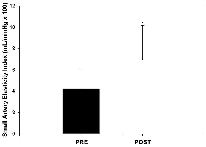
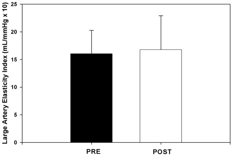
There was no significant change in large artery compliance after FES-LCE (16.0 ± 4.2 to 16.8 ± 6.1 mL/mmHg × 10, p = n.s.). There was a marked (63%) increase in small artery compliance after the FES training program (4.2 ± 1.8 to 6.9 ± 3.2 mL/mmHg × 100, p < 0.05).
Conclusion
It appears that FES-LCE is effective in improving small artery compliance in females with SCI.
Keywords: spinal cord injury, arterial compliance, functional electrical stimulation, exercise
INTRODUCTION
With medical advancements in treatment, many persons with spinal cord injury (SCI) now have the potential for a life expectancy approaching normal.1 As such, they may experience the same chronic conditions as able-bodied persons.2 Over the past several decades cardiovascular disease (CVD) has become a leading cause of death in the SCI population.2 The increase in CVD with aging in SCI individuals reflects that of able bodied individuals, though at an accelerated rate, higher prevalence, and earlier onset.3
In recent years, researchers have used pulse wave analysis to show that arterial stiffness is associated with CVD, specifically atherosclerotic burden.4 Arterial compliance decreases as the severity of atherosclerosis increases.4 Importantly, changes in the compliance of small and large arteries precede (by years) the presence of plaques characteristic of atherosclerosis.5 These changes are important early predictors of the risk of CVD and related events.5,6
It is well known that a sedentary lifestyle is associated with premature mortality and increased morbidity.3 This is a significant risk for persons with SCI who, because of loss of motor function, are often extremely sedentary.3 Indeed, individuals with SCI show an increased prevalence for a number of cardiovascular risk factors associated with reduced vascular health (including decreased arterial compliance).7 A recent study suggests normotensive individuals with paraplegia exhibit decreased arterial compliance compared to able-bodied controls.7
Exercise rehabilitation using the upper extremities has clearly been shown to lead to marked health benefits in persons with SCI.3 Recent evidence also shows lower extremity exercise through functional electrical stimulation leg cycle ergometry (FES-LCE) to be a valuable clinical rehabilitation tool for SCI as it is effective at improving exercise tolerance and measures of cardiovascular health (including aerobic fitness and glucose homeostasis).3 However, the effect of FES-LCE training on systemic arterial compliance has not been evaluated in persons with SCI. Accordingly, the purpose of this study was to examine the effects of FES-LCE training on systemic arterial compliance. We hypothesized that FES-LCE would improve systemic arterial compliance in persons with SCI.
METHODS
Participants
All study participants (Table 1) had sustained a traumatic SCI and were at least one year post-injury. Exclusion included: (1) other neurological conditions in addition to SCI, (2) CVD (e.g., cardiac arrhythmia, high blood pressure (systolic blood pressure >160 mmHg; diastolic blood pressure >90 mmHg)) (3) unhealed pressure ulcers or wounds, (4) previous fragility fracture(s) and/or, (5) abnormal bone formation, spasm or contracture in the lower extremity limiting hip or knee range of motion. Eligible participants provided written informed consent. Following consent, participants were also excluded if there was insufficient response to the FES-LCE (stimulation did not move the leg) or the FES-LCE was not tolerable by the participant (e.g., pain with the lowest level of stimulation). All applicable institutional and governmental regulations concerning the ethical use of human volunteers were followed during the course of this research.
Table 1.
Participant characteristics.
| Participant | Sex | Age (yr) | Years since injury | Lesion level | ASIA Class* |
|---|---|---|---|---|---|
| 1 | F | 29 | 3 | C4 | B |
| 2 | F | 19 | 2.5 | C5 | B |
| 3 | F | 29 | 14 | T4 | A |
| 4 | F | 51 | 16 | T7 | C |
The American Spinal Injury Association (ASIA) score is employed to classify lesion completeness: A, sensory and motor complete; B, sensory incomplete but motor complete; C, sensory and motor incomplete with more than half of key muscles below the neurological level at a muscle grade less than 3.
FES-LCE
Training was performed using a computer controlled leg cycle ergometer (Ergys2, Version H.6, Therapeutic Alliances Inc., Ohio, USA). The ergometer uses six 2×4 inch self-adhesive surface electrodes (Uni-Patch, Wabasha, MN, USA) to provide electrical stimulation to muscles. Electrodes were placed over the hamstrings (proximal electrode on upper 1/3, distal on lower 1/3 of muscle), gluteals (proximal electrode below iliac crest and distal electrode above ischial tuberosity), and quadriceps (proximal and distal electrodes shifted 2/3 of quadriceps width laterally and medially from the midline, respectively),
Cycling was elicited via the administration of electrical stimulation to muscles in a specific sequence. The sinusoidal wave pulse was 500 μsec, 60 Hz synchronized for each electrode pair. The ergometer was programmed to increase stimulation current from 0 to 140 mA to achieve the desired pedaling cadence. Threshold settings, used during warm-up and cool-down, were set to palpable muscle contraction. Maximum stimulation current was set depending on participant’s comfort.
Pedaling cadence was defined by a pre-set upper bound and lower bound. If cadence approached the upper bound, pedaling speed was slowed via a decrease in stimulation current. If stimulation was at its maximum current and cadence dropped below the pre-set lower bound, the ergometer automatically stopped and entered the cool-down mode.
Exercise Protocol
Research indicates that benefits from FES-LCE can be achieved with 30 minutes of exercise at a cycling cadence above 35 rpm.8 Thus, a habituation period (averaging 16 weeks) preceded the training phase. During habituation individuals trained to be able to cycle for 30 minutes against no resistance at a minimum of 35 rpm. When able to complete 30 minutes of continuous cycling for 2 consecutive sessions, participants commenced the training phase which included gradually adding resistance. Participants were asked to train 3 times a week for 12 weeks to complete 36 training sessions.
Training sessions began with a passive one-minute warm-up where stimulation was pulsed between 50% and 100% of an individually determined preset threshold setting. Following the warm-up, active FES-LCE training commenced. When participants were able to complete one session with at least 90% of the pre-set pedal resistance for 30 minutes in two consecutive training sessions, pedal resistance was increased with an increment of 1/8 Kp (corresponding with about 6.1 watts at 50 rpm; the maximum resistance is 7/8 Kp) in the next training session. When pedaling rate dropped below 45 rpm and the maximum stimulation reach 100% of the pre-set maximum stimulation level, resistance was automatically reduced to enable a pedaling cadence close to 49 rpm.
Following exercise, the FES-LCE entered a two minute cool-down mode in which the stimulation was pulsed between 50% and 100% of the threshold setting. Including preparation, sessions lasted approximately 60 minutes. One staff member was involved during each training session.
Arterial Compliance Measurement
Large and small arterial compliance were assessed non-invasively using an applanation tonometer (Hypertension Diagnostics/Pulse Wave ™ CR-3000, Eagan, MN, USA) that measures 30-second recordings of signal-averaged arterial pulse waves using a surface-residing transducer over the radial pulse of supine participants. The waveform was calibrated by the oscillometric method with a cuff on the left arm. A computer-based assessment of the diastolic pressure decay using a modified Windkessel model of the circulation separates diastole into two components: large artery elasticity index (mL/mmHg × 10) and small artery elasticity index (mL/mmHg × 100).5 Data was obtained after a 10-minute rest period. Pre and post measures of arterial compliance were obtained 2 – 7 days after completion of the habituation period and the last exercise session. Measurements were taken in duplicate and averaged. The accuracy of this method of obtaining waveforms has been validated in animal and human studies.9 Previous research has revealed that small 5 and large artery compliance measures can serve as biomarkers of arterial dysfunction and disease.10 Reduced small artery compliance has been reported in ageing, hypertension, and congestive heart failure.10
Statistical Analysis
Participant data are reported as means ± standard deviation. A paired T-test was performed to compare arterial compliance measures before and after the exercise intervention. The alpha level was set a priori at p< 0.05. The statistical analyses were performed using Statistica 6.0 (Stats Soft, Tulsa, OK, USA).
Results
Thirteen individuals expressed interest in the study. Seven participants did not meet the inclusion/exclusion criteria and one subject could not commit the time. Consequently, six participants completed habituation and were accepted into the study. Of these, two did not complete training due to transportation difficulties and illness. Thus pre and post data was collected on four females. Participants were asked to compete 36 training sessions training 3 times a week for 12 weeks. Owing to transportation difficulties and illness, individuals underwent this training an average of 1.9 times per week and completed 29 sessions. Three subjects reported mild autonomic dysreflexia (e.g. mild headache or very light sweating) during initial training sessions.
When compared with sex and age matched values from the able-bodied population,11 participants exhibited small artery compliance 53% of normal values whereas large artery compliance values were within normal limits. With training there was no statistically significant change in large artery compliance (Fig. 1). The relative change in large artery compliance across the group was 5% (16.0 ± 4.2 to 16.8 ± 6.1 mL/mmHg × 10, p = n.s.). All four women demonstrated an increase in small artery compliance following training (Fig. 2). This brought small arterial compliance to 88% of normal values 11 and amounts to an average 63% increase in small artery compliance (4.2 ± 1.8 to 6.9 ± 3.2 mL/mmHg × 100, p < 0.05).
Figure 1.

Changes in large artery compliance before training (PRE) and following training (POST). Values are presented as means ± SD.
*, significantly different from PRE (p < 0.05).
Figure 2.

Changes in small artery compliance before training (PRE) and following training (POST). Values are presented as means ± SD.
Discussion
The high amount of subject attrition and low compliance to training observed in this study is consistent with other research in SCI.3 The occurrence of mild adverse effects from FES-LCE training highlights the challenge in applying this modality to the clinical setting. It is recommended that individuals be assessed on a case by case basis and be supervised to minimize potentially adverse effects such as autonomic dysreflexia.
It is known that arterial compliance is reduced in individuals with chronic hypertension.10 However, the potential effects of acute or chronic bouts of autonomic dysreflexia12 on arterial compliance in individuals with SCI have not been elucidated. If the paroxysmal hypertension that characterizes autonomic dysreflexia causes injury to the endothelium, it may be the genesis of plaque formation and atherosclerosis. As mentioned previously, there is a negative relationship between atherosclerotic burden and arterial compliance.4 Indeed, the possibility of endothelial and end organ damage due to chronic unstable blood pressure with ongoing episodes of autonomic dysreflexia has been suggested in previous literature.13
In the present study, a steep increase in blood pressure in those prone to autonomic dysreflexia during training was avoided via gradual progression. As testing of arterial compliance was scheduled to be at least 2 days following the last training session, the potential for mild dysreflexia experienced during exercise training to affect measurements was minimized.
Our findings indicate that, when compared with age and gender matched values from the able-bodied population,11 all participants exhibited below normal small artery compliance. This trend is in agreement with previous research that shows decreased arterial compliance in persons with paraplegia.7 In individuals with SCI the extreme inactivity resulting from paralysis and the loss of supraspinal sympathetic vascular control are both cited as potential factors for poor arterial compliance.14
This investigation revealed for the first time that FES-LCE is effective in improving small artery compliance in women with SCI. This is important because decreased small artery compliance is an independent risk factor for cardiovascular events.5 In post-puberty, the arteries of females are more distensible than those of males which would result in higher arterial compliance.15 However, arterial compliance is also positively correlated with height and therefore tends to be higher in males.15 Previous research reveals that males with SCI have poor arterial compliance.7 This investigation shows that the same holds true for females with SCI.
There are several plausible mechanisms through which the improvements in small artery compliance in response to FES-LCE training may have occurred. Since biochemical changes in the actual composition of the arterial wall are believed to occur over a period of years,16 the changes in arterial compliance observed following close to 4 months of training in this study likely occurred through other means. One possibility is that increased pulse pressure and mechanical distension during exercise sessions stretched collagen fibers and modified their cross-linking, resulting in increased arterial compliance.16
Arterial compliance can also be altered via modulation of sympathetic-adrenergic tone of smooth muscle cells in the arterial wall which has been shown to be preserved below the lesion level in individuals with SCI.17 It is possible that FES-LCE exercise improved arterial compliance by reducing the chronic suppressive influence of the sympathetic-adrenergic tone either directly or by improving the sympathoinhibitory effect of nitric oxide (NO), an important marker of endothelial health.
Endothelial function improves with exercise training in able-bodied individuals. Thus it is somewhat counterintuitive to have research show that with deconditioning flow mediated dilatation (FMD), a method of non-invasively assessing endothelial health, is actually increased in the legs of individuals with SCI and decreases to normal levels with training.14,18 It has been suggested that this results from increased shear stress, a strong stimulator of NO release, in the leg vasculature of individuals with SCI 18 which may increase endothelial nitric oxide synthase (eNOS). With larger arterial diameters following FES-LCE training,19 shear stress is reduced which may decrease eNOS and FMD while concurrently increasing arterial compliance.
The determinants of change in compliance differ between large and small arteries which may explain why changes in the study participants were not seen in large artery compliance while an improvement in small artery compliance occurred in all participants. Generally, small artery compliance is considered a better predictor of early CVD as it often shows a decrease before, and to a greater degree than large artery compliance.11 In large arteries, collagen and elastin are the major determinants of function.6 In smaller arteries and arterioles, NO released from the endothelium plays a significant role in determining calibre and compliance via its actions on smooth muscle.6 Therefore, it follows that a reduction in the compliance of the small arteries and arterioles is (in part) the result of endothelial dysfunction. However, recent evidence shows that arterial compliance is not necessarily correlated with endothelial function 20 despite the fact that both arterial compliance 5 and endothelial function14 decrease with CVD progression. Further research is warranted to investigate the relationship between arterial compliance and endothelial function.
Limitations
We recognize that the small sample size may limit the generalizability of our findings. However, changes in small artery compliance were consistent across all four subjects despite varying lesion levels. The level of change was large, reflecting a true reduction in the risk for vascular dysfunction. A larger sample could enable us to determine the ideal candidate who may have a positive response. We also acknowledge that despite the health benefits of FES-LCE, practical limitations (including cost, increased staffing, and potential for adverse responses) may limit its use in rehabilitation settings. Despite these limitations, FES-LCE clearly has the potential to lead to multiple health benefits.
Conclusions
In this investigation we revealed that FES training has the potential to improve significantly the small artery compliance of women with SCI. As reduced small artery compliance has been shown to be an independent risk marker for cardiovascular events,5 this research has important implications for the long-term cardiovascular health of persons with SCI providing direct support for the utility of FES training in rehabilitation settings. Furthermore, the findings of this study indicate that the assessment of arterial compliance appears to be an important method for the non-invasive, early detection of CVD following SCI.
Acknowledgments
This study was financially supported by the International Collaboration on Repair Discoveries, the Michael Smith Foundation for Health Research (MSFHR), the Canadian Institutes of Health Research (MSH-63617), the Natural Sciences and Engineering Research Council of Canada, Canada Foundation for Innovation, and the British Columbia Knowledge Development Fund. We also acknowledge the Rick Hansen Man-in-Motion Foundation for provision of the Ergys2 leg cycle ergometer.
References
- 1.Strauss DJ, DeVivo MJ, Paculdo DR, Shavelle RM. Trends in life expectancy after spinal cord injury. Archives of Physical Medicine and Rehabilitation. 2006;87:1079–1085. doi: 10.1016/j.apmr.2006.04.022. [DOI] [PubMed] [Google Scholar]
- 2.Garshick E, Kelley A, Cohen S, Garrison A, Tun CG, Gagnon D, Brown R. A prospective assessment of mortality in chronic spinal cord injury. Spinal Cord. 2005;43:408–416. doi: 10.1038/sj.sc.3101729. [DOI] [PMC free article] [PubMed] [Google Scholar]
- 3.Warburton DER, Eng JJ, Krassioukov AV, Sproule S. Cardiovascular health and exercise rehabilitation in spinal cord injury. Topics in Spinal Cord Injury Rehabilitation. 2007;13:98–122. doi: 10.1310/sci1301-98. [DOI] [PMC free article] [PubMed] [Google Scholar]
- 4.van Popele NM, Grobbee DE, Bots ML, Asman R, Topouchian J, Reneman RS, Hoeks APG, van der Kuip DAM, Hofman A, Witteman JCM. Association between arterial stiffness and atherosclerosis - The Rotterdam study. Stroke. 2001;32:454–460. doi: 10.1161/01.str.32.2.454. [DOI] [PubMed] [Google Scholar]
- 5.Grey E, Bratteli C, Glasser SP, Alinder C, Finkelstein SM, Lindgren BR, Cohn JN. Reduced small artery but not large artery elasticity is an independent risk marker for cardiovascular events. Am J Hypertens. 2003;16:265–9. doi: 10.1016/s0895-7061(02)03271-5. [DOI] [PubMed] [Google Scholar]
- 6.Cohn JN, Quyyumi AA, Hollenberg NK, Jamerson KA. Surrogate markers for cardiovascular disease functional markers. Circulation. 2004;109:31–46. doi: 10.1161/01.CIR.0000133442.99186.39. [DOI] [PubMed] [Google Scholar]
- 7.Wecht JM, Weir JP, DeMeersman RE, Spungen AM, Bauman WA. Arterial stiffness in persons with paraplegia. Journal of Spinal Cord Medicine. 2004;27:255–259. doi: 10.1080/10790268.2004.11753757. [DOI] [PubMed] [Google Scholar]
- 8.Wilder RP, Jones EV, Wind TC, Edlich RF. Functional electrical stimulation cycle ergometer exercise for spinal cord injured patients. J Long Term Eff Med Implants. 2002;12:161–74. [PubMed] [Google Scholar]
- 9.Hayward CS, Kraidly M, Webb CM, Collins P. Assessment of endothelial function using peripheral waveform analysis: a clinical application. J Am Coll Cardiol. 2002;40:521–8. doi: 10.1016/s0735-1097(02)01991-5. [DOI] [PubMed] [Google Scholar]
- 10.Cohn JN, Finkelstein SM. Abnormalities of Vascular Compliance in Hypertension, Aging and Heart-Failure. Journal of Hypertension. 1992;10:S61–S64. [PubMed] [Google Scholar]
- 11.Hypertension Diagnostics I; U.S. Members of the Scientific and Clinical Advisory Board of Hypertension Diagnostics I. Clinical Application of the CVProfilor®: The Value of Arterial Elasticity Assessment in Clinical Practice. Eagan, MN: Hypertension Diagnostics, Inc; 2002. [Google Scholar]
- 12.Krassioukov AV, Furlan JC, Fehlings MG. Autonomic dysreflexia in acute spinal cord injury: An under-recognized clinical entity. Journal of Neurotrauma. 2003;20:707–716. doi: 10.1089/089771503767869944. [DOI] [PubMed] [Google Scholar]
- 13.Tolbert G, Tuck ML. Ambulatory blood pressure monitoring in persons with chronic spinal cord injury. Journal of Spinal Cord Medicine. 2004;27:476–480. doi: 10.1080/10790268.2004.11752240. [DOI] [PubMed] [Google Scholar]
- 14.De Groot P, Crozier J, Rakobowchuk M, Hopman M, MacDonald M. Electrical stimulation alters FMD and arterial compliance in extremely inactive legs. Medicine and Science in Sports and Exercise. 2005;37:1356–1364. doi: 10.1249/01.mss.0000174890.13395.e7. [DOI] [PubMed] [Google Scholar]
- 15.Jani B, Rajkumar C. Ageing and vascular ageing. Postgraduate Medical Journal. 2006;82:357–362. doi: 10.1136/pgmj.2005.036053. [DOI] [PMC free article] [PubMed] [Google Scholar]
- 16.Tanaka H, Dinenno FA, Monahan KD, Clevenger CM, DeSouza CA, Seals DR. Aging, habitual exercise, and dynamic arterial compliance. Circulation. 2000;102:1270–1275. doi: 10.1161/01.cir.102.11.1270. [DOI] [PubMed] [Google Scholar]
- 17.Kooijman M, Rongen GA, Smits P, Hopman MTE. Preserved alpha-adrenergic tone in the leg vascular bed of spinal cord-injured individuals. Circulation. 2003;108:2361–2367. doi: 10.1161/01.CIR.0000096480.55857.3C. [DOI] [PubMed] [Google Scholar]
- 18.de Groot PCE, Poelkens F, Kooijman M, Hopman MTE. Preserved flow-mediated dilation in the inactive legs of spinal cord-injured individuals. American Journal of Physiology-Heart and Circulatory Physiology. 2004;287:H374–H380. doi: 10.1152/ajpheart.00958.2003. [DOI] [PubMed] [Google Scholar]
- 19.Gerrits HL, de Haan A, Sargeant AJ, van Langen H, Hopman MT. Peripheral vascular changes after electrically stimulated cycle training in people with spinal cord injury. Archives of Physical Medicine and Rehabilitation. 2001;82:832–839. doi: 10.1053/apmr.2001.23305. [DOI] [PubMed] [Google Scholar]
- 20.Westhoff TH, Schmidt S, Vallbracht-Israng K, Yildirim H, Franke N, Dimeo F, Zidek W, van der Giet M. Small artery elasticity assessed by pulse wave analysis is no measure of endothelial dysfunction. Journal of Hypertension. 2007;25:571–576. doi: 10.1097/HJH.0b013e3280148301. [DOI] [PubMed] [Google Scholar]


