Abstract
Motor unit number was estimated for the human abductor hallucis (AH) muscle in 11 subjects by counting the number of increments in surface electromyographic responses to progressive increases in current-pulse amplitude applied to the muscle-nerve. The average motor unit count for AH (43) was substantially smaller than that estimated for other human muscles. Consequently, motor unit activity should be readily recordable up to high forces in AH, making it well suited for studies of recruitment and rate coding.
Keywords: motor unit, motor unit number estimation, abductor hallucis, EMG, muscle
The human abductor hallucis (AH) is an intrinsic foot muscle that originates on the medial process of the calcaneus and inserts on the medial aspect of the proximal phalanx of the large toe. It is the predominant large toe abductor, and it is relatively easy to record the isometric force associated with large toe abduction. Furthermore, it is accessible for both intramuscular and surface electromyographic (EMG) recordings. From a clinical perspective, AH is susceptible to denervation atrophy associated with chronic nerve entrapment in the medial ankle1.
We have observed that motor unit potentials can be readily recorded using intramuscular microelectrodes in healthy AH up to high isometric force levels2, including that associated with maximum voluntary contractions. The question arises as to why motor unit potentials can be recorded with such relative clarity up to high forces in AH compared to other muscles. One possibility is that AH might be comprised of only a small number of motor units. Also, AH has a relatively large cross-sectional area3 (~2.7 cm2). The combination of relatively few motor units within a muscle with a large cross-sectional area would mean that each motor unit would, on average, be comprised of large numbers of muscle fibers. Overall, this arrangement would have the effect of amplifying the EMG signature of individual motor units while reducing the probability of interference from the activities of other motor units. As far as we are aware, there have been no estimates of motor unit number in AH. Therefore, we carried out a set of experiments to estimate the number of motor units constituting AH in healthy human subjects using a method originally developed by McComas and colleagues4.
METHODS
Motor unit number estimation for AH was performed on 11 healthy human volunteers (6 female, 5 male, ages 25-43 years). The Institutional Review Board at the University of Arizona approved the experimental procedures. Subjects sat in a dental chair with their right foot secured to a footplate and with the large toe immobilized by a leather strap. Current pulses from an isolated constant current stimulus isolator (Model A365, World Precision Instruments, USA) were delivered transcutaneously to the distal tibial nerve using a bipolar electrode (Model 17-94, Dantec Medical, Denmark) consisting of two felt pads (2.3 cm apart) soaked in tap water. Two surface electrodes (4 mm diameter) were attached to the skin overlying the AH approximately 2 cm apart and parallel to the muscle fiber orientation to record whole muscle EMG activity. Surface EMG signals were amplified (x1000, Grass Instruments, USA), band-pass filtered (10-1000 Hz), digitally sampled (2 kHz), and analyzed offline (Spike2, Cambridge Electronics Design, UK).
The stimulating electrode (with cathode distal) was situated on the medial region of the ankle such that the cathode was placed almost directly inferior to the medial malleolus and likely slightly distal to the bifurcation of the medial and lateral plantar nerves. The electrode was then maneuvered by the investigator to identify the position that elicited the largest EMG response for a given amplitude current pulse. The electrode was then fastened in place using a Velcro strap, and the stimulus intensity was reduced to a sub-threshold level. Stimulus intensity (1 Hz pulses) was then increased very gradually to evoke progressively larger EMG responses in AH. Wide stimulus pulses (1 ms) were used to enable sufficient charge to be delivered to maximally activate the nerve, given the limited current capacity of the stimulator (10 mA). Stimulus intensity was increased until no further escalation in EMG amplitude was detected. The stimulating electrode was then removed, and the entire process was repeated 2 – 5 times in each subject.
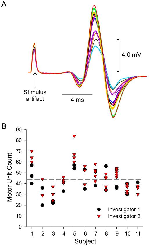
Motor unit numbers were estimated by counting the total number of stepwise increments in EMG amplitude across the entire range of current pulses, where each increment in EMG was assumed to represent activation of a new motor unit4. Figure 1A shows an example set of overlaid EMG responses recorded from AH to a small range of stimulus current pulses. Each trace color indicates an investigator-identified increment, and in this example, there were eleven such increments identified (most easily discerned in the upward phases of the responses) across the upper range of current pulses from 6.1 to 8.9 mA. Usually, there were two or more responses that were categorized as belonging to the same increment. Clearly, there is some degree of subjectivity in these assessments, and this has been one of the criticisms of this method5,6. We indirectly assessed the reliability of this method by having two investigators independently conduct counts on the same data sets.
Figure 1.
A) Example subset of evoked EMG responses in abductor hallucis to graded nerve stimulation. Stimulus range for this set was from 6.1 - 8.9 mA. Each trace color represents the presumed activation of an additional motor unit. B) Estimated number of AH motor units in 11 subjects. Each subject was tested 2 – 5 times. Two investigators performed the analysis on each data set. Dashed line indicates overall average motor unit count.
RESULTS
Across the 11 subjects tested, single-trial motor unit counts in AH ranged from 20 - 84 units. The overall mean count across all subjects was 42.7 (SD ±11) with a median count of 43. While there was some degree of across-trial and across-investigator variability (Fig 1B), all estimates of motor unit number for a particular subject tended to cluster within a reasonable range of values, suggesting a coarse degree of reliability in the measures. Despite the limitation of this method, the relatively low value of estimated motor unit count for AH in comparison to that estimated in other muscles using the same approach (e.g. 203 for extensor digitorum brevis4; 380 for hypothenar7; 125 for first dorsal interosseus8; 421 for abductor pollicis longus9) implies that AH is comprised of a relatively small population of motor units.
DISCUSSION
In general, little is known about how firing rates of motor units change in response to variation in excitatory synaptic drive except for a small range of input associated with weak muscle contractions. This is because the activities of many neighboring motor units typically interfere with the recording of target motor units during stronger contractions. Because AH appears to be comprised of relatively few motor units, it may be possible to record single unit activity over the entire output range of the muscle, as suggested in preliminary studies2. As such, the AH might serve as an ideal muscle model for exploring the recruitment – rate-coding organization of a complete motor unit population.
Acknowledgement
This work was supported by National Institutes of Health Grant NS39489
ACRONYMS
- AH
abductor hallucis
- EMG
electomyogram
REFERENCES
- 1.Dellon AL. The four medial ankle tunnels: a critical review of perceptions of tarsal tunnel syndrome and neuropathy. Neurosurg Clin N Am. 2008;19:629–648. doi: 10.1016/j.nec.2008.07.003. [DOI] [PubMed] [Google Scholar]
- 2.Johns RK, Fuglevand AJ. Limitation of motor unit firing rate during maximum voluntary contraction in human abductor hallucis. Soc Neurosci Abstr. 2003;392:17. [Google Scholar]
- 3.Cameron AF, Rome K, Hing WA. Ultrasound evaluation of the abductor hallucis muscle: reliability study. J Foot Ankle Res. 2008;1:1–12. doi: 10.1186/1757-1146-1-12. [DOI] [PMC free article] [PubMed] [Google Scholar]
- 4.McComas AJ, Fawcett PR, Campbell MJ, Sica RE. Electrophysiological estimation of the number of motor units within a human muscle. J Neurol Neurosurg Psychiatry. 1971;34:121–131. doi: 10.1136/jnnp.34.2.121. [DOI] [PMC free article] [PubMed] [Google Scholar]
- 5.Bromberg MB, Abrams JL. Sources of error in the spike-triggered averaging method of motor unit number estimation (MUNE) Muscle Nerve. 1995;18:1139–1146. doi: 10.1002/mus.880181010. [DOI] [PubMed] [Google Scholar]
- 6.Shefner JM, Gooch CL. Motor unit number estimation. Phys Med Rehabil Clin N Am. 2003;14:243–260. doi: 10.1016/s1047-9651(02)00130-4. [DOI] [PubMed] [Google Scholar]
- 7.Sica RE, McComas AJ, Upton AR, Longmire D. Motor unit estimations in small muscles of the hand. J Neurol Neurosurg Psychiatry. 1974;37:55–67. doi: 10.1136/jnnp.37.1.55. [DOI] [PMC free article] [PubMed] [Google Scholar]
- 8.Milner-Brown HS, Brown WF. New methods of estimating the number of motor units in a muscle. J Neurol Neurosurg Psychiatry. 1976;39:258–265. doi: 10.1136/jnnp.39.3.258. [DOI] [PMC free article] [PubMed] [Google Scholar]
- 9.Defaria CR, Toyonaga K. Motor unit estimation in a muscle supplied by the radial nerve. J Neurol Neurosurg Psychiatry. 1978;41:794–797. doi: 10.1136/jnnp.41.9.794. [DOI] [PMC free article] [PubMed] [Google Scholar]


