Abstract
Previously, it has been shown that in Drosophila steroid hormones are required for progression of oogenesis during late stages of egg maturation. Here, we show that ecdysteroids regulate progression through the early steps of germ cell lineage. Upon ecdysone signalling deficit germline stem cell progeny delay to switch on a differentiation programme. This differentiation impediment is associated with reduced TGF-β signalling in the germline and increased levels of cell adhesion complexes and cytoskeletal proteins in somatic escort cells. A co-activator of the ecdysone receptor, Taiman is the spatially restricted regulator of the ecdysone signalling pathway in soma. Additionally, when ecdysone signalling is perturbed during the process of somatic stem cell niche establishment enlarged functional niches able to host additional stem cells are formed.
Keywords: Drosophila , ecdysone signalling, germline stem cell, stem cell niche
Introduction
One of the key characteristics of adult stem cells is their ability to divide for a long period of time in an environment where most other cells are quiescent. Typically, stem cells divide asymmetrically where a mother cell gives rise to two daughter cells with different fates, another stem cell and a differentiated progeny (Gonczy, 2008).
Adult stem cells also require niches. The niche itself is as significant as stem cell autonomous functions and its environment has the potential to reprogramme somatic cells and to transform them into stem cells (Brawley and Matunis, 2004; Kai and Spradling, 2004; Boulanger and Smith, 2009). The niche includes all cellular and non-cellular components that interact in order to control the adult stem cell. These interactions can be divided into one of two main mechanistic types—physical contacts and diffusible factors. Diffusible factors travel over varying distances from a cell source to instruct the stem cell, often affecting transcription (Walker et al, 2009). Stem cells must be anchored to the niche through cell-to-cell interactions so they will stay both close to niche factors that specify self-renewal and far from differentiation stimuli. While multiple studies focused on the aspects of how the niche regulates stem cells, the question of how the niche is established itself has not been addressed in depth.
The Drosophila ovarian stem cell niche model is an exemplary system where two different stem cell types, germline stem cells (GSCs) and somatic escort stem cells (ESCs) share the same niche and coordinate their development. Niche cells contact GSCs via E-cadherin and Integrin-mediated cell adhesion complexes that bind to the extracellular matrix and connect to the cytoskeleton and this physical docking of stem cells to the niche is essential for GSC maintenance (Xie and Spradling, 2000; Tanentzapf et al, 2007). In addition, the stem cell niche sends short-range signals that specify and regulate stem cell fate by maintaining the undifferentiated state of GSCs next to the niche. Not only does the niche have an effect on stem cells, but also the stem cells communicate with the niche. A feedback loop exists between the stem cells and niche cells: Delta from the GSC can activate Notch in the somatic cells that maintains a functional niche and in turn controls GSC maintenance (Ward et al, 2006). While the management of GSCs within the niche is relatively well understood, the control of the other present stem cell type, ESCs is not clear. An ESC, like a GSC divides asymmetrically producing another ESC and a daughter, escort cell (EC) that will differentiate into a squamous cell that envelops the GSC progeny once disconnected from the niche. It is believed that developing cyst encapsulation by ECs protects from TGF-β signalling that maintains GSC identity (Decotto and Spradling, 2005). The ESC and GSC cycles have to be tightly coordinated, so a sufficient number of ECs will be produced in response to GSC division. However, the pathway used for GSC and ESC communication is unknown.
Adult stem cell division mostly is activated locally in response to tissue demands to replace lost cells. In addition, stem cells can be regulated via more general stimuli in response to systemic needs of the whole organism. Hormones are systemic regulators that regulate a variety of processes in different organs in response to the body’s status. Even though the effects of hormonal signalling have been extensively studied, the specific roles for hormones in stem cell biology remain complex, poorly defined and difficult to study in vivo.
Drosophila is a great system to study the role of endocrine signalling as it contains only one major steroid hormone, ecdysone (20-hydroxyecdysone, 20E) that synchronises the behavioural, genetic and morphological changes associated with developmental transitions and the establishment of reproductive maturity (Shirras and Bownes, 1987; Riddiford, 1993; Buszczak et al, 1999; Kozlova and Thummel, 2003; Gaziova et al, 2004; Schubiger et al, 2005; Terashima and Bownes, 2005; McBrayer et al, 2007). Ecdysteroids act through the heterodimeric nuclear receptor complex consisting of the ecdysone receptor, EcR (Koelle et al, 1991) and its partner ultraspiracle (USP), the Drosophila retinoid X receptor homologue (Shea et al, 1990; Oro et al, 1992; Yao et al, 1992). The ecdysone/EcR/USP receptor/ligand complex binds to ecdysone response elements (EcREs) to coordinate gene expression in diverse tissues (Riddihough and Pelham, 1987; Cherbas et al, 1991; Dobens et al, 1991). Ecdysone signalling is patterned spatially as well as temporally; depending on the tissue type and the developmental stage, the EcR/USP complexes with different co-activators or co-repressors including Taiman, Alien, Rig, SMRTER, Bonus, Trithorax-related protein and DOR (Dressel et al, 1999; Tsai et al, 1999; Bai et al, 2000; Beckstead et al, 2001; Sedkov et al, 2003; Gates et al, 2004; Jang et al, 2009; Francis et al, 2010; Mauvezin et al, 2010). These co-factors can have other binding partners that are themselves regulated by different signalling pathways. For example, Abrupt controlled by JAK/STAT attenuates ecdysone signalling by binding to its co-activator Taiman (Jang et al, 2009). In addition, other signalling pathways (insulin, TGF-β) interact with ecdysone pathway components to further modulate cell type-specific responses (Zheng et al, 2003; Jang et al, 2009; Francis et al, 2010). This offers an additional level of combinatorial possibilities and suggests a model of gene expression regulation that is highly managed by this global endocrine signalling.
Data presented here show that ecdysone signalling is involved in control of early germline differentiation. When ecdysone signalling is perturbed, the strength of TGF-β signalling in GSCs and their progeny is modified resulting in a differentiation delay. Moreover, soma-specific disruption of ecdysone signalling affects germline differentiation cell non-autonomously. Ecdysteroids act in somatic ESCs and their daughters to regulate cell adhesion complexes and cytoskeletal proteins important for soma–germline communication. Misexpression of ecdysone signalling components during developmental stages leads to the formation of the enlarged GSC niche that can facilitate more stem cells.
Results
Taiman, a Drosophila homologue of a steroid receptor co-activator amplified in breast and ovarian cancer (AIB1) influences the size of the niche and GSC number
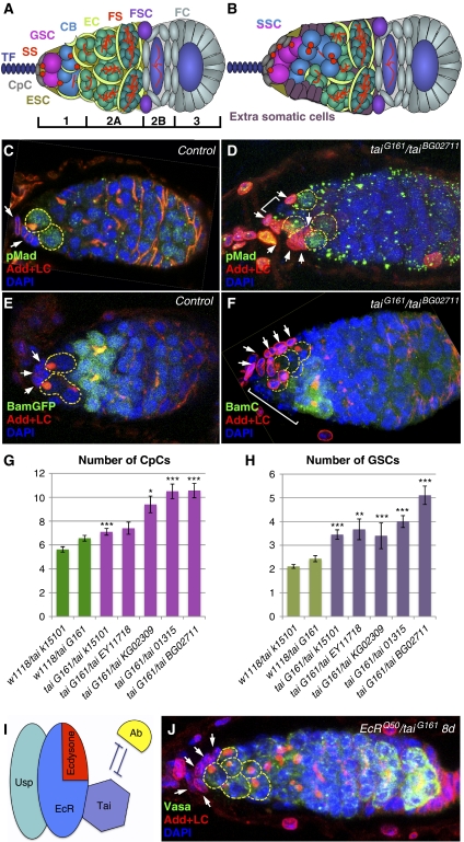
The Drosophila ovary contains distinct populations of stem cells: GSCs, which give rise to the gametes, and two types of somatic stem cells: ESCs and follicle stem cells (FSCs) (Figure 1A). These stem cells reside in stereotyped positions inside the germarium, a specialised structure at the anterior end of the Drosophila ovary. Both GSCs and ESCs are adjacent to somatic signalling centres or niches consisting of the terminal filament (TF) and cap cells (CpCs), which promote stem cell identity. ESCs produce squamous daughters with long processes that encase developing cysts to protect them from niche signalling and allow differentiation. These different cell types have distinct morphologies and molecular markers (Figure 1C and E).
Figure 1.
The ecdysone receptor co-activator Taiman controls the number of ovarian germline stem cell niche cells. (A) Schematic view of a wild-type germarium: germline stem cells (GSCs, pink) marked by anterior spectrosomes (SS, red dots) are located at the apex of the germarium next to the niche cap cells (CpCs, grey). Further noted are terminal filament (TF; dark blue), escort stem cells (ESCs, olive), differentiating cystoblasts (CBs, blue), escort cells (ECs, lime), 4, 8 (bright green) and 16 cell (green) cysts in region 2A, indicated by the presence of fusomes (FS, red branched structures), follicle stem cells (FSCs, violet) and follicle cells (FC, light grey) in regions 2B and 3. (B) Schematic view of a tai mutant germarium with an increased number of single spectrosome containing cells (SSCs, pink and blue), CpCs (grey) and additional somatic cells (plum). (C, E) In wild-type germaria, two GSCs marked by the presence of the stem cell marker pMad (C), spectrosomes (stained with Adducin) and the absence of the differentiation factor BamC (E) are directly attached to the niche (marked with LaminC, arrows). (D, F) In the tai61G1/taiBG02711 transheterozygous mutant germarium, the enlarged niche is coupled with an increased number of GSCs that are pMad positive (D) and BamC negative (F). In addition, extra somatic cells are present at the anterior (marked with brackets). CpC (G) and GSC (H) numbers are increased in tai mutant germaria. (I) Scheme illustrating that Tai is a co-activator of the EcR/USP nuclear receptor complex that is activated upon binding of its ligand ecdysone; Ab negatively regulates the ecdysone signalling by direct binding to Tai (based on Bai et al (2000) and Jang et al (2009)). (J) EcRQ50st/tai61G1 transheterozygous germaria also contain an increased number of GSCs and CpCs, indicating that tai genetically interacts with EcR (see Supplementary Table S1). (D–F, J) Projections of optical sections assembled through the germarial tissue; GSCs are outlined with yellow dashed lines, niche cells are marked with white arrows; Red, Adducin+LaminC; blue, DAPI; and green, pMad (C, D), BamGFP (E), BamC (F) and Vasa (J); Error bars represent s.e.m. *P<0.05, **P<0.005, ***P<0.0005.
We performed a pilot genetic screen where clonal germaria of hsFlp;FRT40A lethals (DGRC) were analysed in order to find novel genes that affect stem cell niche architecture. One of the genes found in our screen encoding a Drosophila homologue of a human steroid receptor co-activator amplified in breast cancer taiman (tai) was of a particular interest. Downregulation of Tai using different combinations of tai amorphic and hypomorphic mutant alleles resulted in increased GSC number and an enlarged niche (Figure 1D and F). The GSC average number ranged from 3.2 to 5.1 depending on the genotype, which was significantly higher than in heterozygous control flies (2.1–2.4, Figure 1D, F and H; Supplementary Table S1). This increase in GSC number coincided with stem cell niche enlargement. While control germaria contained on average 6 niche cells, tai mutant niches consisted of 7–10 CpCs (Figure 1D, F and G; Supplementary Table S1). These observations imply that Tai participates in niche formation and/or GSC maintenance or differentiation.
As it has been shown that in Drosophila Taiman is a co-activator of the ecdysone transcription-activating complex (Figure 1I; Bai et al, 2000), we tested if tai and ecdysone pathway components genetically interact in the process. Transheterozygous germaria (tai/EcR and tai/usp) also showed additional GSCs and enlarged niches (Figure 1J; Supplementary Table S1), suggesting that the ecdysone pathway regulates early germline progression and GSC niche assembly.
The steroid hormone ecdysone controls GSC progeny differentiation
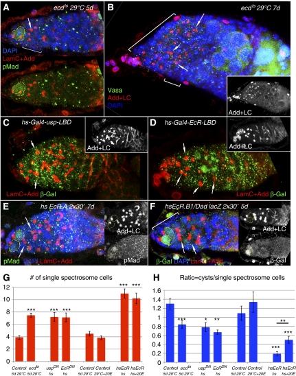
To further test the role of the endocrine pathway in the germline, we used the ecdysoneless1 temperature-sensitive mutation (ecd1ts) that blocks biosynthesis of the mature ecdysteroid hormone, 20-hydroxyecdysone. ecd1ts animals were allowed to develop normally at the permissive temperature and transferred to restrictive temperature conditions as 3-day-old adults. When ecdysone production was disrupted during adulthood, GSCs continued to divide increasing the germarium size, however, their progeny delayed progression through differentiation (Figure 2A and B). Similar phenotypes were obtained upon ecdysone signalling disruption using dominant-negative mutants for the ecdysone receptor, EcR and its dimerisation partner USP (hs–Gal4-EcR-LBD (EcRDN) and hs-Gal4-usp-LBD (uspDN); Kozlova and Thummel, 2002), (Figure 2C and D; Supplementary Table S2). Instead of progressively developed cysts, mutant germaria were filled with germline cells containing a single spectrosome (single spectrosome containing cells (SSCs)), on average seven SSCs per ecd1ts or EcRDN and uspDN germarium were detected in comparison to four in control (Figure 2G; Supplementary Table S2). After longer ecdysone deprivation germaria look even more abnormal; a slightly decreased GSC number and additional follicle cell defects along with abnormal cyst pinching off from the germarium, not shared by tai mutants, were observed (Figure 2B; Supplementary Table S2). The differentiation index or the ratio between developing fusome-containing cells and SSCs in the region 1–2A was decreased 1.5–2-fold in ecdysone mutant germaria (Figure 2H; Supplementary Table S2). Disruption of ecdysone signalling via overexpression of EcR (hsEcR.A and hsEcR.B1) also resulted in the appearance of germaria filled with supernumerary SSCs (on average 11 in comparison to 4 in control, Figure 2E–H; Supplementary Table S3).
Figure 2.
Disrupted ecdysone signalling during adulthood results in delayed germline differentiation. (A) At the restrictive temperature (29°C) ecd1ts adult animals contain germaria filled with supernumerary SSCs. (B) Extended depletion of ecdysone furthermore increases the undifferentiated SSC number and causes somatic cell defects affecting cyst pinching off from the germarium. (C, D) Heat shock induced expression of USP and EcR dominant-negative forms (uspDN (hs-Gal-4-usp-LBD) and EcRDN (hs-Gal-4-usp-LBD)) also lead to the appearance of supernumerary SSCs. (E, F) Similarly to the effects that are caused by disturbing the ecdysone pathway via ecd1ts or dominant-negative EcRDN and uspDN mutations, expression of the EcR isoforms EcR.A or EcR.B1 induced by heat shock (twice per day for 30 min 4 days in a row) increases the number of SSCs, but not GSCs and influences CB differentiation. Note the presence of dumbbell-shaped fusomes in (A–F). (G) In control conditions around four SSCs per germarium are detected. Ecdysone withdrawal via ecd1ts mutation as well as heat shock-induced expression of uspDN or EcRDN and overexpression of EcR led to a 2- or 2.5-fold increase in SSC number, whereas external supply of ecdysone does not change the amount of SSCs within the germarium. (H) The ratio of differentiating cysts to SSCs is about 1.5-fold decreased in ecd1ts, uspDN and EcRDN mutant germaria. This decrease is even more pronounced (seven times) in hsEcR flies. Providing 20E externally can partially, but significantly alleviate this early germline differentiation delay. (A–F) Projections of optical sections assembled through the germarial tissue. GSCs are outlined with yellow dashed lines, dumbbell-shaped fusomes are marked with arrows and additional somatic cells are marked with brackets. Red, LaminC+Adducin; blue, DAPI; and green, pMad (A, E); Vasa (B) and β-galactosidase (C, D, F) Error bars represent s.e.m. *P<0.05, **P<0.005, ***P<0.0005.
The described phenotypes show that ecdysone signalling loss of function (by disruption of ecdysone biosynthesis or by expression of EcR and USP dominant-negative forms) and overexpression of the main receptor of the pathway, EcR cause similar abnormalities. Previously, it has been shown that the EcR can form homodimers in the absence of its binding partners in vitro (Elke et al, 1997), moreover the un-liganded receptor complex is repressive and this repression is relieved as the hormone titre increases (Schubiger and Truman, 2000; Schubiger et al, 2005).
To test if the latter can be the case in our system, we performed experiments where adult flies were fed with 20E. Feeding flies with ecdysone alone had no significant effect on the number of SSCs or germline differentiation measured by the ratio of differentiated cysts to SSCs within one germarium (Figure 2G and H; Supplementary Table S3). Interestingly, feeding of ecdysone to the animals that overexpressed EcR moderately, but significantly rescued the cyst/SSC ratio (Figure 2H; Supplementary Table S3), indicating that EcR overexpression when the ecdysone receptor is abundant and the ligand is limited is unfavourable for germline differentiation.
Ecdysone signalling disturbance affects the intensity of TGF-β signalling
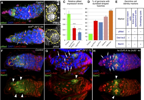
Next, we attempted to analyse the identity of supernumerary SSCs. If they are GSCs, they should express appropriate markers. However, we found that additional SSCs are negative for the stem cell markers, phosphorylated Mad and Dad (Figures 2A, E, F, 3B and E). We also noticed that levels of pMad in GSCs were significantly reduced upon ecdysone deficit (Figure 3A–C), suggesting that ecdysone signalling can modulate pMad levels.
Figure 3.
Ecdysone signalling affects the TGF-β pathway. (A) Wild-type germarium containing two GSCs, marked by pMad staining. (B) Upon blocked ecdysone production the relative pMad expression levels in GSCs are decreased (C, compare the pMad levels measured by grey value in A and B). (D) Disruption of ecdysone signalling results in the increase of dumbbell-shaped fusome quantity. In ecd1ts4210 mutant flies that were at the restrictive temperature for up to 7 days, 55% (n=33) of the germaria have dumbbell-shaped fusomes (ecd1ts218 51%, n=37) whereas in equally treated w1118 germaria, only 18% (n=11) of the germaria contain dumbbell-shaped fusomes. After overexpression of EcR.A or EcR.B1 for 7 days 67% or 84% (n=15, 19, respectively) of the analysed germaria contain dumbbell-shaped fusomes. (E) The characteristics of GSCs, SSCs and developing cysts are compared schematically. GSCs express the stem cell markers pMad and Dad lacZ and developing cysts the differentiation factor BamC, whereas additional SSCs in germaria deficient of ecdysone signalling are pMad, Dad lacZ and BamC negative, showing that they do not maintain stem cell identity and are delayed in development. (F) In wild-type germarium, BamC is present in developing CBs adjacent to GSCs, while in ecdysone pathway mutants, ecd1ts (G) and hsEcR (H), the anterior part of the germarium is filled with cells that do not express the differentiation marker BamC and contain a single spectrosome or a dumbbell-shaped fusome. (A, B, F–H) Projections of optical sections assembled through the germarial tissue. GSCs are outlined with yellow dashed lines, dumbbell-shaped fusomes are marked with arrows and BamC-positive differentiating cysts with arrowheads. Red, Adducin+LaminC; blue, DAPI; and green, pMad (A, B); BamC (F–H). Error bars represent s.e.m. Significance calculated using the t-test (C), χ2-test (D). *P<0.05, ***P<0.0005.
As supernumerary SSCs did not express the stem cell markers, we next analysed if the increased number of SSCs can be explained by abnormal organisation of fusomes, the structures that connect daughter cells within one cyst. Cysts are formed by a process of mitosis with incomplete cytokinesis, and all cells forming one cyst divide simultaneously (de Cuevas and Spradling, 1998). If ecdysone signalling affects fusome stability leading to the appearance of dot-like instead of branched fusomes, then SSCs are really cells within a differentiating cyst and should have synchronised divisions. However, staining with a mitotic marker phosphohistone H3 (PH3) showed that the cell cycle was not coordinated in SSCs, which shows that single spectrosomes are not the result of fusome breakage in pursuit of cyst de-differentiation into single stem cell-like cells (Supplementary Figure S1).
We also noticed that many fusomes had a dumbbell shape, which is a characteristic of perturbed Bam, a TGF-β signalling target (McKearin and Ohlstein, 1995) (Figures 2B–F, 3B, G and H). The amount of germaria with dumbbell-shaped fusomes increased from 18% in control to 51–84% in animals with exogenous EcR expression and ecdysone deficit (Figure 3D). Interestingly, SSCs in germaria mutant for ecdysone signalling, unlike wild-type differentiating cystoblasts do not express Bam, a factor essential for germline differentiation (Figure 3F–H). Taken together, these analyses show that additional SSCs resulting from ecdysone signalling disruption are ‘undecided’ cells that express neither stem cell nor differentiation markers (Figure 3E).
These data suggest that ecdysone signalling affects early germline differentiation possibly by modulation of the TGF-β signalling strength causing a developmental delay. Eventually some germline differentiation takes place implying that ecdysone signalling is at least partially redundant with other pathways for germline progression.
Ecdysone signalling is predominantly active in ESCs and Taiman, an EcR/USP co-activator is spatially limited to the soma
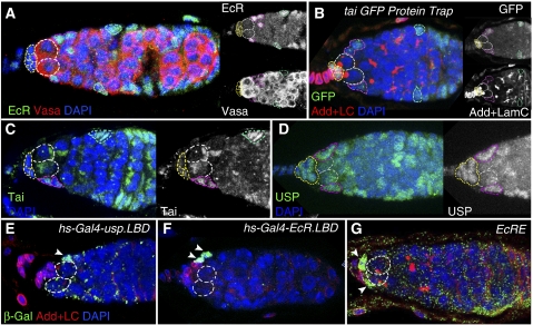
Previous studies show that ecdysone signalling in Drosophila has a role in egg maturation and vitellogenesis (Shirras and Bownes, 1987; Riddiford, 1993; Buszczak et al, 1999; Kozlova and Thummel, 2003; Gaziova et al, 2004; Schubiger et al, 2005; Terashima and Bownes, 2005; McBrayer et al, 2007), now our data indicate that it is also required for differentiation of developing germline cysts. As germline differentiation can be regulated cell autonomously or cell non-autonomously, we decided to test what goes awry in the GSC niche community when the ecdysone pathway is perturbed. We began with analysing the expression pattern of ecdysone signalling pathway components to find out in which cell types ecdysone signalling is working. The EcR protein measured by a specific antibody was detected mostly in ESCs and ECs, thin cells which envelop the differentiating cystoblast to assist in differentiation by protecting it from the niche signals (Figure 4A). Next, we used a GFP protein trap line inserted in the tai gene and detected levels of GFP expression in CpCs that form the niche and also to a lesser amount in ESCs (Figure 4B). Similarly, staining with Tai and USP-specific antibodies (Figure 4C and D; Supplementary Figure S2) showed that these proteins are expressed predominantly in somatic cells, however, some low levels are also present in the germline indicative of a possible dual role of this endocrine pathway in the germline and the soma.
Figure 4.
Expression pattern of the ecdysone pathway components in the Drosophila germarium. (A) The anti-EcR (common region) antibody detects high levels of EcR in ESCs and FCs. (B) In the tai G00308 protein trap line where GFP is expressed under the control of the endogenous tai promoter, high GFP levels were detected in CpCs, ESCs and FSCs. (C) Comparable expression pattern is observed with the anti-Tai antibody. (D) The nuclear receptor USP detected by the anti-USP antibody shows identical expression pattern to its binding partner EcR. (E, F) Spatial patterns of ecdysone signalling activation identified via β-Gal staining of heat-treated hs-Gal4-usp.LBD/+; UAS-lacZ/+ (E) and hs-Gal4-EcR.LBD/+; UAS-lacZ/+ (D) germaria prove ecdysone signalling being mainly active in the ESCs (marked with arrowheads). (E) The ecdysone signalling reporter EcRE-lacZ shows the presence of active ecdysone transcription complex in ESCs as well (marked with arrowheads). Different cell types are marked as follows: GSCs, white dashed lines; CpCs, yellow dashed lines; ESCs/ECs, red dashed lines; FCs, green dashed lines. Red, Vasa (A), Adducin+LaminC (B, E–G); blue, DAPI; and green, anti-EcR (common region) (A); GFP (B); anti-Tai (C), anti-USP (D) and β-galactosidase (E–G).
After determining protein expression we wanted to confirm that the ecdysone signalling pathway was active. For this, we used reporters with a Gal4 transcription factor fused to the ligand-binding domain of USP or EcR (hs-Gal4-uspLBD, hs-Gal4-EcRLBD; Kozlova and Thummel, 2002). The ecdysone pathway activity was detected mainly in ESCs and ECs analysed using a somatically expressed UASt lacZ transgene (Figure 4E and F). The EcRE-lacZ construct that senses the presence of the active ecdysone receptor transcription complex (Koelle et al, 1991) also validated the pathway activity in ESCs and random CpCs (Figure 4G).
Ecdysone signalling is required cell non-autonomously for progression through the early steps of germ cell lineage
Our expression data demonstrate that ecdysone signalling components are expressed in somatic cells within the GSC niche and the signalling is active predominantly in ESCs, leading to the hypothesis that ecdysone signalling controls germline cell differentiation extrinsically. This idea is further supported by the analysis of tai loss-of-function germline clones (Supplementary Figure S3) that show that Tai is not essential for germline progression: tai mutant GSCs were normally maintained (Supplementary Table S4; Supplementary Figure S3B) and in general germline differentiation was not affected (Supplementary Figure S3A). Together with spatially restricted somatic Tai expression this provides evidence that the ecdysone co-activator Taiman can act as a cell-specific co-activator of ecdysone signalling in niche and ECs.
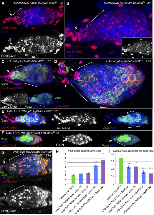
To identify specific cellular processes regulated by the ecdysone pathway in somatic cells proximal to the ovarian stem cell niche, we downregulated ecdysone signalling using transgenic UAS tai RNAi, UAS EcR RNAi and UAS ab lines crossed to ovarian soma-specific drivers (bab1Gal4 and ptcGal4, for expression patterns see Supplementary Figure S4) combined with the temperature-sensitive Gal80 system to avoid the lethality caused by downregulation of ecdysone pathway components during developmental stages.
When the co-activator of ecdysone signalling Tai was downregulated or the co-repressor Abrupt overexpressed in soma, mutant germaria contained multiple SSCs (Figure 5A–C); this mutant phenotype became even more pronounced over time (Figure 5B and D) resembling older ecd1ts (Figure 2B) as well as JAK/STAT mutant germaria (Decotto and Spradling, 2005). Similar phenotypes were observed when EcR RNAi flies were kept at the restrictive temperature; the development of germline cysts was retarded (Figure 5E–G), and the ratio of fusome-containing cysts to SSCs was reduced 2–3 times (Figure 5I; Supplementary Table S5). Downregulation of EcR for longer periods (15, 21 days) led to an increase in the number of SSCs (from 5 to 9–11 SSCs per germarium, Figure 5H; compare 5F and 5G). In addition, in proximity to undeveloped cysts mutant germaria contained extra somatic cells, most likely improperly differentiated ECs (Figure 5, brackets).
Figure 5.
Ecdysteroids act from the soma to regulate the progression of germline development in the germarium. (A, B) The EcR co-activator Tai is downregulated specifically in the somatic cells of the germarium using ptcGal4 and bab1Gal4 in combination with tubGal80ts system to avoid lethality. Upon downregulation of tai in the soma, the number of developmentally delayed SSCs increases dramatically. (C, D) Overexpression of the Tai repressor, Abrupt using UAS ab with the same drivers causes similar phenotypes as seen with downregulation of tai. (B, D) The tai and ab mutant germaria are filled with undifferentiated SSCs, cysts are not pinching off and additional somatic cells (brackets) are in the vicinity. Note the similarity of phenotypes caused by ecdysteriod deficit (ecd1ts, Figure 2B) and disruption of ecdysone signalling pathway components just in germarial soma. (E–G) The downregulation of the EcR in the somatic cells of the germarium via expression of UAS EcR RNAi97 under control of ptcGal4 (E) and bab1Gal4 (F, G) leads to an increase of SSCs at the expense of developing cysts. Note the presence of dumbbell-shaped spectrosomes and additional somatic cells. (H) Bar graph showing extra quantities of SSCs upon EcR downregulation via expression of UAS EcR RNAi97 or UAS EcR RNAi104 with the somatic drivers ptcGal4 or bab1Gal4. This phenotype gets more pronounced with longer duration of EcR abolition. (I) The ratio of differentiating cysts to SSCs is also decreased correspondingly to the increase in the SSC number. (A–G) Projections of optical sections assembled through the germarial tissue are shown. CpCs are marked with arrows, additional somatic cells with brackets. Red, Adducin+LaminC (A–G); blue, DAPI; and green, Vasa (C–F), Cadherin (G). Error bars represent s.e.m. *P<0.05, **P<0.005, ***P<0.0005.
These data provide evidence that the soma-specific disruption of the ecdysone pathway is causing germline differentiation defects, indicating a cell non-autonomous role of this steroid hormone signalling.
Ecdysone signalling regulates turnover of cell adhesion proteins
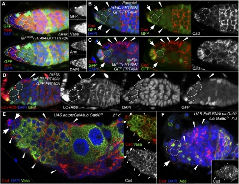
In order to analyse how mutant somatic cells cause a block in germline cyst maturation, we used an FRT recombination system to compare ecdysone pathway deficient and wild-type somatic cells within one germarium. Detailed analysis of tai mutant ESCs and their progeny showed that they lose their squamous shape, and form a layer resembling columnar epithelium (Figure 6A). Interestingly, these mutant cells expressed higher levels of the cell adhesion molecules β-Catenin/Armadillo, DE-Cadherin and a cytoskeleton component Adducin (Figure 6A, C and D). DE-Cadherin was also upregulated in abnormal somatic cells resulting from somatic overexpression of Abrupt or downregulation of EcR (UAS ab or UAS EcR RNAi crossed to ptcGal4/tubGal80ts; Figure 6E and F) pointing towards possible defects in cell–cell contacts, shape rearrangement and signalling transduction processes. These data imply that in our system the ecdysone pathway has a specific role in EC differentiation via regulation of cell adhesion complexes that are required for establishment of correct germline–soma communications. Perhaps, when connections between germline cysts and surrounding soma are perturbed, signalling cascades that initiate germline differentiation are also perturbed causing a developmental delay.
Figure 6.
Cell adhesion and cytoskeleton proteins are misregulated in escort cells mutant for ecdysone signalling pathway components. (A) The progeny of taik15101-deficient ESCs marked by the absence of GFP (hsFlp; taik15101FRT40A/UbiGFP FRT40A) formed a columnar epithelium-like somatic tissue adjacent to the stem cell niche. These cells also express higher levels of β-catenin/Armadillo than normal. (B) In the control germarium (hsFlp; FRT40A/UbiGFP FRT40A) ESCs (marked by arrows) and EC (marked by arrowheads) show moderate levels of DE-Cadherin, while niche–GSC cell contacts have higher DE-Cadherin levels. (C) tai61G1 deficiency (hsFlp; taiG161FRT40A/UbiGFP FRT40A) led to the upregulation of the cell adhesion protein DE-Cadherin in ECs (arrowheads) and ESCs (arrows). Note also that the number of abnormally shaped tai mutant escort cells is increased in (A, C). (D) tai mutant escort cells (arrowheads, hsFlp; taiG161FRT40A/UbiGFP FRT40A) do not properly change their morphology and show higher levels of the cytoskeletal protein Adducin and nuclear envelope marker LaminC. (E) The overexpression of the ecdysone signalling inhibitor Abrupt leads to a strongly mutant germarial structure. Somatic cells (marked by the absence of the germline marker Vasa) are forming layers all along the germarium and show high DE-Cadherin levels. (F) Somatic abolition of EcR (UAS EcR RNAi; ptcGal4/tubGal80ts) also increases levels of cell adhesion and cytoskeleton proteins, DE-Cadherin and Adducin. Wide arrows, ESCs; arrowheads, ECs; Red, Vasa, Armadillo (A), Cadherin (B, C, E, F), Adducin+LaminC (D); blue, DAPI; and green, GFP (A–E).
Ecdysone signalling controls the stem cell niche formation
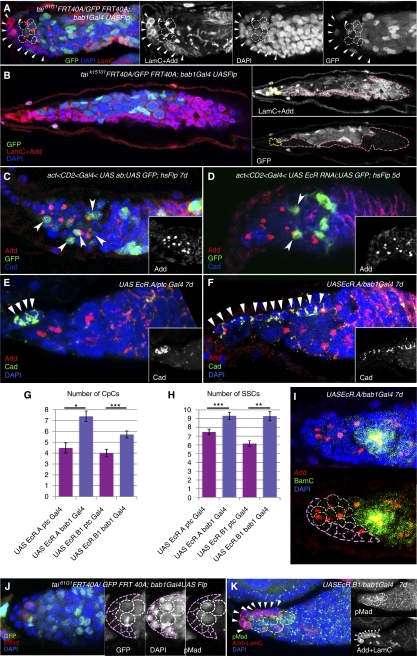
Another process in the germarium that should require a very accurate regulation of cell adhesion is the niche establishment. If ecdysone signalling is essential to control this process as well, we would expect to see abnormalities in niche formation in ecdysone pathway mutants. Recall that mutant tai animals indeed had enlarged niches and extra GSCs (Figure 1C and D), a phenotype not seen in other cases analysed here. This discrepancy can be explained by the time during the animal's development when the mutation was introduced. In the tai experiment, animals were tai deficient during all developmental stages, including the period of niche establishment. In other cases in this study the ecdysone pathway was misregulated during adulthood after the niche was already formed and CpCs had stopped division. Also, in tai heterozygouts both the soma and the germline were mutant and the germline can affect via Notch signalling the size of the niche (Ward et al, 2006). To prove that the niche expansion is a soma-originated phenotype, we knocked down tai in somatic pre-adult cells that contribute to niches using the FRT/bab1Gal4/UASFlp system that allows to induce mutant CpC clones during niche formation. As expected, germaria with tai clonal CpCs had substantially enlarged niches (Figure 7A and B), which provides evidence that the ecdysone pathway co-activator Tai is required during developmental stages specifically in the pre-niche cells to control the GSC niche assembly. Possibly in tai mutant somatic cells within the larval ovary, like in ECs in adults, increased levels of cell adhesion molecules allow them to adhere better to germline cells and receive more signalling (Notch for example) which makes them adopt the niche cell fate.
Figure 7.
Ecdysone signalling is required for niche formation. (A) Downregulation of tai61G1 before the niche is established (taiG161FRT40A/UbiGFP FRT40A; bab1Gal4 UASFlp) causes significant niche enlargement (CpCs marked with arrowheads) that allows to anchor more GSC-like cells (marked with white dashed lines). (B) In some extreme cases taik15101 mutant somatic cells (marked with pink dashed lines) encapsulate the whole germarium that is filled with SSCs. CpCs are marked with yellow dashed lines. (C) Clonal overexpression of the Tai repressor Ab (UAS<CD2<Gal4 UAS ab; UAS GFP; hsFlp) in somatic cells results in the appearance of supernumerary SSCs that are anchored to UAS ab cells marked by GFP. (D) The same can be observed in somatic clonal EcR mutant cells (UAS<CD2<Gal4 UAS EcR RNAi; UAS GFP; hsFlp). (E, F) The pre-adult expression of exogenous EcR only in the niche progenitor cells (bab1Gal4, F), but not in other somatic cells (ptcGal4, E) results in the appearance of enlarged niches marked by DE-Cadherin (arrowheads). The average numbers of CpCs (G) and SSCs (H) are significantly increased when UAS EcR.A or UAS EcR.B1 are overexpressed during the niche establishment in most anterior pre-niche somatic cells (bab1Gal4), but not in other intermingled somatic cells (ptcGal4) within the larval ovary. (I) The niche expansion increases the number of SSCs that are also negative for the differentiation marker BamC. Niche is outlined with pink and GSCs with white dashed lines. (J, K) The enlarged tai clonal niches (tai61G1FRT 40A/Ubi GFP FRT 40A; bab1 Gal4 Flp) and niches overexpressing EcR bear a higher number of GSCs whose identity is confirmed by the stem cell marker pMad. Niche is outlined with pink dashed lines in (J) and arrowheads in (K), GSCs are marked with white dashed lines. (A–F, I–K) Projections of optical sections assembled through the germarial tissue are shown. Red, Adducin+LaminC (A, B, K), Adducin (C–F, I), pMad (J); blue, DAPI; and green, GFP (A–D, J), Cadherin (E, F), BamC (I), pMad (K). Error bars represent s.e.m. *P<0.05, **P<0.005, ***P<0.0005.
To confirm that the niche enlargement is an ecdysone signalling-reliant phenotype and is not associated with Tai-independent function, we introduced other ecdysone pathway component mutations during the period of niche development. As most of the tested mutant combinations affected viability, we could disrupt ecdysone signalling during development only via induction of single cell clones using the act<CD2<Gal4, hsFlp system and via EcR overexpression. Mutant single somatic clonal cells expressing UAS ab or UAS EcR RNAi resembled niche cells by their shape and ability to hold SSCs (Figure 7C and D). On average, mutant germaria contained 7.5–8.5 germline SSCs oriented either towards ab or EcR mutant or niche cells. UAS EcR.A and UAS EcR.B1 expressed by the niche cell-specific driver bab1Gal4 also caused formation of an enlarged niche (on average 10 CpCs in comparison to 6 in control, Figure 7F, G and K; Supplementary Table S6) and appearance of supernumerary SSCs (Figure 7H and I; Supplementary Table S6). To test if these excessive niches were able to host extra stem cells, we analysed the number of GSCs per germarium by staining mutant germaria with specific markers. We observed that in tai and EcR mutants additional SSCs that are touching expanded niches are positive for the stem cell marker pMad and do not stain positively for the differentiation factor Bam (Figure 7H–K). The number of pMad-positive GSCs per germarium significantly increased in clonal tai mutants (4.47±0.26 (P=4.29 × 10⁻⁷, n=15) in tai61G1FRT40A/UbiGFP FRT40A;bab1Gal4Flp in comparison to 2.18±0.26 (n=12) in control) and ecdysone mutants (3.50±0.43 (P=0.02, n=6) in UAS EcR.A bab1Gal4 and 3.33±0.29 (P=0.01, n=9) in UAS EcR.B1 bab1Gal4 in comparison to 2.36±0.20 (n=11) in UASlacZ, bab1Gal4 control). These observations infer that additional cells in enlarged niches are functional and can facilitate extra GSCs. We assume that during development the ecdysone signalling pathway has a role in the establishment of the stem cell niche.
Discussion
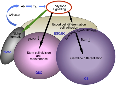
Here, we show for the first time that in Drosophila ecdysone signalling regulates differentiation of a GSC daughter and modulates ovarian stem cell niche size (Figure 8). The delay in GSC progeny differentiation correlates with reduced expression levels of TGF-β pathway components. Based on expression patterns it appears that germarial somatic cells, niche and ECs are the critical sites of ecdysteroid action and a co-activator of ecdysone receptor, Taiman is the spatially restricted regulator of ecdysone signalling in soma. During adulthood the ecdysone pathway has a specific role in EC differentiation and soma–germline cell contact establishment. In addition, during development the ecdysone signalling pathway has a role in somatic niche formation (Figure 8).
Figure 8.
Model showing the role of the ecdysone signalling in Drosophila ovarian stem cell niche. During development (green arrow) ecdysone signalling participates in defining the stem cell niche size. During adulthood (black arrows) this hormonal pathway has a dual role in regulation of early germline differentiation: regulation of cell contacts and cell shape rearrangements via adjustment of adhesion complexes and cytoskeletal proteins in ESCs and their progeny and control of the potency of TGF-β signalling.
Ecdysteroids in general control major developmental transformations such as metamorphosis and morphogenesis in Drosophila. Different tissues and even different cell types within the same tissue respond to this broad signalling in a specific fashion and in a timely manner. In the developing Drosophila ovary steroid hormone receptors are expressed in a well-timed mode, high levels coinciding with proliferative and immature stages and low levels preceding reduced DNA replication and differentiation (Hodin and Riddiford, 1998). Mutations in ecdysone pathway components affect ovarian morphogenesis, including heterochronic delay or acceleration in the onset of TF differentiation. During the niche establishment the levels of both ecdysone receptors, EcR and USP are greatly downregulated in anterior somatic cells that will contribute to the niche per se (Hodin and Riddiford, 1998). Now, we show that perturbation of ecdysone signalling in pre-adult ovarian soma leads to the formation of enlarged niches. The specific response to systemic hormonal signalling in niche precursors is achieved by a specific function of the ecdysone receptor co-activator Taiman. When timely regulation of ecdysone signalling does not occur, more cells are recruited to become niche cells resulting in enlarged niches that are capable to host more stem cells. These data first show that ecdysone steroid hormonal signalling regulates the formation of the adult stem cell niche and suggest that a developmental tuning of ecdysone signalling controls the number of anterior somatic cells that will differentiate into CpCs.
It is logical that stem cell division and germline differentiation are regulated by some systemic signalling depending on the general state of the organism, which depends on age, nutrition, environmental conditions and so on. Hormones are great candidates for this type of regulation as they act in a paracrine fashion and their levels are changing in response to ever-changing external and internal conditions. Steroid binding to nuclear receptors in vertebrates triggers a conformational switch accompanied by increased histone acetylation that permits transcriptional co-activators binding and the transcription initiation complex assembly (Collingwood et al, 1999; Privalsky, 2004). In Drosophila, the trithorax-related protein, a histone H3 methyltransferase that like Taiman belongs to the p160 class of co-activators, and an ISWI-containing ATP-dependent chromatin remodelling complex (NURF), that regulates transcription by catalysing nucleosome sliding, both bind EcR in an ecdysone-dependent manner (Sedkov et al, 2003; Badenhorst et al, 2005), showing that chromatin modifications can mediate response to this general signalling. Transcriptional regulation has a key role in GSC maintenance and differentiation, for example, the TGF-β ligand dpp secreted by niche cells induces phosphorylation of the transcription factor Mad in GSCs that in turn suppresses transcription of the differentiation factor Bam (McKearin and Ohlstein, 1995; Xie and Spradling, 1998; Chen and McKearin, 2003; Song et al, 2004). In addition, it has been shown recently that in Drosophila adult GSC ecdysone modulates the strength of TGF-β signalling through a functional interaction with the chromatin remodelling factors ISWI and Nurf301, a subunit of the ISWI-containing NURF chromatin remodelling complex (Ables and Drummond-Barbosa, 2010). Therefore, it is plausible that ecdysone regulates Mad expression cell autonomously via chromatin modifications. As pMad directly suppresses a differentiation factor Bam, it is expected that Bam would be expressed in pMad-negative cells. Interestingly, our findings show that ecdysone deficit decreases amounts of phosphorylated Mad in GSCs and also cell non-autonomously suppresses Bam in SSCs. As SSCs that express neither pMad nor Bam are accumulated when the ecdysone pathway is perturbed it suggests that there should be an alternative mechanism of Bam regulation. Even though eventually this still can be done on the level of chromatin modification, our data suggest that the origin of this soma-generated signal may be associated with cell adhesion protein levels. Further understanding of the nature of this signalling is of a great interest.
The progression of oogenesis within the germarium requires cooperation between two stem cell types, germline and somatic (escort) stem cells. In Drosophila, reciprocal signals between germline and escort (in female) or somatic cyst (in male) cells can inhibit reversion to the stem cell state (Brawley and Matunis, 2004; Kai and Spradling, 2004) and restrict germ cell proliferation and cyst growth (Matunis et al, 1997). Therefore, the non-autonomous ecdysone effect can be explained by the necessity of two stem cell types that share the same niche (GSC and ESC) to coordinate their division and progeny differentiation. This coordination is most likely achieved via adhesive cues, as disruption of ecdysone signalling affects turnover of adhesion complexes and cytoskeletal proteins in somatic ECs: mutant cells exhibited abnormal accumulation of DE-Cadherin, β-catenin/Armadillo and Adducin.
Cell adhesion has a crucial role in Drosophila stem cells; GSCs are recruited to and maintained in their niches via cell adhesion (Song et al, 2002). Two major components of this adhesion process, DE-Cadherin and Armadillo/β-catenin, accumulate at high levels in the junctions between GSCs and niche cells, while in the developing CB and ECs levels of these proteins are strongly reduced. Levels of DE-Cadherin in GSCs are regulated by various signals, for example, nutrition activation of insulin signalling or chemokine activation of STAT (Hsu and Drummond-Barbosa, 2009; Leatherman and Dinardo, 2010), and here we show that in ESCs it is regulated by steroid hormone signalling. Possibly, these two stem cell types respond to different signals but then differentiation of their progeny is synchronised via cell contacts. While hormones, growth factors and cytokines certainly manage stem cell maintenance and differentiation, our evidence also reveals that the responses to hormonal stimuli are strongly modified by adhesive cues.
Specificity to endocrine signalling can be achieved via availability of co-factors in the targeted tissue. Tai is a spatially restricted co-factor that cooperates with the EcR/USP nuclear receptor complex to define appropriate responses to globally available hormonal signals. Tai-positive regulation of ecdysone signalling can be alleviated by Abrupt via direct binding of these two proteins that prevents Tai association with EcR/USP (Jang et al, 2009). Abrupt has been shown to be downregulated by JAK/STAT signalling (Jang et al, 2009). Interestingly, JAK/STAT signalling also has a critical role in ovarian niche function and controls the morphology and proliferation of ESCs as well as GSCs (Decotto and Spradling, 2005). JAK/STAT signalling may interact with ecdysone pathway components in ECs to further modulate cell type-specific responses to global endocrine signalling. A combination of regulated by different signalling pathway factors that are also spatially and timely restricted builds a network that ensures the specificity of systemic signalling.
Knowledge of how steroids regulate stem cells and their niche has a great potential for stem cell and regenerative medicine. Our findings open the way for a detailed analysis of a role for steroid hormones in niche development and regulation of germline differentiation via adjacent soma.
Materials and methods
Fly stocks
Drosophila melanogaster stocks were raised on standard cornmeal-yeast-agar-medium at 25°C unless otherwise stated. Clones were induced using the hsFlp/FRT system for mitotic recombination. The following stocks were used: yd2w1118;taik15101 FRT40A/CyO (DGRC Kyoto), dpovtai61G1FRT40A/CyO, tai01351cn1/CyO;ry506, w1118;taiBG02711, taiKG02309/CyO, w1118;y1w67c23;taiEY11718/CyO, w1118;pUASt tai, EcRM554fs/SM6b, EcRQ50st/SM6b, w1118;hs-GAL4-EcR.LBD, w1118;hs-GAL4-usp.LBD, w1118;EcRE.lacZ, w1118;hs-EcR.B1, w1118,hs-EcR.A, w1118;UAS-EcR-RNAi97, w1118,UAS-EcR-RNAi104, usp4/FM7a, uspEP1193, w1118;UAS-EcR.A, w[*];UAS-EcR.B1, w1118;UAS-ab.B, UAS-lacZ, ecd1218, ecd4210 (Bloomington Stock Centre), tai G00308/CyO (Carnegie GFP trap line), tai RNAi (w1118; P{GD4265}, VDRC), BamGFP (Dennis McKearin), w1118 was used for wild-type analysis.
Transheterozygous interaction
We used the amorph and hypomorph tai alleles and ecdysone pathway mutants EcRQ50st/SM6b or usp4/FM7a, uspEP1193. Both the number of GSCs (single spectrosome cells that are touching the CpCs) and the number of CpCs itself were counted. As a control, dpovtai61G1FRT40A/CyO and yd2w1118; taik15101FRT40A/CyO were crossed to w1118 flies.
Disruption of EcR in soma
To specifically disrupt the ecdysone signalling in the somatic cells of the germarium, w1118;UAS EcR RNAi97, w1118,UAS EcR RNAi104, w1118;UAS ab.B or tai RNAi (w1118; P{GD4265}), females were crossed to ptcGal4; tubGal80ts or tubGal80ts; bab1Gal4/TM6 males at 18°C. The hatched flies were then transferred to 29°C and aged for 7, 14 and 21 days. Controls were treated the same.
Clonal analysis
Germline and somatic cell clones were done as described previously (Shcherbata et al, 2004, 2007) using hsFlp/FRT system for mitotic recombination. Early formation of clones in CpCs and ESCs were obtained via crossing yd2w1118; taik15101FRT40A/CyO and dpovtai61G1FRT40A/CyO to Ubi-GFP FRT40A/CyO; bab1Gal4:UAS-Flp/TM2 flies (gift from A González-Reyes). Mutant clones were identified by the absence of GFP.
To induce adult clones yd2w1118; taik15101FRT40A/CyO and dpovtai61G1FRT40A/CyO males were crossed to hsFlp; FRT40A GFP/CyO females. 2–4-day-old adult F1 females were heat shocked in empty vials for 60 min 2 days in a row in a 37°C water bath and analysed 5, 7, and 12, 14 days after heat shock. CpC and ESC clones were identified by the absence of GFP.
For generation of somatic ovarian clones we crossed hsFlp;;UASt GFPact>FRT-CD2-FRT>Gal4/TM3 males to w1118;UAS ab.B or w1118,UAS EcR RNAi females. Third instar larvae were heat shocked 2 days in a row for 2 h. Clonal cells expressing ab.B or EcR RNAi were identified by GFP expression.
Overexpression analysis
For overexpression of EcR isoforms in adult flies, w1118;hsEcR.A flies were crossed to w1118 flies. The offspring with one copy of the transgene were heat shocked (37°C) twice per day for 30 min 4 or 7 days in a row. Controls were heat shocked as well. Furthermore, flies with a copy of the hsEcR.A transgene were kept at 25°C without heat treatment.
To overexpress the different EcR isoforms specifically in the soma w1118;UAS EcR.A and w1118;UAS EcR.B1 (Bloomington Stock Center) were crossed to bab1Gal4/TM6 or ptcGal4.
Alteration of ecdysone signalling
To supply more ecdysone hormone, 20-Hydroxyecdysone (20E, Sigma-Aldrich) was diluted in 5% ethanol to a 1 μM concentration and mixed with dry yeast to reach a dough-like consistency. The mixture was then placed on top of agar juice plates to culture flies. In all, 5% ethanol was used for controls.
The ecd1ts temperature-sensitive mutation is known to reduce ecdysone levels at the non-permissive temperature. Fly stocks were kept at the permissive temperature (18°C) and adult flies were shifted to the restrictive temperature (29°C) in order to block ecdysone synthesis. As a control, wild-type flies were kept at 29°C for the same time and ecd1ts flies that had not been shifted to 29°C were analysed.
w1118;hs-GAL4-EcR.LBD and w1118;hs-GAL4-usp.LBD (Kozlova and Thummel, 2002) animals were heat shocked 30 min/day, 1–3 days in a row.
Ecdysone signalling pattern
To analyse the ecdysone signalling in the germarium, w1118;hs-GAL4-EcR.LBD and w1118;hs-GAL4-usp.LBD (Kozlova and Thummel, 2002) females were crossed to UAS-lacZ males. Flies were heat shocked for 60 min in a water bath before they were fixed and stained. EcRE-lacZ, a homozygous viable stock with seven EcREs inserted into a lacZ promoter was used to determine the pattern of ecdysone signalling (Koelle et al, 1991). Adult flies were stained for β-galactosidase. Taiman expression was identified with the tai G00308/CyO enhancer-trap line (Morin et al, 2001).
Immunofluorescence and antibodies
Ovaries were fixed in 5% formaldehyde (Polysciences, Inc.) for 10 min and the staining procedure was performed as described (Shcherbata et al, 2004). We used the following mouse monoclonal antibodies: anti-Armadillo (1:40); anti-Adducin (1:50), anti-LaminC (1:50), anti-EcR Ag10.2 (1:20, EcR common region) (Developmental Studies Hybridoma Bank), anti-usp (1:50, RB Olano, F Kafatos), rat anti-DE-Cadherin (1:50, DSHB), anti-BamC (1:1000, D McKearin) and rat anti-Vasa (1:1000, P Lasko), rabbit anti-pMad (1:5000, D Vasiliauskas, S Morton, T Jessell and E Laufer), anti-β-Gal (1:1000), rabbit anti-tai (1:1000, D Montell), rabbit anti-PH3 (1:3000, Upstate Biotechnology) and anti-GFP-directly conjugated with AF488 (1:3000, Invitrogen), Alexa 488, 568 or 633 goat anti-mouse, anti-rabbit (1:500, Molecular Probes), goat anti-rat Cy5 (1:250, Jackson Immunoresearch). Images were obtained with a confocal laser-scanning microscope (Leica SPE5) and processed with Adobe Photoshop.
Analysis and statistics
To determine the number of CpCs, LaminC-positive cells on the tip of the germarium were counted. Single spectrosome cells that were touching the niche cells were counted as GSCs. Single spectrosome cells that were not touching the niche were counted separately. In addition, the number of fusomes (indicating the number of cysts) until region 2B, where follicle cells start cyst encapsulation, was counted. To describe the differentiation in a given germarium the number of cysts was divided by the number of SSCs (ratio=cysts/SSCs). The percentage of germaria-containing dumbbell-shaped fusomes (McKearin and Ohlstein, 1995) was counted. The intensity of the pMad-positive area was determined via measuring the grey value in at least 10 GSCs with Leica LAS AF Lite software, the background levels were measured by the intensity of the pMad-negative area in the germarium. Background levels were subtracted to normalise the levels of antibody staining in different germaria. Intensity levels relative to control were calculated. GSC maintenance was determined by comparison of the percentage of germaria with clonal GSCs between two different time points after clonal induction.
χ2-test was used to determine if the percentage of dumbbell-shaped fusomes was significantly increased. For all other statistical analyses, the two-tailed Student’s t-test was performed.
Supplementary Material
Acknowledgments
We thank Hannele Ruohola-Baker, Acaimo González-Reyes, Dennis McKearin, Ed Laufer, Denise Montell, Rosa Barrio Olano and Fotis Kafatos for flies and reagents, Department of Stefan Hell for use of confocal microscope, April Marrone and Mariya Kucherenko for comments on the manuscript and Department of Herbert Jäckle for discussion. The Max Planck Society supported this work.
Footnotes
The authors declare that they have no conflict of interest.
References
- Ables ET, Drummond-Barbosa D (2010) The steroid hormone ecdysone functions with intrinsic chromatin remodeling factors to control female germline stem cells in Drosophila. Cell Stem Cell 7: 581–592 [DOI] [PMC free article] [PubMed] [Google Scholar]
- Badenhorst P, Xiao H, Cherbas L, Kwon SY, Voas M, Rebay I, Cherbas P, Wu C (2005) The Drosophila nucleosome remodeling factor NURF is required for ecdysteroid signaling and metamorphosis. Genes Dev 19: 2540–2545 [DOI] [PMC free article] [PubMed] [Google Scholar]
- Bai J, Uehara Y, Montell DJ (2000) Regulation of invasive cell behavior by taiman, a Drosophila protein related to AIB1, a steroid receptor coactivator amplified in breast cancer. Cell 103: 1047–1058 [DOI] [PubMed] [Google Scholar]
- Beckstead R, Ortiz JA, Sanchez C, Prokopenko SN, Chambon P, Losson R, Bellen HJ (2001) Bonus, a Drosophila homolog of TIF1 proteins, interacts with nuclear receptors and can inhibit betaFTZ-F1-dependent transcription. Mol Cell 7: 753–765 [DOI] [PMC free article] [PubMed] [Google Scholar]
- Boulanger CA, Smith GH (2009) Reprogramming cell fates in the mammary microenvironment. Cell Cycle 8: 1127–1132 [DOI] [PMC free article] [PubMed] [Google Scholar]
- Brawley C, Matunis E (2004) Regeneration of male germline stem cells by spermatogonial dedifferentiation in vivo. Science 304: 1331–1334 [DOI] [PubMed] [Google Scholar]
- Buszczak M, Freeman MR, Carlson JR, Bender M, Cooley L, Segraves WA (1999) Ecdysone response genes govern egg chamber development during mid-oogenesis in Drosophila. Development 126: 4581–4589 [DOI] [PubMed] [Google Scholar]
- Chen D, McKearin DM (2003) A discrete transcriptional silencer in the bam gene determines asymmetric division of the Drosophila germline stem cell. Development 130: 1159–1170 [DOI] [PubMed] [Google Scholar]
- Cherbas L, Lee K, Cherbas P (1991) Identification of ecdysone response elements by analysis of the Drosophila Eip28/29 gene. Genes Dev 5: 120–131 [DOI] [PubMed] [Google Scholar]
- Collingwood TN, Urnov FD, Wolffe AP (1999) Nuclear receptors: coactivators, corepressors and chromatin remodeling in the control of transcription. J Mol Endocrinol 23: 255–275 [DOI] [PubMed] [Google Scholar]
- de Cuevas M, Spradling AC (1998) Morphogenesis of the Drosophila fusome and its implications for oocyte specification. Development 125: 2781–2789 [DOI] [PubMed] [Google Scholar]
- Decotto E, Spradling AC (2005) The Drosophila ovarian and testis stem cell niches: similar somatic stem cells and signals. Dev Cell 9: 501–510 [DOI] [PubMed] [Google Scholar]
- Dobens L, Rudolph K, Berger EM (1991) Ecdysterone regulatory elements function as both transcriptional activators and repressors. Mol Cell Biol 11: 1846–1853 [DOI] [PMC free article] [PubMed] [Google Scholar]
- Dressel U, Thormeyer D, Altincicek B, Paululat A, Eggert M, Schneider S, Tenbaum SP, Renkawitz R, Baniahmad A (1999) Alien, a highly conserved protein with characteristics of a corepressor for members of the nuclear hormone receptor superfamily. Mol Cell Biol 19: 3383–3394 [DOI] [PMC free article] [PubMed] [Google Scholar]
- Elke C, Vogtli M, Rauch P, Spindler-Barth M, Lezzi M (1997) Expression of EcR and USP in Escherichia coli: purification and functional studies. Arch Insect Biochem Physiol 35: 59–69 [DOI] [PubMed] [Google Scholar]
- Francis VA, Zorzano A, Teleman AA (2010) dDOR is an EcR coactivator that forms a feed-forward loop connecting insulin and ecdysone signaling. Curr Biol 20: 1799–1808 [DOI] [PubMed] [Google Scholar]
- Gates J, Lam G, Ortiz JA, Losson R, Thummel CS (2004) Rigor mortis encodes a novel nuclear receptor interacting protein required for ecdysone signaling during Drosophila larval development. Development 131: 25–36 [DOI] [PubMed] [Google Scholar]
- Gaziova I, Bonnette PC, Henrich VC, Jindra M (2004) Cell-autonomous roles of the ecdysoneless gene in Drosophila development and oogenesis. Development 131: 2715–2725 [DOI] [PubMed] [Google Scholar]
- Gonczy P (2008) Mechanisms of asymmetric cell division: flies and worms pave the way. Nat Rev Mol Cell Biol 9: 355–366 [DOI] [PubMed] [Google Scholar]
- Hodin J, Riddiford LM (1998) The ecdysone receptor and ultraspiracle regulate the timing and progression of ovarian morphogenesis during Drosophila metamorphosis. Dev Genes Evol 208: 304–317 [DOI] [PubMed] [Google Scholar]
- Hsu HJ, Drummond-Barbosa D (2009) Insulin levels control female germline stem cell maintenance via the niche in Drosophila. Proc Natl Acad Sci USA 106: 1117–1121 [DOI] [PMC free article] [PubMed] [Google Scholar]
- Jang AC, Chang YC, Bai J, Montell D (2009) Border-cell migration requires integration of spatial and temporal signals by the BTB protein Abrupt. Nat Cell Biol 11: 569–579 [DOI] [PMC free article] [PubMed] [Google Scholar]
- Kai T, Spradling A (2004) Differentiating germ cells can revert into functional stem cells in Drosophila melanogaster ovaries. Nature 428: 564–569 [DOI] [PubMed] [Google Scholar]
- Koelle MR, Talbot WS, Segraves WA, Bender MT, Cherbas P, Hogness DS (1991) The Drosophila EcR gene encodes an ecdysone receptor, a new member of the steroid receptor superfamily. Cell 67: 59–77 [DOI] [PubMed] [Google Scholar]
- Kozlova T, Thummel CS (2002) Spatial patterns of ecdysteroid receptor activation during the onset of Drosophila metamorphosis. Development 129: 1739–1750 [DOI] [PubMed] [Google Scholar]
- Kozlova T, Thummel CS (2003) Essential roles for ecdysone signaling during Drosophila mid-embryonic development. Science 301: 1911–1914 [DOI] [PubMed] [Google Scholar]
- Leatherman JL, Dinardo S (2010) Germline self-renewal requires cyst stem cells and stat regulates niche adhesion in Drosophila testes. Nat Cell Biol 12: 806–811 [DOI] [PMC free article] [PubMed] [Google Scholar]
- Matunis E, Tran J, Gonczy P, Caldwell K, DiNardo S (1997) punt and schnurri regulate a somatically derived signal that restricts proliferation of committed progenitors in the germline. Development 124: 4383–4391 [DOI] [PubMed] [Google Scholar]
- Mauvezin C, Orpinell M, Francis VA, Mansilla F, Duran J, Ribas V, Palacin M, Boya P, Teleman AA, Zorzano A (2010) The nuclear cofactor DOR regulates autophagy in mammalian and Drosophila cells. EMBO Rep 11: 37–44 [DOI] [PMC free article] [PubMed] [Google Scholar]
- McBrayer Z, Ono H, Shimell M, Parvy JP, Beckstead RB, Warren JT, Thummel CS, Dauphin-Villemant C, Gilbert LI, O’Connor MB (2007) Prothoracicotropic hormone regulates developmental timing and body size in Drosophila. Dev Cell 13: 857–871 [DOI] [PMC free article] [PubMed] [Google Scholar]
- McKearin D, Ohlstein B (1995) A role for the Drosophila bag-of-marbles protein in the differentiation of cystoblasts from germline stem cells. Development 121: 2937–2947 [DOI] [PubMed] [Google Scholar]
- Morin X, Daneman R, Zavortink M, Chia W (2001) A protein trap strategy to detect GFP-tagged proteins expressed from their endogenous loci in Drosophila. Proc Natl Acad Sci USA 98: 15050–15055 [DOI] [PMC free article] [PubMed] [Google Scholar]
- Oro AE, McKeown M, Evans RM (1992) The Drosophila retinoid X receptor homolog ultraspiracle functions in both female reproduction and eye morphogenesis. Development 115: 449–462 [DOI] [PubMed] [Google Scholar]
- Privalsky ML (2004) The role of corepressors in transcriptional regulation by nuclear hormone receptors. Annu Rev Physiol 66: 315–360 [DOI] [PubMed] [Google Scholar]
- Riddiford LM (1993) Hormone receptors and the regulation of insect metamorphosis. Receptor 3: 203–209 [PubMed] [Google Scholar]
- Riddihough G, Pelham HR (1987) An ecdysone response element in the Drosophila hsp27 promoter. EMBO J 6: 3729–3734 [DOI] [PMC free article] [PubMed] [Google Scholar]
- Schubiger M, Carre C, Antoniewski C, Truman JW (2005) Ligand-dependent de-repression via EcR/USP acts as a gate to coordinate the differentiation of sensory neurons in the Drosophila wing. Development 132: 5239–5248 [DOI] [PubMed] [Google Scholar]
- Schubiger M, Truman JW (2000) The RXR ortholog USP suppresses early metamorphic processes in Drosophila in the absence of ecdysteroids. Development 127: 1151–1159 [DOI] [PubMed] [Google Scholar]
- Sedkov Y, Cho E, Petruk S, Cherbas L, Smith ST, Jones RS, Cherbas P, Canaani E, Jaynes JB, Mazo A (2003) Methylation at lysine 4 of histone H3 in ecdysone-dependent development of Drosophila. Nature 426: 78–83 [DOI] [PMC free article] [PubMed] [Google Scholar]
- Shcherbata HR, Althauser C, Findley SD, Ruohola-Baker H (2004) The mitotic-to-endocycle switch in Drosophila follicle cells is executed by Notch-dependent regulation of G1/S, G2/M and M/G1 cell-cycle transitions. Development 131: 3169–3181 [DOI] [PubMed] [Google Scholar]
- Shcherbata HR, Ward EJ, Fischer KA, Yu JY, Reynolds SH, Chen CH, Xu P, Hay BA, Ruohola-Baker H (2007) Stage-specific differences in the requirements for germline stem cell maintenance in the Drosophila ovary. Cell Stem Cell 1: 698–709 [DOI] [PMC free article] [PubMed] [Google Scholar]
- Shea MJ, King DL, Conboy MJ, Mariani BD, Kafatos FC (1990) Proteins that bind to Drosophila chorion cis-regulatory elements: a new C2H2 zinc finger protein and a C2C2 steroid receptor-like component. Genes Dev 4: 1128–1140 [DOI] [PubMed] [Google Scholar]
- Shirras AD, Bownes M (1987) Separate DNA sequences are required for normal female and ecdysone-induced male expression of Drosophila melanogaster yolk protein 1. Mol Gen Genet 210: 153–155 [DOI] [PubMed] [Google Scholar]
- Song X, Wong MD, Kawase E, Xi R, Ding BC, McCarthy JJ, Xie T (2004) Bmp signals from niche cells directly repress transcription of a differentiation-promoting gene, bag of marbles, in germline stem cells in the Drosophila ovary. Development 131: 1353–1364 [DOI] [PubMed] [Google Scholar]
- Song X, Zhu CH, Doan C, Xie T (2002) Germline stem cells anchored by adherens junctions in the Drosophila ovary niches. Science 296: 1855–1857 [DOI] [PubMed] [Google Scholar]
- Tanentzapf G, Devenport D, Godt D, Brown NH (2007) Integrin-dependent anchoring of a stem-cell niche. Nat Cell Biol 9: 1413–1418 [DOI] [PMC free article] [PubMed] [Google Scholar]
- Terashima J, Bownes M (2005) A microarray analysis of genes involved in relating egg production to nutritional intake in Drosophila melanogaster. Cell Death Differ 12: 429–440 [DOI] [PubMed] [Google Scholar]
- Tsai CC, Kao HY, Yao TP, McKeown M, Evans RM (1999) SMRTER, a Drosophila nuclear receptor coregulator, reveals that EcR-mediated repression is critical for development. Mol Cell 4: 175–186 [DOI] [PubMed] [Google Scholar]
- Walker MR, Patel KK, Stappenbeck TS (2009) The stem cell niche. J Pathol 217: 169–180 [DOI] [PubMed] [Google Scholar]
- Ward EJ, Shcherbata HR, Reynolds SH, Fischer KA, Hatfield SD, Ruohola-Baker H (2006) Stem cells signal to the niche through the Notch pathway in the Drosophila ovary. Curr Biol 16: 2352–2358 [DOI] [PubMed] [Google Scholar]
- Xie T, Spradling AC (1998) Decapentaplegic is essential for the maintenance and division of germline stem cells in the Drosophila ovary. Cell 94: 251–260 [DOI] [PubMed] [Google Scholar]
- Xie T, Spradling AC (2000) A niche maintaining germ line stem cells in the Drosophila ovary. Science 290: 328–330 [DOI] [PubMed] [Google Scholar]
- Yao TP, Segraves WA, Oro AE, McKeown M, Evans RM (1992) Drosophila ultraspiracle modulates ecdysone receptor function via heterodimer formation. Cell 71: 63–72 [DOI] [PubMed] [Google Scholar]
- Zheng X, Wang J, Haerry TE, Wu AY, Martin J, O’Connor MB, Lee CH, Lee T (2003) TGF-beta signaling activates steroid hormone receptor expression during neuronal remodeling in the Drosophila brain. Cell 112: 303–315 [DOI] [PubMed] [Google Scholar]
Associated Data
This section collects any data citations, data availability statements, or supplementary materials included in this article.


