



Abstract
The mechanisms underlying the efficacy of the ketogenic diet (KD) remain unknown. Recently, we showed that the KD increased glutathione (GSH) biosynthesis. Since the NF E2-related factor 2 (Nrf2) transcription factor is a primary responder to cellular stress and can upregulate GSH biosynthesis, we asked whether the KD activates the Nrf2 pathway. Here we report that rats consuming a KD show acute production of H2O2 from hippocampal mitochondria, which decreases below control levels by 3 weeks, suggestive of an adaptive response. 4-hydroxy-2-nonenal (4-HNE), an electrophilic lipid peroxidation end product known to activate the Nrf2 detoxification pathway was also acutely increased by the KD. Nrf2 nuclear accumulation was evident in both the hippocampus and liver, and the Nrf2 target, NAD(P)H:quinone oxidoreductase (NQO1), exhibited increased activity in both the hippocampus and liver after 3 weeks. We also found chronic depletion of liver tissue GSH, while liver mitochondrial antioxidant capacity was preserved. These data suggest that the KD initially produces mild oxidative and electrophilic stress which may systemically activate the Nrf2 pathway via redox signaling leading to chronic cellular adaptation, induction of protective proteins, and improvement of the mitochondrial redox state.
Keywords: ketogenic diet, epilepsy, glutathione, mitochondria, oxidative stress, Nrf2, 4-hydroxy-2-nonenal
Introduction
The ketogenic diet (KD) has been successfully utilized since the 1920s as a treatment for intractable seizures in children. The diet consists of a regimen of high-fat and low-carbohydrate, most often in a 4:1 ratio, fat to nonfat. Despite its lengthy history, the mechanisms by which it confers an anticonvulsant effect remain unknown. It has been noted in the clinic, as well as in animal studies, that there is a lag between initiation of the diet and decrease in seizure frequency (Uhlemann and Neims, 1972; Appleton and DeVivo, 1974; Rho et al., 1999; Kossoff et al., 2008). To date, the mechanisms underlying this phenomenon are poorly understood. Recent evidence suggests that chronic consumption of a KD may alter mitochondrial function by chronically decreasing production of reactive oxygen species (ROS), increasing the expression of uncoupling proteins, promoting mitochondrial biogenesis, and stimulating glutathione (GSH) biosynthesis (Sullivan et al., 2004; Bough et al., 2006; Jarrett et al., 2008), all of which can be components of a cellular stress response (Shi et al., 1994; Tian et al., 1997; Sagara et al., 1998; Woods et al., 1999).
The primary transcription factor responsible for initiating the response to oxidative stress is NF E2-related factor 2 (Nrf2). Nrf2 is a member of the cap ‘n’ collar family of basic leucine zipper proteins. Under normal conditions, Nrf2 is sequestered in the cytosol through its interaction with an inhibitory binding partner, Keap1. At steady state, Keap1 acts as an adaptor for an E3 ubiquitin ligase, thereby targeting Nrf2 to the proteasome for degradation (Kobayashi et al., 2004). Oxidants and electrophiles can alter the binding interaction between Keap1 and Nrf2, allowing Nrf2 to dissociate and translocate into the nucleus where it binds DNA at a consensus sequence known as the antioxidant or electrophilic response element (ARE/EpRE) (Rushmore et al., 1991; Venugopal and Jaiswal, 1996). In this way, Nrf2 regulates transcription of a large group of detoxification genes, among which are the subunits of glutamate cysteine ligase (GCL), the rate-limiting enzyme in GSH biosynthesis, which we previously found to be upregulated in the hippocampus of rats fed a KD for 3 weeks (Jarrett et al., 2008). The goal of this study was to determine whether this cellular detoxification pathway was activated by the KD, as the Nrf2 pathway is a primary mechanism by which to induce GSH biosynthesis. Here we report that the KD produced a biphasic change in hippocampal mitochondrial H2O2 production, increases in 4-hydroxy-2-nonenal (4-HNE) levels, and Nrf2 activation, which may play a role in the protection afforded by the KD. Furthermore, in addition to the brain, we examined the possibility that the same pathways may be activated in the liver, due to the systemic effects of a dietary treatment. We found increased nuclear Nrf2 protein in the liver, as well as increased Nrf2 targets NAD(P)H:quinone oxidoreductase (NQO1) and heme oxygenase-1 (HO-1). Since the liver is known to supply extrahepatic tissues with GSH, we examined the effects of the KD on liver GSH levels. A profound depletion of liver tissue GSH, accompanied by improved mitochondrial antioxidant capacity, was observed.
Materials and Methods
Reagents
All reagents were obtained from Sigma (St. Louis, MO) unless otherwise stated.
Animals and Diet Protocol
Animal housing and all animal studies were conducted in compliance with University of Colorado Denver IACUC procedures and protocols. Adolescent male Sprague Dawley rats (P28) were housed under a 12 h light/dark cycle with water ad lib. Rats were fasted 8-10 h prior to initiation of KD or control diet (F3666 or F3517, respectively, Bio-Serv, Frenchtown, NJ). Diets were fed isocalorically, calorie-restricted to 90% of the recommended daily requirement, and maintained for 1 day, 3 days, 1 week or 3 weeks. The KD was comprised of 78% fat and 0.76% carbohydrate, while the control diet was 5% fat and 65% carbohydrate. Diets were matched for vitamins and minerals. KD-fed rats weighed significantly less than controls by the end of the 3 week time period (146.05 ± 4.37 g in KD vs. 201.33 ± 12.44 g in controls, values represent mean ± SEM, n=6 rats per group, two-tailed t-test revealed ***p ≤ 0.001). This weight difference is consistent with animal studies, as well as children on the KD (Likhodii et al., 2002; Thio et al., 2006; Neal et al., 2008).
Mitochondrial Isolation
Hippocampal mitochondria were isolated via a Percoll density gradient centrifugation procedure as previously described (Castello et al., 2007). Briefly, hippocampi homogenized with a Dounce tissue grinder (Wheaton, Millville, NJ) in isolation buffer (70 mM sucrose, 210 mM mannitol, 5 mM Tris-HCl, 1 mM EDTA; pH 7.4) and then diluted 1:1 in 24% Percoll. Homogenates were centrifuged at 30,700 × g at 4°C for 10 minutes. The sediment was subjected to Percoll density gradient (19% on 40%) centrifugation at 30,700 × g at 4°C for 10 minutes. The material located at the interface of the lowest two layers was slowly diluted 1:4 with mitochondrial isolation buffer containing 1 mg/ml bovine serum albumin and centrifuged at 6,700 × g at 4°C for 10 minutes to obtain final pellets consisting of respiring mitochondria. The final pellet was resuspended in fresh isolation buffer and used immediately.
Measurement of Hydrogen Peroxide (H2O2) Production
H2O2 production in pure mitochondria was measured by fluorometric detection using the horseradish peroxidase-linked Amplex Ultra Red assay (Molecular Probes, Carlsbad, CA) as previously described (Castello et al., 2007). Briefly, cellular fractions (10 μg) were added to a 96-well plate containing 100 μl of reaction buffer containing 0.1 units/ml horseradish peroxidase, 50 μM Amplex UltraRed, and 10 mM succinate. Resorufin fluorescence was monitored on a microplate reader equipped for excitation in the range of 530-560 nm and emission detection at 590 nm. Production of fluorescence was monitored continuously for 30 minutes.
Preparation of Nuclear Fractions
Nuclear fractions were prepared using the Pierce NE-PER reagent kit (Pierce Biotechnology, Rockford, IL). Samples were prepared according to the protocol provided. All samples were stored at -80°C until use.
Western Blot Analysis
Tissue or subcellular fractions were run on 10% acrylamide gels for immunoblot analysis. Antibodies used were anti-Nrf2 (1:500, Santa Cruz Biotechnology, Santa Cruz, CA), anti-actin (1:500, Sigma, St. Louis, MO), anti-HO-1 (1:1000, Stressgen, Ann Arbor, MI) and goat anti-rabbit (1:20000 or 1:25000). Antibodies were detected using enhanced chemiluminescence (Amersham Biosciences, Piscataway, NJ) and bands were imaged on a Storm Optical Scanner (Molecular Dynamics Inc, Sunnyvale, CA).
NQO1 Activity Assay
NQO1 enzyme activity was measured as described by Dehn et al (Dehn et al., 2006). Brain or liver tissue was homogenized and sonicated in cold sample buffer (25 mM Tris-HCl pH 7.4, 250 mM sucrose, 5 μM FAD) and centrifuged at 13,000 × g for 15 min at 4°C. Supernatant was transferred to a new tube and centrifuged at 16,000 × g for 60 min at 4°C. The supernatant was collected and used in the assay. Protein concentrations were determined using the Bradford method. Absorbance was read for 1 min on a spectrophotometer set at 600 nm. NQO1 activity was defined as the dicumarol-inhibitable reduction of 2,6-dichlorophenol-indophenol at 600 nm at 30°C.
HPLC Analysis of 4-hydroxy-2-nonenal (4-HNE)
Measurement of 4-HNE was performed by HPLC with UV detection as previously described (Murphy et al., 2003), with minor modifications. Briefly, tissue was sonicated in 40% acetonitrile with 100 μg BHT. Samples were incubated at room temperature for 30 min, vortexing every 10 min to facilitate extraction of 4-HNE. 4 N PCA was then added to each sample at a ratio of 1:40 and then mixed well. Samples were then centrifuged at 16,000 × g for 15 min at 4°C. Supernatant was analyzed for levels of 4-HNE using a CoulArray system (Model 5600, ESA) equipped with a UV detector set at 221 nm and a 5 μM, 150×4.6 mm YMC ODS-A C18 column. The mobile phase was composed of 35% acetonitrile with 0.3% phosphoric acid, and the flow rate was set at 0.7 ml/min.
HPLC Analysis of GSH
GSH was measured by HPLC equipped with electrochemical detection as previously described (Liang and Patel, 2006). Analytes were detected using a CoulArray system (Model 5600, ESA) on two coulometric array cell modules, each containing four electrochemical sensors attached in series. Electrochemical detector potentials were 120/355/480/560/680/800 mV (vs Pd). Liver tissue was sonicated in 0.1 N perchloric acid (1:20, w/v) before thawing to prevent artificial formation of thiol disulfide during sample preparation. The homogenates were centrifuged at 13,000 × g at 4°C for 15 minutes. Aliquots of the supernatant were injected into the HPLC and separated on a 5 μm 4.6×150mm YMC ODS-A column (Waters Com., Milford, MA). The mobile phase was composed of 125 mM sodium phosphate, 1% methanol, pH 2.8, and a flow rate of 0.7 ml/min was maintained. GSH peaks were verified by adding standard solutions of known concentrations to the samples.
HPLC Analysis of CoASH
Reduced Coenzyme A (CoASH) was measured by HPLC equipped with UV detection as previously described (Liang and Patel, 2006). Briefly, liver tissue was sonicated in 0.1 M sodium phosphate containing 25 mM N-ethylmaleimide (NEM) buffer (pH 7.4) prior to thawing. The tissue was acidified with equal volumes of 4% HClO4 and centrifuged at 12,000 × g 4°C for 20 minutes. Aliquots of supernatant were separated on a Zorbax SB-C18 column using the following mobile phases: (A) 10% methanol and 10 mM tetrabutylammonium hydrogen sulfate (pH 5.0), (B) 85% methanol and 10 mM tetrabutylammonium hydrogen sulfate (pH 5.0). The gradient was 5 minutes at 100% A, 0% B, followed by a 30 minute linear gradient to 40% A and 60% B. Peak height was quantified by UV detection at 254 nm. The mixture was analyzed by HPLC using individual standard solutions of known concentrations.
Statistical Analyses
All data are presented as mean ± standard error of the mean. Statistical analysis was performed using GraphPad Prism 5.0 (San Diego, CA). Experimental groups were compared using a two-tailed t-test, one-way ANOVA with Bonferroni’s post hoc test, or two-way ANOVA. A value of p ≤ 0.05 was considered statistically significant.
Results
Evidence of Acute Oxidative Stress in Rats on a KD
Previously we reported that rats fed a KD for 3 weeks exhibited increased hippocampal GSH biosynthesis (Jarrett et al., 2008). In an effort to determine the mechanism by which this may occur, we hypothesized a role for the Nrf2 pathway, as this transcription factor is the primary means of inducing transcription of both subunits of GCL, the rate-limiting enzyme in GSH biosynthesis (Wild et al., 1999). Since low levels of ROS, such as H2O2, can activate the Nrf2 pathway, we asked whether the KD altered mitochondrial H2O2 production. A previous study reported uncoupled brain mitochondria and decreased mitochondrial ROS production in mice fed a KD for at least 1 week (Sullivan et al., 2004). We hypothesized that an increase in mitochondrial H2O2 production may be an early consequence of switching to a KD, thus activating the Nrf2 pathway and a chronic adaptive response. Rats were fed a control or KD for 1 day, 3 days, 1 week, or 3 weeks. At each time point, we isolated hippocampal mitochondria and assessed succinate-driven H2O2 production. As hypothesized, we observed that hippocampal mitochondria from KD-fed rats produced greater substrate-driven H2O2 compared to controls after only 1 day (Fig. 1). The time course of hippocampal mitochondrial H2O2 production revealed increased production in the KD group after 1 day, followed by a significant decrease after 3 weeks (Fig. 1). The latter is in agreement with a previous study in which ROS production was decreased in animals maintained on a KD for more than 1 week (Sullivan et al., 2004). Thus, we observed a biphasic change in mitochondrial H2O2 production following initiation of the KD: increased production at early time points followed by decreased production at chronic time points.
Figure 1.

Mitochondrial H2O2 production as a function of time on KD or control diet. Hippocampal mitochondria were isolated from rats fed a control or KD for 1 day, 3 days, 1 week, or 3 weeks. Substrate-driven H2O2 production was assessed using a fluorometric method. Bars represent mean ± standard error of the mean. *p ≤ 0.05, **p ≤ 0.01 by one-way ANOVA.
As further evidence of acute oxidative stress, we asked whether mitochondrial GSH was preferentially oxidized to GSSG following initiation of the KD, prior to increasing at chronic time points, which we have already reported (Jarrett et al., 2008). After 3 days on the KD, we found a significant increase in GSSG levels in hippocampal mitochondria compared to controls (0.0488 ± 0.004 nmol/mg protein in KD vs. 0.0390 ± 0.003 nmol/mg protein in control, values represent mean ± SEM, *p ≤ 0.05), while GSH levels remained unchanged. Increased GSSG levels suggest a more oxidized state compared to controls. These data, together with H2O2 production, suggest that an early low level of oxidative stress occurs in response to the KD.
Finally, since the Nrf2 pathway can be activated by electrophiles in addition to ROS, we assessed the levels of 4-HNE, an electrophilic byproduct of lipid peroxidation. This is particularly important given the high fat composition of a KD. Hippocampal homogenates were analyzed for 4-HNE levels at 3 days, 1 week and 3 weeks after initiation of either a control or KD. 4-HNE levels were higher in KD hippocampi compared to controls at the 3 day time point, further suggesting that mild oxidative stress occurs shortly following initiation of a KD (Fig. 2).
Figure 2.
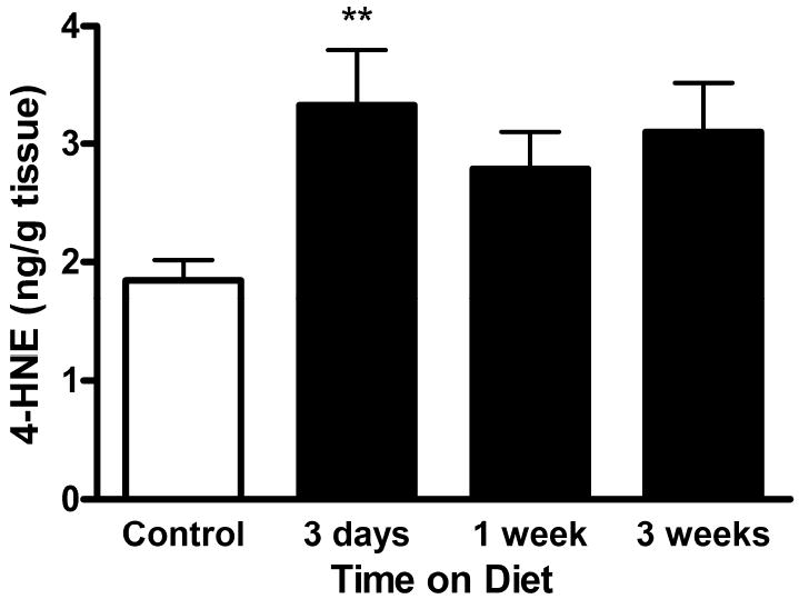
Levels of 4-HNE in hippocampus of rats fed KD or control diet. 4-HNE levels were assessed in the hippocampus of rats fed a control or KD for 3 days, 1 week, or 3 weeks by HPLC with UV detection. Bars represent mean ± standard error of the mean. **p ≤ 0.01 by one-way ANOVA.
Nrf2 Pathway Activation in Hippocampus of KD-fed Rats
Oxidative and electrophilic stresses are known to activate the Nrf2 pathway to mount an adaptive response (Numazawa et al., 2003; Levonen et al., 2004; Kobayashi et al., 2006; Satoh et al., 2006). Since we observed acute increases in H2O2 and 4-HNE, we asked whether consumption of a KD activates Nrf2 by assessing nuclear expression of the protein which is indicative of nuclear translocation. We fractionated hippocampal tissue from rats fed control or KD for 3 days, 1 week or 3 weeks and assessed nuclear levels of Nrf2 protein by Western blot analysis. Nuclear Nrf2 protein levels in KD-fed rat hippocampus were significantly greater than controls after 1 week on the KD and remained chronically elevated at 3 weeks (Fig. 3).
Figure 3.

Nrf2 nuclear expression in the hippocampus of rats fed KD or control diet. Nuclear fractions were prepared from rats fed a control or KD for 3 days, 1 week, or 3 weeks. (A) Western blot analysis of Nrf2 protein in nuclear fractions of rat hippocampus. Actin was used as a loading control. (B) Densitometry of Nrf2 protein expression exhibiting a time-dependent increase in the hippocampus of rats fed a KD (black bars) compared to control (open bars). Bars represent mean ± standard error of the mean. **p ≤ 0.01.
To further confirm activation of the Nrf2 pathway in the brains of KD-fed rats, we assessed activity of the Nrf2-dependent gene product, NQO1. NQO1 is an enzyme that detoxifies quinones by reducing them to their corresponding hydroquinones and is a prototypical Nrf2 target (Venugopal and Jaiswal, 1996). After 3 weeks on either diet, NQO1 activity was greater in hippocampus of rats fed a KD compared to controls, further suggesting an activation of the Nrf2 detoxification pathway (Fig. 4).
Figure 4.

Activity of the Nrf2 target, NQO1, in hippocampus of rats fed KD or control diet. NQO1 activity was assessed in the hippocampus of rats fed a control or KD for 3 weeks. Activity was assessed with a spectrofluorometric assay. Bars represent mean ± standard error of the mean. *p ≤ 0.05 by two-tailed t-test.
Improved Mitochondrial Antioxidant Capacity in the Liver Despite Tissue GSH Depletion
To determine whether the Nrf2 pathway was activated systemically by the KD or specific to the brain, we assessed key indices of this pathway in the livers of control and KD-fed rats. Interestingly, liver tissue GSH levels were significantly depleted in KD rats compared to controls at all time points examined (Fig. 5A). In previous work in the hippocampus, we found increased mitochondrial GSH and tissue CoASH but no significant changes in hippocampal tissue GSH (Jarrett et al., 2008). Therefore, to determine if liver mitochondrial fractions showed improved mitochondrial antioxidant capacity in a manner similar to the brain, we measured CoASH levels at 3 days, 1 week, and 3 weeks following diet initiation. CoASH is primarily localized within mitochondria, so mitochondrial antioxidant capacity can be assessed without extensive sample preparation, thus decreasing the possibility of artificial oxidation occurring while processing the tissue. CoASH was significantly depleted after 3 days on a KD compared to control but was significantly increased after 3 weeks on a KD (Fig. 5B), despite the chronic depletion of tissue GSH. These data suggest that acute mitochondrial oxidative stress occurs in the liver, much like the brain, and that mitochondrial antioxidant capacity improves above control levels by 3 weeks on a KD.
Figure 5.

GSH and CoASH levels in liver of rats fed KD or control diet. (A) Liver GSH levels in rats fed KD or control diets for 3 days, 1 week, and 3 weeks. (B) Liver CoASH levels in rats fed KD or control diets for 3 days, 1 week, and 3 weeks. Bars represent mean ± standard error of the mean. *p ≤ 0.05, ***p ≤ 0.001 by one-way ANOVA.
Systemic Activation of the Nrf2 Pathway by the KD
To determine whether the Nrf2 pathway was also activated in the liver, we examined Nrf2 accumulation in the liver. Nuclear fractions were prepared from liver tissue of rats fed control or KD for 3 days, 1 week, and 3 weeks. Nrf2 protein increased significantly throughout the time course, implying Nrf2 was activated and translocated to the nucleus (Fig. 6). Additionally, we assessed two Nrf2 targets, HO-1 and NQO1. HO-1 is highly inducible in the liver and a regularly utilized marker of Nrf2 activation. After 3 weeks on a KD, HO-1 protein levels were significantly greater in KD-fed rats compared to controls (Fig. 7A). Similarly, NQO1 activity was significantly increased in liver of KD-fed rats compared to controls after 3 weeks (Fig. 7B). These data suggest that the Nrf2 pathway is systemically activated by the KD and not specific to the brain.
Figure 6.

Nrf2 nuclear expression in the liver of rats fed KD or control diet. Nuclear fractions were prepared from liver of rats fed a control or KD for 3 days, 1 week, or 3 weeks. (A) Western blot analysis of Nrf2 protein in nuclear fractions of rat liver. Actin was used as a loading control. (B) Densitometry of Nrf2 protein expression exhibiting a time-dependent increase in the liver of rats fed a KD (black bars) compared to control (open bars). Bars represent mean ± standard error of the mean. **p ≤ 0.01, ***p ≤ 0.001 by one-way ANOVA.
Figure 7.
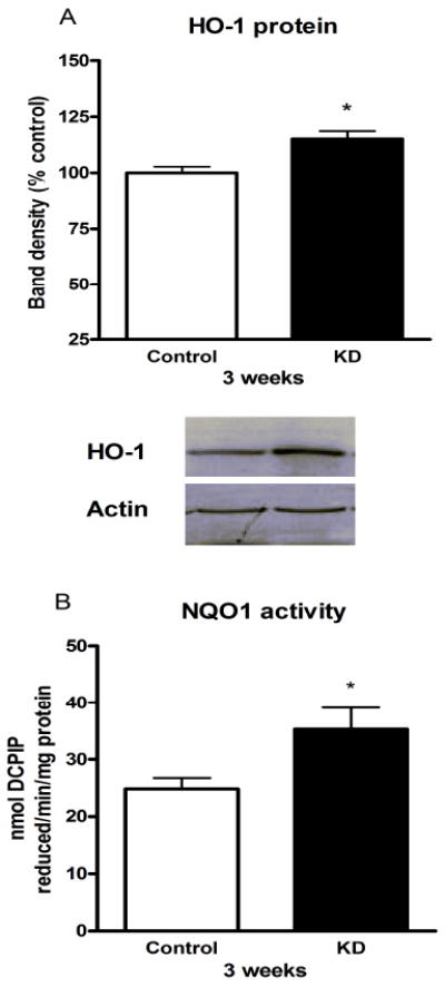
Nrf2 target upregulation in liver of rats fed KD or control diet for 3 weeks. (A) HO-1 protein was assessed in liver of rats fed a control or KD for 3 weeks. Western blot densitometry analysis of HO-1 protein expression in liver, normalized to actin. (B) NQO1 activity was assessed in liver of rats fed a control or KD for 3 weeks. Bars represent mean ± standard error of the mean. *p ≤ 0.05 by two-tailed t-test.
Discussion
We have previously shown that the KD increases glutathione biosynthesis (Jarrett et al., 2008). The rate-limiting enzyme in GSH biosynthesis is GCL, which is comprised of two subunits, one catalytic (GCLC) and one modulatory (GCLM). Protein levels of both GCL subunits were increased in the hippocampus of rats fed a KD for 3 weeks, which is suggestive of adaptation to cellular stress (Shi et al., 1994; Tian et al., 1997). One of the primary means by which GSH biosynthesis is stimulated is through induction of the Nrf2 transcription factor pathway, as the promoters of both Gclc and Gclm contain consensus sequences to which Nrf2 can bind to initiate transcription (Moinova and Mulcahy, 1999; Wild et al., 1999). In response to cellular stress Nrf2 releases from its cytoplasmic inhibitory binding partner, Keap1, translocates to the nucleus, and activates transcription of protective target genes, such as Gclc, Gclm, Ho-1 and Nqo1, thus initiating an adaptive response.
Here we present novel data suggesting that consumption of a KD initially causes production of low levels of ROS, which may serve a redox signaling role, thereby activating one of the major cellular detoxification pathways, the Nrf2 pathway. Consistent with this, shortly following diet initiation, we observed increased mitochondrial H2O2 production, GSSG, and 4-HNE levels. In contrast, by 3 weeks on the diet, all indices decreased, concomitant with the previously reported increase in mitochondrial GSH (Jarrett et al., 2008). This biphasic response to consumption of a KD suggests the activation of protective gene products as part of an adaptive response to cellular stress. Acute elevation of H2O2 and 4-HNE, together with our previous report of increased GSH biosynthesis is suggestive of activation of the Nrf2 pathway. In vitro pretreatment with 4-HNE has been shown to protect against subsequent H2O2-induced toxicity via a Nrf2-dependent mechanism and to be sufficient to induce Nrf2 dissociation from Keap1, thereby allowing nuclear translocation and target gene transcription (Numazawa et al., 2003; Chen et al., 2005). H2O2 itself has also been shown to increase DNA binding of Nrf2 to the ARE (Wilson et al., 2005). In the KD, acute increases in 4-HNE and H2O2 may assist in activating the release of Nrf2 from Keap1 to initiate the detoxification response, but the question persists as to how this pathway remains activated if it is important for the chronic protection afforded by the KD.
While the functional significance of Nrf2 activation by the KD remains to be established, literature suggests Nrf2 may play a role in seizure susceptibility in addition to its documented neuroprotective effects. It has been shown that mice lacking Nrf2 are more sensitive to kainic acid (Kraft et al., 2006), and there is substantial evidence to support the idea that activation of Nrf2 is neuroprotective. In vitro studies have demonstrated that Nrf2 activation or overexpression protects neurons against toxicity from NMDA, glutamate, or H2O2 (Shih et al., 2003; Satoh et al., 2006). Additionally, Nrf2-dependent protection against glutamate-mediated toxicity is dependent on GSH release from Nrf2-overexpressing glia, suggesting that increased GSH may play a central role in neuroprotection by Nrf2. Several groups have utilized the Nrf2 inducers tert-butylhydroquinone (tBHQ) and sulforaphane to demonstrate the protective effects of activating this pathway in vivo. Mice supplemented with tBHQ in their diets demonstrated attenuated toxicity to 3-nitropropionic acid, a mitochondrial complex II inhibitor (Shih et al., 2005b). This effect was lost when examined in Nrf2-/- mice, suggesting that the observed neuroprotection was Nrf2-dependent. Rats pretreated with tBHQ prior to an ischemia-reperfusion injury demonstrated decreased cortical damage at both 24 hours and 1 month post-injury (Shih et al., 2005a). Recently, the isothiocyanate sulforaphane, an extract of broccoli, has been found to potently activate Nrf2 in vivo and to protect post-injury loss of neurons in traumatic brain injury models (Zhao et al., 2006; Zhao et al., 2007). These studies suggest that augmenting the Nrf2 pathway may prove beneficial as a new drug target.
The bulk of KD research has focused on the brain, as the diet’s clinical application is primarily for the control of intractable epilepsies. This has left a void in the literature on the systemic effects of such a diet. One of the primary sources of brain GSH is export from the liver, which is consistent with our observation of depletion of liver GSH levels. This suggests that the liver may be exporting GSH in order to sustain GSH levels for other organs such as the brain. Even more striking was the finding that CoASH, a reduced mitochondrial thiol, was significantly increased in liver of KD-fed rats, suggesting a highly compartment specific effect of the KD. These data suggest that mitochondria are specifically increasing their thiol pools and thereby maintaining a reduced state, despite ongoing GSH depletion in non-mitochondrial compartments, such as the cytosol. To our knowledge the only other instance in which this has been reported is during fasting. It was found that during a 48-hour fast, hepatic GSH concentrations were depleted, while CoASH concentrations were increased (Jenniskens et al., 2002). This is particularly interesting given that the KD was initially designed to metabolically mimic the fasted state. With respect to Nrf2 activation in the liver, our results strikingly parallel those of acetaminophen toxicity studies in which liver GSH is depleted, concomitant with nuclear translocation of Nrf2 and increased transcription of Gclc and HO-1 (Goldring et al., 2004). Thus, the health effects of chronically depleting liver GSH need to be addressed in future studies of the KD.
While the KD is generally considered protective, here we show for the first time that this protection may be the result of redox signaling. This is not unreasonable given the extensive role of redox signaling as a preconditioning effect. In the traditional preconditioning paradigm, brief periods of sub-lethal stress lead to delayed protection against what would normally be a toxic insult. Importantly, it has been shown that preconditioning requires the production of ROS (Facundo et al., 2006), and that the ROS involved in this process are mitochondrially-derived (Vanden Hoek et al., 1998). Our model fits within this paradigm as we see initial increases in mitochondrial H2O2 production, followed closely by nuclear translocation of the Nrf2 transcription factor. Furthermore, in vitro evidence suggests that treatment with sub-lethal concentrations of H2O2 is sufficient to increase GSH content (Seo et al., 2004). While H2O2 was initially recognized as a toxin, recent evidence suggests that at sub-toxic levels, it can function as a redox signaling molecule.
One important question that remains to be addressed is whether these biochemical changes occur in an epileptic animal. Depletion of brain GSH has been reported following kainic acid-induced status epilepticus (Liang and Patel, 2006). As we have previously reported KD-induced maintenance of brain tissue GSH in conjunction with increased mitochondrial GSH, it is important to establish whether the KD can reverse these deficits in an epileptic animal. Additionally, the novel data that chronic consumption of a KD depletes liver GSH makes it essential for the medical community to recognize the importance of systemic and brain GSH and how they are affected by the KD.
Whether activation of the Nrf2 pathway, and thus GSH biosynthesis, contributes to the anticonvulsant effects of the KD remains to be determined. We have identified systemic activation of a primary cellular detoxification pathway, presumably initiated and maintained by low levels of cellular stress. These findings can suggest strategies for turning this nonpharmacological dietary treatment into a pharmacological therapy for the treatment of medically refractory epilepsies.
Acknowledgments
This work was supported by NIH RO1NS039587 (M.P.) and American Epilepsy Society pre-doctoral fellowship (J.B.M.).
Footnotes
Publisher's Disclaimer: This is a PDF file of an unedited manuscript that has been accepted for publication. As a service to our customers we are providing this early version of the manuscript. The manuscript will undergo copyediting, typesetting, and review of the resulting proof before it is published in its final citable form. Please note that during the production process errors may be discovered which could affect the content, and all legal disclaimers that apply to the journal pertain.
References
- Appleton DB, DeVivo DC. An animal model for the ketogenic diet. Epilepsia. 1974;15:211–227. doi: 10.1111/j.1528-1157.1974.tb04943.x. [DOI] [PubMed] [Google Scholar]
- Bough KJ, Wetherington J, Hassel B, Pare JF, Gawryluk JW, Greene JG, Shaw R, Smith Y, Geiger JD, Dingledine RJ. Mitochondrial biogenesis in the anticonvulsant mechanism of the ketogenic diet. Ann Neurol. 2006;60:223–235. doi: 10.1002/ana.20899. [DOI] [PubMed] [Google Scholar]
- Castello PR, Drechsel DA, Patel M. Mitochondria are a major source of paraquat-induced reactive oxygen species production in the brain. J Biol Chem. 2007;282:14186–14193. doi: 10.1074/jbc.M700827200. [DOI] [PMC free article] [PubMed] [Google Scholar]
- Chen ZH, Saito Y, Yoshida Y, Sekine A, Noguchi N, Niki E. 4-Hydroxynonenal induces adaptive response and enhances PC12 cell tolerance primarily through induction of thioredoxin reductase 1 via activation of Nrf2. J Biol Chem. 2005;280:41921–41927. doi: 10.1074/jbc.M508556200. [DOI] [PubMed] [Google Scholar]
- Dehn DL, Siegel D, Zafar KS, Reigan P, Swann E, Moody CJ, Ross D. 5-Methoxy-1,2-dimethyl-3-[(4-nitrophenoxy)methyl]indole-4,7-dione, a mechanism-based inhibitor of NAD(P)H:quinone oxidoreductase 1, exhibits activity against human pancreatic cancer in vitro and in vivo. Mol Cancer Ther. 2006;5:1702–1709. doi: 10.1158/1535-7163.MCT-06-0105. [DOI] [PubMed] [Google Scholar]
- Facundo HT, Carreira RS, de Paula JG, Santos CC, Ferranti R, Laurindo FR, Kowaltowski AJ. Ischemic preconditioning requires increases in reactive oxygen release independent of mitochondrial K+ channel activity. Free Radic Biol Med. 2006;40:469–479. doi: 10.1016/j.freeradbiomed.2005.08.041. [DOI] [PubMed] [Google Scholar]
- Goldring CE, Kitteringham NR, Elsby R, Randle LE, Clement YN, Williams DP, McMahon M, Hayes JD, Itoh K, Yamamoto M, Park BK. Activation of hepatic Nrf2 in vivo by acetaminophen in CD-1 mice. Hepatology. 2004;39:1267–1276. doi: 10.1002/hep.20183. [DOI] [PubMed] [Google Scholar]
- Jarrett SG, Milder JB, Liang LP, Patel M. The ketogenic diet increases mitochondrial glutathione levels. J Neurochem. 2008;106:1044–1051. doi: 10.1111/j.1471-4159.2008.05460.x. [DOI] [PubMed] [Google Scholar]
- Jenniskens FA, Jopperi-Davis KS, Walters LC, Schorr EN, Rogers LK, Welty SE, Smith CV. Effects of fasting on tissue contents of coenzyme A and related intermediates in rats. Pediatr Res. 2002;52:437–442. doi: 10.1203/00006450-200209000-00022. [DOI] [PubMed] [Google Scholar]
- Kobayashi A, Kang MI, Watai Y, Tong KI, Shibata T, Uchida K, Yamamoto M. Oxidative and electrophilic stresses activate Nrf2 through inhibition of ubiquitination activity of Keap1. Mol Cell Biol. 2006;26:221–229. doi: 10.1128/MCB.26.1.221-229.2006. [DOI] [PMC free article] [PubMed] [Google Scholar]
- Kobayashi A, Kang MI, Okawa H, Ohtsuji M, Zenke Y, Chiba T, Igarashi K, Yamamoto M. Oxidative stress sensor Keap1 functions as an adaptor for Cul3-based E3 ligase to regulate proteasomal degradation of Nrf2. Mol Cell Biol. 2004;24:7130–7139. doi: 10.1128/MCB.24.16.7130-7139.2004. [DOI] [PMC free article] [PubMed] [Google Scholar]
- Kossoff EH, Laux LC, Blackford R, Morrison PF, Pyzik PL, Hamdy RM, Turner Z, Nordli DR., Jr When do seizures usually improve with the ketogenic diet? Epilepsia. 2008;49:329–333. doi: 10.1111/j.1528-1167.2007.01417.x. [DOI] [PubMed] [Google Scholar]
- Kraft AD, Lee JM, Johnson DA, Kan YW, Johnson JA. Neuronal sensitivity to kainic acid is dependent on the Nrf2-mediated actions of the antioxidant responseelement. J Neurochem. 2006;98:1852–1865. doi: 10.1111/j.1471-4159.2006.04019.x. [DOI] [PubMed] [Google Scholar]
- Levonen AL, Landar A, Ramachandran A, Ceaser EK, Dickinson DA, Zanoni G, Morrow JD, Darley-Usmar VM. Cellular mechanisms of redox cell signalling: role of cysteine modification in controlling antioxidant defences in response to electrophilic lipid oxidation products. Biochem J. 2004;378:373–382. doi: 10.1042/BJ20031049. [DOI] [PMC free article] [PubMed] [Google Scholar]
- Liang LP, Patel M. Seizure-induced changes in mitochondrial redox status. Free Radic Biol Med. 2006;40:316–322. doi: 10.1016/j.freeradbiomed.2005.08.026. [DOI] [PubMed] [Google Scholar]
- Likhodii SS, Musa K, Cunnane SC. Breath acetone as a measure of systemic ketosis assessed in a rat model of the ketogenic diet. Clin Chem. 2002;48:115–120. [PubMed] [Google Scholar]
- Moinova HR, Mulcahy RT. Up-regulation of the human gamma-glutamylcysteine synthetase regulatory subunit gene involves binding of Nrf-2 to an electrophile responsive element. Biochem Biophys Res Commun. 1999;261:661–668. doi: 10.1006/bbrc.1999.1109. [DOI] [PubMed] [Google Scholar]
- Murphy TC, Amarnath V, Picklo MJ., Sr Mitochondrial oxidation of 4-hydroxy-2-nonenal in rat cerebral cortex. J Neurochem. 2003;84:1313–1321. doi: 10.1046/j.1471-4159.2003.01628.x. [DOI] [PubMed] [Google Scholar]
- Neal EG, Chaffe HM, Edwards N, Lawson MS, Schwartz RH, Cross JH. Growth of children on classical and medium-chain triglyceride ketogenic diets. Pediatrics. 2008;122:e334–340. doi: 10.1542/peds.2007-2410. [DOI] [PubMed] [Google Scholar]
- Numazawa S, Ishikawa M, Yoshida A, Tanaka S, Yoshida T. Atypical protein kinase C mediates activation of NF-E2-related factor 2 in response to oxidative stress. Am J Physiol Cell Physiol. 2003;285:C334–342. doi: 10.1152/ajpcell.00043.2003. [DOI] [PubMed] [Google Scholar]
- Rho JM, Kim DW, Robbins CA, Anderson GD, Schwartzkroin PA. Age-dependent differences in flurothyl seizure sensitivity in mice treated with a ketogenic diet. Epilepsy Res. 1999;37:233–240. doi: 10.1016/s0920-1211(99)00068-6. [DOI] [PubMed] [Google Scholar]
- Rushmore TH, Morton MR, Pickett CB. The antioxidant responsive element. Activation by oxidative stress and identification of the DNA consensus sequence required for functional activity. J Biol Chem. 1991;266:11632–11639. [PubMed] [Google Scholar]
- Sagara Y, Dargusch R, Chambers D, Davis J, Schubert D, Maher P. Cellular mechanisms of resistance to chronic oxidative stress. Free Radic Biol Med. 1998;24:1375–1389. doi: 10.1016/s0891-5849(97)00457-7. [DOI] [PubMed] [Google Scholar]
- Satoh T, Okamoto SI, Cui J, Watanabe Y, Furuta K, Suzuki M, Tohyama K, Lipton SA. Activation of the Keap1/Nrf2 pathway for neuroprotection by electrophilic [correction of electrophillic] phase II inducers. Proc Natl Acad Sci U S A. 2006;103:768–773. doi: 10.1073/pnas.0505723102. [DOI] [PMC free article] [PubMed] [Google Scholar]
- Seo YJ, Lee JW, Lee EH, Lee HK, Kim HW, Kim YH. Role of glutathione in the adaptive tolerance to H2O2. Free Radic Biol Med. 2004;37:1272–1281. doi: 10.1016/j.freeradbiomed.2004.07.012. [DOI] [PubMed] [Google Scholar]
- Shi MM, Kugelman A, Iwamoto T, Tian L, Forman HJ. Quinone-induced oxidative stress elevates glutathione and induces gamma-glutamylcysteine synthetase activity in rat lung epithelial L2 cells. J Biol Chem. 1994;269:26512–26517. [PubMed] [Google Scholar]
- Shih AY, Li P, Murphy TH. A small-molecule-inducible Nrf2-mediated antioxidant response provides effective prophylaxis against cerebral ischemia in vivo. J Neurosci. 2005a;25:10321–10335. doi: 10.1523/JNEUROSCI.4014-05.2005. [DOI] [PMC free article] [PubMed] [Google Scholar]
- Shih AY, Imbeault S, Barakauskas V, Erb H, Jiang L, Li P, Murphy TH. Induction of the Nrf2-driven antioxidant response confers neuroprotection during mitochondrial stress in vivo. J Biol Chem. 2005b;280:22925–22936. doi: 10.1074/jbc.M414635200. [DOI] [PubMed] [Google Scholar]
- Shih AY, Johnson DA, Wong G, Kraft AD, Jiang L, Erb H, Johnson JA, Murphy TH. Coordinate regulation of glutathione biosynthesis and release by Nrf2-expressing glia potently protects neurons from oxidative stress. J Neurosci. 2003;23:3394–3406. doi: 10.1523/JNEUROSCI.23-08-03394.2003. [DOI] [PMC free article] [PubMed] [Google Scholar]
- Sullivan PG, Rippy NA, Dorenbos K, Concepcion RC, Agarwal AK, Rho JM. The ketogenic diet increases mitochondrial uncoupling protein levels and activity. Ann Neurol. 2004;55:576–580. doi: 10.1002/ana.20062. [DOI] [PubMed] [Google Scholar]
- Thio LL, Erbayat-Altay E, Rensing N, Yamada KA. Leptin contributes to slower weight gain in juvenile rodents on a ketogenic diet. Pediatr Res. 2006;60:413–417. doi: 10.1203/01.pdr.0000238244.54610.27. [DOI] [PubMed] [Google Scholar]
- Tian L, Shi MM, Forman HJ. Increased transcription of the regulatory subunit of gamma-glutamylcysteine synthetase in rat lung epithelial L2 cells exposed to oxidative stress or glutathione depletion. Arch Biochem Biophys. 1997;342:126–133. doi: 10.1006/abbi.1997.9997. [DOI] [PubMed] [Google Scholar]
- Uhlemann ER, Neims AH. Anticonvulsant properties of the ketogenic diet in mice. J Pharmacol Exp Ther. 1972;180:231–238. [PubMed] [Google Scholar]
- Vanden Hoek TL, Becker LB, Shao Z, Li C, Schumacker PT. Reactive oxygen species released from mitochondria during brief hypoxia induce preconditioning in cardiomyocytes. J Biol Chem. 1998;273:18092–18098. doi: 10.1074/jbc.273.29.18092. [DOI] [PubMed] [Google Scholar]
- Venugopal R, Jaiswal AK. Nrf1 and Nrf2 positively and c-Fos and Fra1 negatively regulate the human antioxidant response element-mediated expression of NAD(P)H:quinone oxidoreductase1 gene. Proc Natl Acad Sci U S A. 1996;93:14960–14965. doi: 10.1073/pnas.93.25.14960. [DOI] [PMC free article] [PubMed] [Google Scholar]
- Wild AC, Moinova HR, Mulcahy RT. Regulation of gamma-glutamylcysteine synthetase subunit gene expression by the transcription factor Nrf2. J Biol Chem. 1999;274:33627–33636. doi: 10.1074/jbc.274.47.33627. [DOI] [PubMed] [Google Scholar]
- Wilson LA, Gemin A, Espiritu R, Singh G. ets-1 is transcriptionally up-regulated by H2O2 via an antioxidant response element. Faseb J. 2005;19:2085–2087. doi: 10.1096/fj.05-4401fje. [DOI] [PubMed] [Google Scholar]
- Woods JS, Kavanagh TJ, Corral J, Reese AW, Diaz D, Ellis ME. The role of glutathione in chronic adaptation to oxidative stress: studies in a normal rat kidney epithelial (NRK52E) cell model of sustained upregulation of glutathione biosynthesis. Toxicol Appl Pharmacol. 1999;160:207–216. doi: 10.1006/taap.1999.8774. [DOI] [PubMed] [Google Scholar]
- Zhao J, Kobori N, Aronowski J, Dash PK. Sulforaphane reduces infarct volume following focal cerebral ischemia in rodents. Neurosci Lett. 2006;393:108–112. doi: 10.1016/j.neulet.2005.09.065. [DOI] [PubMed] [Google Scholar]
- Zhao J, Moore AN, Redell JB, Dash PK. Enhancing expression of Nrf2-driven genes protects the blood brain barrier after brain injury. J Neurosci. 2007;27:10240–10248. doi: 10.1523/JNEUROSCI.1683-07.2007. [DOI] [PMC free article] [PubMed] [Google Scholar]