
Figure 3.
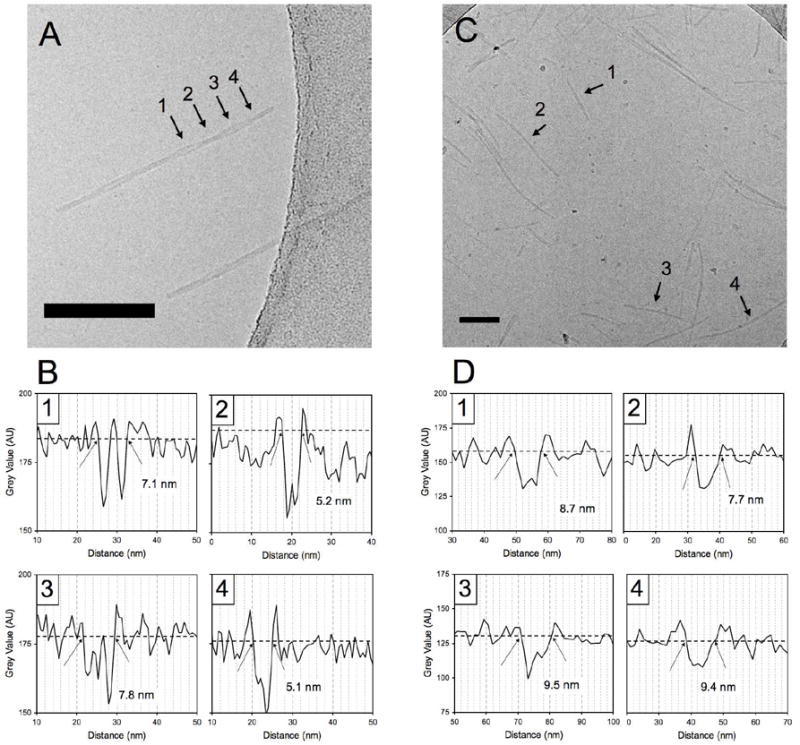
Cryo-TEM micrographs of C16p53 (A) and C16A4p53 (C) in PBS 10mM. C16p53 structures self-assembled into twisted ribbon structures, whereas C16A4p53 appeared as core-shell worm-like micelles. Pixel intensity profiles to determine aggregate width at positions indicated by the arrows in (A) and (C) are shown in (B) for C16p53 and (D) for C16A4p53, respectively. Scale bars: 100 nm