Abstract

Spectrophotometric procedures allow rapid and precise measurements of the pH of natural waters. However, impurities in the acid–base indicators used in these analyses can significantly affect measurement accuracy. This work describes HPLC procedures for purifying one such indicator, meta-cresol purple (mCP), and reports mCP physical–chemical characteristics (thermodynamic equilibrium constants and visible-light absorbances) over a range of temperature (T) and salinity (S). Using pure mCP, seawater pH on the total hydrogen ion concentration scale (pHT) can be expressed in terms of measured mCP absorbance ratios (R = λ2A/λ1A) as follows:
 |
where −log(K2Te2) = a + (b/T) + c ln T – dT; a = −246.64209 + 0.315971S + 2.8855 × 10⁻⁴S2; b = 7229.23864 – 7.098137S – 0.057034S2; c = 44.493382 – 0.052711S; d = 0.0781344; and mCP molar absorbance ratios (ei) are expressed as e1 = −0.007762 + 4.5174 × 10⁻⁵T and e3/e2 = −0.020813 + 2.60262 × 10⁻⁴T + 1.0436 × 10⁻⁴ (S – 35). The mCP absorbances, λ1A and λ2A, used to calculate R are measured at wavelengths (λ) of 434 and 578 nm. This characterization is appropriate for 278.15 ≤ T ≤ 308.15 and 20 ≤ S ≤ 40.
Introduction
Spectrophotometric measurements of seawater pH, based on methods developed in the late 1980s,1−5 are simple, rapid, and precise. Observations obtained during global surveys (e.g., http://cdiac.ornl.gov/oceans/) have demonstrated shipboard measurement precisions on the order of ±0.0004 pH units. At this level of precision, pH measurements can play an important role in CO2-system characterizations and quality control assessments.(6)
Spectrophotometric pH values obtained via measurements of absorbance ratios are directly grounded on indicator molecular properties: molar absorptivity ratios and protonation characteristics. Indicators can therefore serve as molecular standards. These indicators have been used, for example, to monitor and evaluate the quality of aging pH standards.(7) (As buffers age, they absorb atmospheric CO2 and their pH declines.) Furthermore, archived spectrophotometric pH data can be quantitatively revised should improved indicator equilibrium constants and molar absorptivity ratios later become available.(3) However, as noted by Yao et al.,(8) indicator impurities can introduce systematic errors in reported spectrophotometric pH even though measurement precision remains quite good.
Yao et al.(8) pointed out that indicator impurities vary with manufacturer and can also differ among batches from a single manufacturer. Systematic differences in reported pH obtained using indicators from different sources were as large as 0.01 pH units. Consequently, in order to fully realize the advantages of spectrophotometric pH measurements—ensuring accuracy as well as precision—the issue of indicator impurities must be carefully addressed.
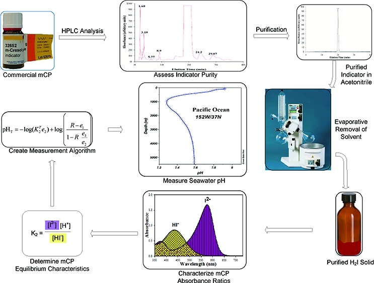
This work focused on the physical–chemical characteristics of the pH indicator meta-cresol purple (mCP), and endeavored to provide (a) an efficient procedure for indicator purification, and (b) a procedure for oceanic seawater pH measurements that is free of vendor-specific pH indicator impurities. Purification via high-performance liquid chromatography (HPLC) was performed, and the characteristics of purified mCP are reported for a wide range of seawater salinity and temperature. Using the methods described here, independent investigators should be able to produce pH measurements that are directly comparable through time, independent of dye source.
Analytical Procedures
Spectrophotometric pH measurements involving the use of sulfonephthalein indicators (H2I) are based on observations of the relative absorbance contributions of protonated (HI–) and unprotonated (I2–) species1−3,7,9,10 in the sample of interest. Solution pH can be calculated using the following equation:
where K2T is the dissociation constant of HI– on the total hydrogen ion concentration scale,(3)
and [ ] denotes concentration in mol/kg-solution. The parameter R in eq 1 is the ratio of sulfonephthalein absorbances at wavelengths λ2 and λ1
For mCP, λ2 = 578 nm and λ1 = 434 nm.(3)
The symbols e1, e2, and e3 are ratios of molar absorptivities of the HI– and I2– indicator forms
where λεI is the molar absorptivity of I2– at wavelength λ and λεHI is the molar absorptivity of HI– at wavelength λ.
Equation 1 can be alternatively written in a form with fewer parameters:(11)
 |
The e3/e2 term involves determinations of I2– molar absorptivity ratios and is directly obtained via measurements in synthetic solutions at a pH sufficiently high that species other than I2– are negligible (i.e., pH ∼12). The e1 term involves determinations of HI– molar absorptivity ratios and is obtained, as a very good approximation, through measurements at low pH (i.e., pH ∼4.5), where HI– is the strongly predominant form of mCP. The K2Te2 term can be determined as a single parameter by measuring the absorbance ratio R in a solution of known pHT (e.g., tris seawater buffer). Finally, an iterative procedure is applied to refine, in an internally consistent manner, the e1 initial approximation and resulting K2e2 term.
Methods
Reagents
The m-cresol purple (mCP) indicator, in acid form or as a sodium salt, was obtained from the following vendors: Acros (batch A0182569), Aldrich (batches 11517KC and 07005HH), Alfa Aesar (batch H11N06), JT Baker (batch B40626), MP Bio (batch 1426K), Ricca (batch 4003124), and TCI (batch FDP01). Kodak mCP is no longer in production and was not used in this study. Sodium chloride (ReagentPlus), potassium chloride (99.999%), sodium sulfate (SigmaUltra), magnesium chloride hexahydrate (SigmaUltra), calcium chloride dihydrate (SigmaUltra), and trifluoroacetic acid were obtained from Sigma and were used without further purification. Tris (tris (hydroxymethyl)-aminomethane) was obtained from NIST (SRM 723D). Tris and all salts except MgCl2 and CaCl2 were stored in a desiccator over P2O5 until the weight of each substance stabilized. Solutions of MgCl2 and CaCl2, each with a concentration of 1 mol/kg solution, were prepared, and the final concentration of each was measured via ICP-MS analysis. Acetonitrile (AN, HPLC grade) was obtained from Fisher Scientific. Hydrochloric acid with a concentration of 1 mol/kg solution was prepared by dilution of concentrated HCl (Fisher Scientific). The final concentration of the acid was determined by spectrophotometric titration with phenol red.
HPLC Purification of m-Cresol Purple
The mCP used in this study was characterized and purified using a Waters PrepLC HPLC system. This system includes a Prep LC controller, two HPLC pumps capable of flow rates between 1 and 150 cm(3)/min, a fraction collector, and a model 2998 Photodiode Array Detector.
The HPLC columns were from SIELC Technologies. The Primesep B2 column used in this work has a mixed-mode resin that interacts with analytes via hydrophobic and ion exchange mechanisms. An analytical column (Part B2-46-250.0510, 4.6 × 250 mm, particle size 5 μm) was used to optimize separation conditions. Subsequently, a preparative column (Part B2-220.250.0510, 22 × 250 mm, particle size 5 μm) was used to purify the mCP. The Primesep columns were housed in a Shimadzu column oven (model CTO-10A) at 30 °C.
The HPLC mobile phase composition was 70% AN plus 30% H2O; 0.05% trifluoroacetic acid (TFA) was present as a mobile phase modifier. The pump rate was 1.5 cm3/min for the analytical column and 31.26 cm3/min in preparative mode. The injection loop volume was 0.020 cm3 in analytical mode and 7 cm3 in preparative mode. Separations were begun by preparing a 70 mM (10–3 mol dm–3) solution of mCP in the mobile phase solution. Pure mCP component was collected at its characteristic retention time (approximately 20 min). The solvent was separated from the mCP using a rotary evaporator (Buchi Rotavapor-R). The evaporation flask was submerged in a 35 °C water bath, with the contents of the flask under partial vacuum. Evaporation of the mobile phase to dryness produced solid mCP in acid form (H2I), free of salts. Each injection produced approximately 0.2 g of purified mCP, resulting in a daily yield of about 2 g. Thymol blue from the original batch of Zhang and Byrne(10) was also examined on the analytical column to assess its levels of impurities.
Comparisons of mCP from Different Vendors
Batches of mCP direct from seven vendors were characterized relative to a reference (purified) mCP via paired pH measurements in strongly buffered solutions over a pH range of 7.2 to 8.2. The direct-from-vendor mCPs were then purified and again compared to the reference mCP.
The reference mCP consisted of Acros Organics mCP that had been purified via the HPLC method outlined above. All indicator stock solutions were adjusted to the same R ratio before use in the pH comparisons. The buffered solutions were prepared by adding 0.08 mol tris, EPPS, or HEPES to 0.04 mol either HCl or NaOH, depending on the form of the buffering agent. The solutions were brought to 0.7 m (mol/kg solution) ionic strength by addition of NaCl. Because measured pH differed slightly among different batches of the same buffer, the purified Acros mCP was always used as a reference, thereby generating paired pH (difference) observations for each mCP comparison.
For each pH measurement, the buffered solution was weighed (140 g) into a custom-made quartz open-top spectrophotometric cell of 10 cm path length. After the stirred sample reached the target temperature (25 °C), a blank was recorded. Indicator solution (0.05 cm3 of 10 mM indicator) was then injected into the sample and the absorbance ratio, R, was measured. Addition of indicator to the strongly buffered solutions had a negligible effect on solution pH, so “pH perturbation” calculations were not necessary.
Absorbance Measurements for Characterization of Purified mCP
Absorbance measurements were obtained using a Varian Cary 400 spectrophotometer. The wavelength accuracy of the instrument was verified using a holmium oxide standard (NIST SRM 2034), and the linearity of the spectrophotometer was verified with NIST SRM 930D glass filters. The spectral bandwidth of the instrument was 1.5 nm. Sample solutions were thermostatted in a custom-made, water-jacketed spectrophotometric cell holder that was connected via insulated tubing to a Neslab refrigerating circulator. Solution temperatures were monitored with a VWR digital thermometer or an Ertco model 4400 thermometer. Thermometer measurement accuracy was ±0.01 °C. For data obtained at 5 °C, a dry N2 stream was directed to the optical windows of the spectrophotometric cell to prevent condensation of water vapor.
Determination of Molar Absorptivity Ratios (ei)
The molar absorptivity ratio (ei) measurements were obtained using a 10 cm open-top quartz cell (NSG Precision Cells, Inc.). A cover for this cell had openings for insertion of a custom-made motor-driven stirring rod and a digital thermometer to ensure well-mixed solutions and accurately measured temperatures. Solutions were thermostatted at temperatures between 5 and 35 °C. Absorbance measurements for a given solution of mCP were taken against a baseline solution that contained no mCP.
Values of e1 were obtained by measuring 578A and 434A in a solution that contained 0.02 m acetate buffer in 0.7 m NaCl solution. The pH of the buffer solution, determined with a Ross combination electrode, was adjusted to pH 4.50 by addition of NaOH or HCl. The pH electrode was calibrated on the free hydrogen ion concentration scale by titrating unbuffered 0.7 m NaCl solutions with 1 M (mol dm–3) standard HCl. The absorbance measurements were corrected by an iterative procedure (described below) to produce e1 values. Ancillary experimental data showed that e1 has a negligible dependence on ionic strength and composition, indicating that this buffer solution is adequately seawater-like for the purpose of e1 determination.
In contrast, e3/e2 was found to be significantly influenced by ionic strength and, to a lesser extent, ionic composition. Consequently, e3/e2 values were obtained in solutions with compositions similar to seawater, over a range of temperatures (278.21–308.37 K) and salinities (20–40). The synthetic seawater contained 0.01 m NaOH. To avoid precipitation of magnesium and sulfur salts at high pH (pH ∼12), Na2SO4 was replaced by NaCl, and MgCl2 was replaced by CaCl2.
Titration of solutions of mCP in 0.7 m NaCl with 1 M NaOH demonstrated that absorbance contributions from HI– were negligible in solutions containing 0.01 m NaOH. Titrations of acetate buffer solutions with HCl demonstrated that minima in e1 ratios were obtained near pH 4.5.
Determination of K1 as a Function of Temperature
Equilibrium constants (K1) for dissociation of H2I (H2I ↔ HI– + H+) are required to estimate the absorbance contributions of H2I and I2– in the course of e1 determination. K1 values over the temperature range 279.45–307.95 K were obtained from observations of mCP absorbances in aqueous HCl–NaCl mixtures (1 ≤ pH ≤ 2) at ionic strength 0.70 ± 0.02 m. At each temperature, [H+] and absorbance at λ = 529 nm (the H2I absorbance maximum) were recorded, along with isosbestic point absorbances, which were used to account for dilution due to the acid additions. Hydrogen ion concentrations were calculated via the mixing ratios of the HCl and NaCl solutions.
Determination of −log(K2Te2)
To determine the −log(K2Te2) term in eq 5, tris seawater buffer was used as a calibrating medium over a range of salinities between 20 and 40 and temperatures between 278.15 and 303.15 K. Solutions of 0.04 m tris buffer were prepared at different salinities according to the recipe of DelValls and Dickson.(12) The solutions consisted of synthetic seawater in which 0.04 mol/kg H2O of NaCl was replaced with an identical molality of HCl, plus tris at a total concentration of 0.08 mol/kg H2O. The gravimetric procedure was facilitated using an Excel file recipe provided by Dr. A. Dickson of UCSD. The tris buffers at salinity 35 prepared in our experiments produced an R ratio identical to the measured R value of tris buffer provided by Dr. A. Dickson (batch 2, August, 2008). The pH of each tris buffer was calculated on the total hydrogen ion concentration scale(12) with the following equation:
 |
To determine the −log(K2Te2) values, sulfonephthalein absorbance ratios (R) were obtained in the tris buffers, and eq 5 was used to calculate the −log(K2e2) term for each T – S condition.
Iterative Refinement of e1 and K2Te2
Subsequent to the measurements of K2Te2, small refinements were made to the initial estimates of e1. These quantitatively small refinements were necessary because the molar absorptivities of I2– and H2I at 578 nm are very large relative to that of HI–. As such, even though HI– is the strongly predominant form of mCP at pH ∼4.5, the absorbance contributions of I2– and H2I are significant and must be quantitatively taken into account.
The directly measured absorbances of mCP at low pH (578A and 434A) are summations of the major contribution from HI– and minor contributions from I2– and H2I:
Using eqs 7a and 7b, the parameter e1 can be written as
 |
where b is path length. The absorbances of H2I and I2– in eq 8 can then be expressed in terms of molar absorptivities (λεi) and path length (b):
Employing eq 9, the 578Ai and 434Ai absorbance terms in eq 8 can be expressed in terms of molar absorptivities and mCP species concentrations. Concentrations of H2I and I2– can be expressed in terms of free hydrogen ion concentrations ([H+]), total concentrations of mCP (IT), and H2I and HI– dissociation constants on the free hydrogen ion concentration scale (K1 = [HI–][H+][H2I]−1 and K2 = [I2–][H+][HI–]−1). The 578Ai and 434Ai absorbance terms in eq 8 can then be written as
 |
 |
 |
 |
The molar absorptivity terms 578εH2I and 434εH2I were calculated from measurements in 1 M HCl where H2I is a strongly predominant species; the molar absorptivity terms 578εI and 434εI were calculated from experiments at pH 12 where I2– strongly predominates. The data used for K1 determinations consisted of coupled λA and [H+] observations at λ= 529 nm (i.e., at the H2I absorbance maximum). Absorbance observations were fitted with the following equation, from which K1 can be obtained:
K2 values used for determinations of I2– concentrations on the free hydrogen ion concentration scale were calculated from the K2Te2 term in eq 5. These calculations utilized e2 values obtained from paired observations of indicator absorbances for HI– at λ = 434 nm in acetate buffer (pH ∼4.5) and I2– at λ = 578 nm in 0.01 m NaOH. Measurements of e2 were obtained over a range of temperatures in 0.7 m NaCl. The concentrations of mCP, IT, in these experiments were calculated through observations of isosbestic point absorbances.
Refined values of e1 were obtained iteratively. Initial e1 estimates were used in eq 5 to calculate initial K2T values. K2 values on the free H+ scale were then calculated using the following relationship:(13)
where KS = [H+][SO42–]/[HSO4–], ST is the total sulfate concentration at a given salinity, and KS is the dissociation constant of HSO4–.(14)
Using calculated values of K1 (eq 11) and K2 (eq 12), eqs 8 through 10 were then used to refine e1 by accounting for small absorbance contributions from I2– and H2I. The refined e1 value was then used in eq 5 to obtain a refined K2T value. Iterations were repeated until −log(K2e2) values were in agreement to 0.0001 and e1 values were in agreement to within 0.000001. This process produced extremely small refinements in K2T but moderate refinements in e1.
For the salinity-dependent terms in eq 5 (K2T and e2), the salinity values are consistent with the characterizations of DelValls and Dickson.(12) For e1, which is not salinity-dependent, the ionic strength of the solution was maintained at 0.7 mol/kg solution. For the purposes of these calculations, it is appropriate to regard the activity coefficients of H+, I2–, and HI– as functions of ionic strength independent of medium composition.
Natural Seawater pH Measurements
Seawater samples were collected 20 miles offshore in the Gulf of Mexico. The salinity of this seawater, measured with a Seabird model 59 CTD, was 35.90 (practical salinity scale). Total alkalinity (AT), total CO2 (CT), and pHT of the seawater samples were measured to assess the internal consistency of these three carbonate-system parameters. AT was measured using the method of Yao and Byrne.(15)CT was measured with a UIC CM 5104 Coulometer with the UIC 5130 acidification module. Sample seawater was drawn into a gastight syringe and then injected into an acidification flask. A stream of CO2-free air carried evolved CO2 from the acidification flask to the cathode solution for subsequent coulometric measurement. The certified reference material (CRM) used to calibrate the coulometer was provided by Dr. Andrew Dickson. For pH determinations, seawater samples were collected in 10-cm cylindrical cells (Hellma Cells), and pHT was measured using the procedures outlined in SOP 6b of Dickson et al.(13)
Results and Discussion
HPLC Purification of mCP
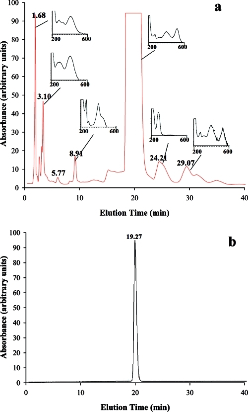
Yao et al.(8) demonstrated that mCP batches obtained from different vendors have variable amounts and types of impurities. Figure 1a shows an HPLC chromatograph of an unpurified Aldrich mCP sample on a Primesep B2 column. Several major peaks were identified both before and after the pure mCP peak, which had a retention time of approximately 20 min. The pure mCP fraction had spectrophotometric absorption peaks at 406 and 529 nm in the 70% AN mobile phase (see inset graph). The component with an HPLC retention time of 3.10 min had a peak absorbance at 421 nm. This peak overlaps with the mCP HI– peak in aqueous solution, which is potentially problematic. Components eluted at 1.68, 8.91, and 29.07 min all had nonzero absorbances at wavelengths greater than 400 nm, which could also affect spectrophotometric pH measurements. In contrast, the components that eluted at 2.90 and 24.21 min had no absorbances at wavelengths greater than 350 nm and so would likely not affect mCP absorbance readings and pH measurements in aqueous solutions.
Figure 1.
HPLC chromatographs of mCP. (a) mCP direct from vendor (Aldrich). Absorbances versus elution time were obtained using a broadband light source. The small inset panels show the UV–vis spectra (200 to 600 nm) of indicator components eluted at different times. (b) Purified mCP.
The analytical HPLC procedure, which characterizes the relative absorbance contributions of components and establishes effective separation conditions, was successfully scaled up to a preparative-scale procedure, which separates components and allows for production of purified mCP. The injection volume was increased to 7 cm3 at an mCP concentration of 70 mM. Because salt components were eluted at the beginning of the HPLC run and the rotary evaporation procedure eliminated AN and TFA, pure mCP in acid form was obtained in the flask.
Figure 1b is the chromatograph of a purified mCP. Only the mCP peak is present, indicating that the contamination components were successfully separated out. As will be shown below, the differences among purified mCPs produced from various batches/vendors are insignificant.
As shown in Figure 1a (see small inset panels), components with absorbances at wavelengths greater than 400 nm are present, which may affect spectrophotometric pH measurements in seawater. At higher pH, where concentrations of the acid form of mCP, HI–, are expected to be low, the effects of the impurities that absorb at λ = 434 nm are likely enhanced relative to low-pH samples, where 434A is large. Therefore, it is expected that the effects of mCP impurities on spectrophotometric pH measurements would be more pronounced for high-pH surface waters (pH ∼8) than for low-pH waters (pH ∼7.2).
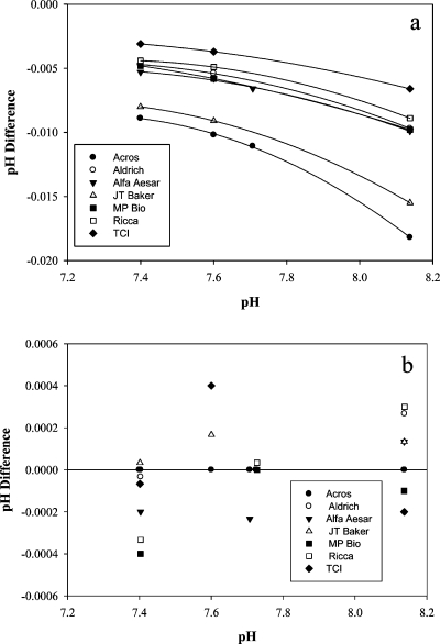
Using mCP from seven different vendors, spectrophotometric pH values were measured in a range of strongly buffered solutions. Figure 2a compares pH values obtained using unrefined (unpurified) mCP to corresponding measurements obtained using purified mCP (Acros). Using unrefined mCP yields pH deviations, relative to values obtained with purified mCP, as large as 0.010 pH units at pH 7.4 and as large as 0.018 pH units at pH 8.2. As expected from eluate absorbances (Figure 1a), the effects of using unpurified mCP are greatest at higher pH values.
Figure 2.
Comparison of pH values obtained using mCP from seven different vendors in strongly buffered solutions: (a) unpurified mCP, and (b) purified mCP. All pH differences are expressed relative to the pH measured with the reference mCP (purified Acros).
Figure 2b compares pH values obtained using purified mCP from the seven different vendors. The pH differences (ΔpH = pH(purified sample) – pH(reference)) are within ±0.0004 pH units over a pH range between 7.4 and 8.2. At any given pH, the maximum spread among all vendors (pH(max) – pH(min)) is 0.0006. The maximum pH deviation is approximately equal to the precision of spectrophotometric pH measurements at sea,3−5 indicating that the purification procedure described here eliminates indicator impurities to an extent that pH measurements made with purified mCP are independent of vendor and batch.
mCP Isosbestic Point Wavelengths As a Function of Temperature
The absorption spectrum for HI– was obtained at pH 4.5, where that form predominates. The I2– spectrum was obtained at pH 12, where I2– predominates. Wavelengths of 434 nm for HI– and 578 nm for I2– were chosen for our analyses, consistent with the choices of Clayton and Byrne.(3) The temperature dependence of the isosbestic point wavelength for HI–/I2– can be summarized as
where t is temperature in degrees Celsius. The isosbestic point wavelength as a function of temperature for H2I/HI– is summarized as
Isosbestic point wavelengths decrease by 0.1 nm with each 1 °C increase in temperature.
e3/e2 as a Function of Temperature and Salinity
At pH ∼12, absorbance contributions of the H2I and HI– species are negligible. Therefore e3/e2 can be obtained at this high pH directly from observations of the absorbance characteristics of I2– (i.e., at pH ∼12, A434/A578 = e3/e2). The temperature and salinity dependences of e3/e2 can be summarized as
K1 as a Function of Temperature
The temperature dependence of K1 in 0.7 m NaCl is given by
e1 as a Function of Temperature
The variation of A578/A434 with temperature is small but well-defined:
To determine e1, the contributions of H2I and I2– to A578/A434 (a first approximation of e1) were accounted for through the iterative procedure outlined in the Methods section. The resulting e1 term can be summarized as
Values of e1 are approximately 0.0008 units smaller than A578/A434 values. Approximately two-thirds of the difference is due to the absorbance contributions of I2– and one-third is attributable to H2I. The small difference between e1 and A578/A434 has very minor implications for pH calculations at 8.2 (Δ ∼0.0002 pH units) but a larger influence at pH 7.5 (Δ ∼0.0008 pH units).
Determination of pH Using Purified mCP
The final equation for spectrophotometric pH measurement using purified mCP is
where
a = −246.64209 + 0.315971S + 2.8855 × 10⁻⁴S2
b = 7229.23864 – 7.098137S – 0.057034S2
c = 44.493382 – 0.052711S
d = 0.0781344
e1 = −0.007762 + 4.5174 × 10⁻⁵T
e3/e2 = −0.020813 + 2.60262 × 10⁻⁴T + 1.0436 × 10⁻⁴ (S – 35)
for 20 ≤ S ≤ 40 and 278.15 ≤ T ≤ 308.15 K.
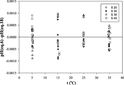
The results of pH comparisons in tris buffers over a wide range of S and T—pH(tris) from eq 6 vs. pH(mCP) from eq 18—are summarized in Figure 3. The average absolute residual is 0.0004, and the maximum residual is 0.0011. Consequently, we regard the overall uncertainty of our calibration technique relative to tris buffer standards as ±0.001 or better.
Figure 3.
Residuals for pH (expected pH (eq 6) minus measured pH (eq 18)) determined in synthetic tris seawater buffer.
Although e3/e2 is significantly dependent on salinity, the dependence is sufficiently slight that an assumption of independence has only small consequences for pH calculations. For samples of S = 20 or S = 40, assuming that e3/e2 is independent of salinity (i.e., that e3/e2 = 0.05626, as at S = 30) yields deviations from the salinity-dependent calculation (eq 18) of about 0.0002 pH units at pH 7.4 and 0.0015 units at pH 8.2.
A high-performance spectrophotometer was used in this work, but other properly calibrated spectrophotometers (e.g., Agilent or Ocean Optics) should provide pH measurements of comparable quality. To ensure that this expectation is met, we recommend (a) the use of NIST absorbance standards to ensure spectrophotometer absorbance linearity at the wavelengths used in the pH analyses and (b) adherence to the measurement procedures outlined in SOP 6b of Dickson et al.(13) Investigators can check their coding of the pH algorithm (eq 18) by comparing their test calculations with the pHT(mCP) results given for a range of S, T, and R values in Table S4 (Supporting Information).
Comparison with Thymol Blue
HPLC chromatographs of the Kodak thymol blue used by Zhang and Byrne(10) showed no impurity peaks that extended above 400 nm. Because seawater pH characterizations based on this indicator use RTB = 596A/435A, it is unlikely that impurities in the Zhang and Byrne(10) thymol blue impacted their indicator characterizations. As such, it appears that the pH measurement algorithm of Zhang and Byrne(10) is appropriate for pure thymol blue.
Paired pH results for Gulf of Mexico surface seawater (S = 35.9, T = 25.0 °C), comparing purified mCP and the thymol blue used by Zhang and Byrne(10) are given below:
pHT(mCP) = 8.0254 ± 0.0002
pHT(TB) = 8.0252 ± 0.0007.
Each average and standard deviation is based on five measurements. Agreement between the two indicators is better than the typical at-sea precision of pH measurements made with mCP. It should be noted, however, that assessments of thymol blue from batches other than that used by Zhang and Byrne(10) exhibited pH differences as large as 0.01 (not shown in this work). Therefore, indicator-purification procedures similar to those described in the Methods section should be used to ensure the accuracy of pH measurements obtained with thymol blue.
Internal Consistency
Total dissolved inorganic carbon, pH, and total alkalinity were measured in a sample of Gulf of Mexico surface seawater. AT was also calculated from CT and pHT(mCP) measurements using the dissociation constants of Mehrbach et al.(16) as refitted by Dickson and Millero.(17) Measured AT and calculated AT are in excellent agreement:
CT(measured) = 2096.6 ± 0.2 μmol/kg
pHT(measured) = 8.0226 ± 0.0002
AT(measured) = 2400.4 ± 1.3 μmol/kg
AT(calculated) = 2398.6 ± 1.0 μmol/kg.
These results, each based on five sets of measurements, suggest that the algorithm for calculation of pHT (eq 18) provides pH measurements that can be used to reliably link other measured carbon system parameters (AT, CT, and CO2 fugacity).
Correction of pH Measurements Made with Unpurified Indicator
Yao et al.(8) recommended that unpurified indicators used for pH measurements be archived so that comparisons and corrections could eventually be made when purified indicators come available. They also illustrated an empirical procedure for generating batch-specific correction algorithms. To correct older pH measurements obtained using unpurified indicator:
-
(1)
Use the archived unpurified indicator and purified indicator to make paired pH measurements in seawater over a range of pH values (e.g., 7.2–8.2). To calculate apparent pH obtained with the unpurified indicator (pHu), use the pH algorithm originally employed to generate the older data set. To calculate the “true” pH obtained with the purified mCP (pHp), use eq 18.
-
(2)
Plot the measured pH differences (ΔpH = pHp – pHu) as a function of pHu (see Figure 2 of Yao et al.(8)). Figure 2a indicates that pH measurements made with unpurified indicator could have errors as large as 0.02 pH units relative to those made with purified indicator. The greatest differences are likely to be seen at higher pH values.
-
(3)
Fit an equation to the curve that describes the offset between the two measurements (see eq 5 of Yao et al.(8)). Use this equation to correct the older pH measurements to a scale consistent with the use of purified mCP and eq 18: pHp = pHu + ΔpH.
Summary and Future Work
Equation 18 extends the single-temperature characterizations of Clayton and Byrne(3) to cover a wide range of temperature and salinity (278.15 K ≤ T ≤ 308.15 and 20 ≤ S ≤ 40). Use of purified mCP is required to facilitate direct comparisons of cross-batch pH measurements—i.e., measurements by independent investigators and measurements by a single investigator through time. Future work describing the influence of pressure on pH measurements with mCP, similar to that of Hopkins et al.(18) for thymol blue, will enable in situ pH measurements with mCP throughout the oceanic water column.
Acknowledgments
This work was supported by NSF Award OCE-0727082. Thanks are given to Eckerd College student Tianshui Wang, who worked on aspects of this project as a summer intern in 2010. The insightful and helpful comments of the editor and three anonymous reviewers are greatly appreciated.
Supporting Information Available
Data tables and figures showing mCP properties as a function of temperature and salinity; data for the comparison (residuals) shown in Figure 3; pHT(mCP) values that investigators can use to check their coding of eq 18. This information is available free of charge via the Internet at http://pubs.acs.org.
Supplementary Material
References
- Robert-Baldo G.; Morris M. J.; Byrne R. H. Spectrophotometric determination of seawater pH using phenol red. Anal. Chem. 1985, 57, 2564–2567. [Google Scholar]
- Byrne R. H.; Breland J. A. High precision multiwavelength pH determinations in seawater using cresol red. Deep-Sea Res., Part A 1989, 36, 803–810. [Google Scholar]
- Clayton T. D.; Byrne R. H. Spectrophotometric seawater pH measurements: total hydrogen ion concentration scale calibration of m-cresol purple and at-sea results. Deep-Sea Res., Part A 1993, 40, 2115–2129. [Google Scholar]
- Clayton T. D.; Byrne R. H.; Breland J. A.; Feely R. A.; Millero F. J.; Campbell D. J.; Murphy P. P.; Roberts M. L. The role of pH measurements in modern oceanic CO2-system characterizations: precision and thermodynamic consistency. Deep-Sea Res. 1993, 42, 411–429. [Google Scholar]
- McElligott S.; Byrne R. H.; Lee K.; Wanninkhof R.; Millero F. J.; Feely R. A. Discrete water column measurements of CO2 fugacity and pHT in seawater: a comparison of direct measurements and thermodynamic calculations. Mar. Chem. 1998, 60, 63–73. [Google Scholar]
- Byrne R. H.; McElligott S.; Feely R. A.; Millero F. J. The role of pHT measurements in marine CO2-system characterizations. Deep-Sea Res. 1999, 46, 1985–1997. [Google Scholar]
- Byrne R. H. Standardization of standard buffers by visible spectrometry. Anal. Chem. 1987, 59, 1479–1481. [Google Scholar]
- Yao W.; Liu X.; Byrne R. H. Impurities in indicators used for spectrophotometric seawater pH measurements: assessment and remedies. Mar. Chem. 2007, 107 (2), 167–172. [Google Scholar]
- Yao W.; Byrne R. H. Spectrophotometric determination of freshwater pH using bromocresol purple and phenol red. Environ. Sci. Technol. 2001, 35, 1197–1201. [DOI] [PubMed] [Google Scholar]
- Zhang H.; Byrne R. H. Spectrophotometric pH measurements of surface seawater at in situ conditions: absorbance and protonation behavior of thymol blue. Mar. Chem. 1996, 52, 17–25. [Google Scholar]
- Byrne R. H.; Yao W. Procedures for measurement of carbonate ion concentrations in seawater by direct spectrophotometric observations of Pb(II) complexation. Mar. Chem. 2008, 112 (1–2), 128–135. [Google Scholar]
- DelValls T. A.; Dickson A. G. The pH of buffers based on 2-amino-2-hydroxymethyl-1,3-propanediol (tris) in synthetic sea water. Deep-Sea Res., Part I 1998, 45 (9), 1541–1554. [Google Scholar]
- Dickson A. G., Sabine C. L., Christian J. R., Eds. Guide to Best Practices for Ocean CO2 Measurements; PICES Special Publication 3, 2007. [Google Scholar]
- Dickson A. G. Standard potential of the (AgCl(s) + 1/2H2 (g) = Ag(s) + HCl(aq)) cell and the dissociation constant of bisulfate ion in synthetic sea water from 273.15 to 318.15 K. J. Chem. Thermodyn. 1990, 22, 113–127. [Google Scholar]
- Yao W.; Byrne R. H. Simplified seawater alkalinity analysis: use of linear array spectrometers. Deep-Sea Res. 1998, 45, 1383–1392. [Google Scholar]
- Mehrbach C.; Culberson C. H.; Hawley J. E.; Pytkowicz R. M. Measurement of the apparent dissociation constants of carbonic acid in seawater at atmospheric pressure. Limnol. Oceanogr. 1973, 18, 897–907. [Google Scholar]
- Dickson A. G.; Millero F. J. A comparison of the equilibrium constants for the dissociation of carbonic acid in seawater media. Deep-Sea Res. 1987, 34A, 1733–1743. [Google Scholar]
- Hopkins A. E.; Sell K. S.; Byrne R. H. In situ spectrophotometric pH measurements: the effect of pressure on thymol blue protonation and absorbance characteristics. Mar. Chem. 2000, 71 (1–2), 103–109. [Google Scholar]
Associated Data
This section collects any data citations, data availability statements, or supplementary materials included in this article.


