

Abstract
The larval stage of the pork tapeworm (Taenia solium) infects the human nervous system, causing neurocysticercosis. This disease is one of the main causes of epileptic seizures in many less developed countries and is also increasingly seen in more developed countries because of immigration from endemic areas. Little information is available on the natural evolution of taeniasis or cysticercosis. Available therapeutic measures include steroids, treatments for symptoms, surgery, and, more controversially, antiparasitic drugs to kill brain parasites. Efforts to control and eliminate this disease are underway through antiparasitic treatment of endemic populations, development of pig vaccines, and other measures.
Taenia solium infection and the resulting disease neurocysticercosis are endemic in less developed countries where pigs are raised as a food source.1,2 Neurocysticercosis is common throughout Latin America, most of Asia, sub-Saharan Africa, and parts of Oceania, and is the greatest cause of acquired epilepsy worldwide.3 It is now increasingly diagnosed in more developed countries owing to immigration of tapeworm carriers from endemic zones.2,4
T solium has a complex two-host life cycle. Human beings are the only definitive host and harbour the adult tapeworm (taeniasis), whereas both people and pigs can act as intermediate hosts and harbour the larvae or cysticerci (figure 1).
Figure 1.
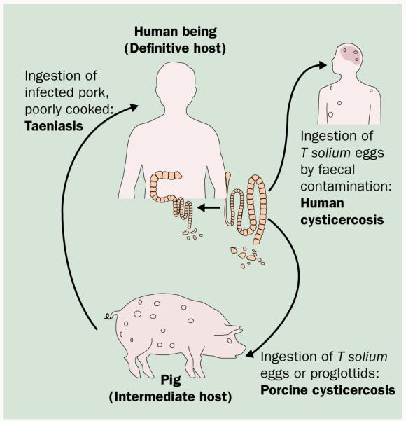
Life cycle of Taenia solium
Taeniasis
Taeniasis occurs only in the human host, after ingestion of undercooked pork infected with cysticerci. Although cysticercosis has been known for ages, its relation to the adult tapeworm was not clear until it was shown by Kuchenmaister in 1855; he fed condemned prisoners with cysticercosis-infected pork and recovered young tapeworms at autopsy.5 The larvae evaginate in the small intestine; the head (scolex) attaches to the mucosa and begins forming segments (proglottids). T solium has a scolex with four suckers and a double crown of hooks, a narrow neck, and a large strobila measuring 2–4 m and consisting of several hundred proglottids (figure 2). About 2 months after infection, gravid proglottids begin to detach from the distal end and are excreted in the faeces; each segment contains 50–60×103 fertile eggs.6
Figure 2. Scolex (A) and strobila (B) of adult tapeworm.

The morphology of the proglottids changes as they mature and become gravid.
The worm attaches strongly to the mucosa of the upper small intestine by means of its suckers and hooks. The adult tapeworm causes only mild inflammation at the implantation site,7 without substantial damage to the intestine.4 Taeniasis is characterised by mild symptoms or none at all.4 Abdominal pain, distension, diarrhoea, and nausea have been attributed to tapeworm infestation, but there are no data from controlled experiments that demonstrate any association, and most patients seem to be free of symptoms.4,6 In community settings many, if not most, carriers of T solium will neither look for medical care nor notice the tapeworm segments in their stools. Conversely, most patients infected with Taenia saginata notice passage of proglottids, which are motile, more numerous, and larger than those of T solium.8 Identification of T solium infections is important because of the risk of cysticercosis in the carrier or the immediate environment.
The frequency of autoinfection in individuals with taeniasis is not known. Dixon and Lipscomb9 noted that nearly 25% of patients with neurocysticercosis either had harboured or were harbouring a tapeworm. Up to 15% of patients harbour a tapeworm at the time of diagnosis of neurocysticercosis,10 and the proportion with tapeworms is directly related to the number of cerebral parasites,10,11 which strongly suggests autoinfection. In other cases the tapeworm carrier can be found in the patient’s household.12 Despite the poor sensitivity of stool examinations, parasitological screening of the patient and household members is recommended so that sources of infection can be detected and eliminated.
The lifespan of the adult T solium is also unknown. Parasitology textbooks and reviews cite 20–25 years, on the basis of anecdotal cases.8,13 Age-specific data from later studies suggest a much shorter lifespan, probably less than 5 years.14-16
Diagnosis of taeniasis
Two problems hamper the diagnosis of infection with T solium: the poor sensitivity of stool microscopy, and the morphological similarity between the eggs of T solium and T saginata. On the rare occasions when the worm scolex is found, the presence of the double crown of hooks (present only in T solium) provides definite species identification. The morphology of the genitalia in mature proglottids or that of the gravid uterus in the distal proglottids can also enable the identification of species.4 Haematoxylin-eosin staining of histological sections of proglottids can help.17 Mature, gravid proglottids are rarely available, and morphological diagnosis of species on the basis of proglottid materials is technically difficult. In our experience, cleansing the intestine with a purge immediately before treatment improves the recovery of parasite material including the scolex, allowing species identification.
Visualisation of taenia eggs by microscopy was the only diagnostic method available until the early 1990s. Perianal scraping with adhesive tape (Graham’s test) is highly sensitive for T saginata but not for T solium.18,19 The best available diagnostic assay for intestinal taeniasis is a coproantigen detection ELISA, which detects taenia-specific molecules in faecal samples, demonstrating current tapeworm infection.20 It has sensitivity of about 95% and specificity greater than 99% and is an effective tool for epidemiological studies.15,20 Coproantigen detection assays confirmed that microscopy was poorly sensitive, missing 60–70% of cases. Diverse DNA-based assays, mainly to discriminate T solium from T saginata infections,17,21 and a serological assay for specific identification of tapeworm carriers22 have been described lately.
Treatment of taeniasis
The two available drugs are niclosamide and praziquantel. Niclosamide is the drug of choice because it is not absorbed from the intestinal lumen.23 With praziquantel there is a small risk that asymptomatic viable brain cysts are affected by the drug in serum, causing neurological symptoms (headache, seizures).24 The usual dose is 2 g orally in a single dose for niclosamide, and 5–10 mg/kg orally in a sole dose for praziquantel. Both drugs have very limited marketing and are difficult to find.
Human cysticercosis
Cysticercosis is infection with the larval stage of the parasite. Human beings acquire cysticercosis through faecal-oral contamination with T solium eggs from tapeworm carriers.1,4,25 Thus, vegetarians and other people who do not eat pork can acquire cysticercosis.26 Water, wind, flies, and other indirect means of infection play little part in transmission.27 Internal autoinfection by regurgitation of proglottids into the stomach in taeniasis has been proposed but not proven.1,27
Clinical presentation
The invasive oncospheres (embryos) in the eggs are liberated by the action of gastric acid and intestinal fluids and actively cross the bowel wall, enter the bloodstream, and are carried to the muscles and other tissues.6,28 At small terminal vessels, they establish and encyst as cysticerci (figure 3), reaching their definitive size of about 1 cm in 2–3 months.29 Clinical manifestations depend on the affected organ; neurocysticercosis and ophthalmic cysticercosis are associated with substantial morbidity.
Figure 3. Cysticerci.

(A): as seen in infected pork. (B): excised into a Petri dish. The white dot in each cyst corresponds to the scolex.
Extraneural cysticercosis
Outside the central nervous system, cysticercosis causes no major symptoms. Subcutaneous cysticercosis presents as small, movable, painless nodules that are most commonly noticed in the arms or chest. After a few months or even years, the nodules become swollen, tender, and inflamed, and then they gradually disappear.30 Subcutaneous cysticercosis is rare in Latin America but very common in Asia and Africa. Biopsy or fine-needle cytology31 of a subcutaneous nodule helps to confirm the diagnosis of cysticercosis infection. Muscular cysticercosis is a casual finding, appearing as dot-shaped or ellipsoidal calcifications following the muscle bundles in the thighs or arms, when radiography is done for an unrelated reason. In a classic series, 75% of patients with neurocysticercosis showed muscular calcifications after several years of radiographic follow-up,9 but this study has not been replicated with more sensitive CT scanning. In rare cases, very massive parasite burdens enlarge the patient’s limbs (muscular pseudohypertrophy; figure 4). The heart is another occasional location of cysticerci, infected in about 5% of patients.32 As far as is known, cardiac cysticercosis is asymptomatic.
Figure 4. Cysticercosis outside the nervous system.

(A): ocular cyst floating in the vitreous humour. (B): calcified cysts on CT (three-dimensional filtered reconstruction). (C) and (D): massive infection causing muscular pseudohypertrophia; thousands of cysts following the muscle planes can be seen.
Ophthalmic cysticercosis
Although ophthalmic cysticercosis is much less common than neurocysticercosis (occurring in 1–3% of all infections), T solium is the most common intraorbital parasite.33 Intraocular cysts are most frequently found floating freely in the vitreous humour or in the subretinal space. Visual disturbance is related to the degree of damage to retinal tissue or the development of chronic uveitis.34 Cysticercosis may also present in the anterior chamber or affect the conjunctiva or extraocular muscles. Some individuals with massive infections have cysts in the retro-ocular space, affecting the optic nerve or causing proptosis.35,36 Orbital ultrasonography is a valuable and harmless diagnostic method.33,34 Visual loss can also be related to cerebral cysticercosis causing papilloedema, hydrocephalus, or chiasm compression.37
Neurocysticercosis
The parasite commonly infects the central nervous system, causing neurocysticercosis, a pleiomorphic clinical disorder. After entering the central nervous system, cysticerci are viable and elicit few inflammatory changes in the surrounding tissues. Cysticerci may remain for a long time in this stage, protected by the blood-brain barrier6,38,39 and active immune-evasion mechanisms by the cysticerci.6 After a variable and unknown time (estimated to be several years on the basis of classic studies in English soldiers returning from India),9,30,40,41 the parasite degenerates with associated immune-mediated inflammation. Cysticerci cause symptoms because of mass effect or by blocking the circulation of cerebrospinal fluid, but most symptoms in neurocysticercosis are the direct result of the inflammatory process that accompanies cyst degeneration. Clinical manifestations are thus related to individual differences in the number, size, and topography of lesions and in the severity of the host’s immune response to the parasites.28 Symptoms and signs are varied and non-specific.
Epileptic seizures are the commonest presentation of neurocysticercosis and generally represent the primary or sole manifestation of the disease. Seizures occur in 50–80% of patients with parenchymal brain cysts or calcifications but are less common in other forms of the disease.3,42,43 In endemic regions, recent onset of seizures in otherwise healthy teenage, young adult, or middle-aged individuals strongly suggests neurocysticercosis.44 Most of these patients are normal on neurological examination. A series including mostly patients with mild forms of infection45 showed that about 50% of patients with neurocysticercosis presenting a seizure have further seizures (epilepsy). The proportion of relapses in patients with more severe forms of disease is expected to be higher.
Neurocysticercosis also presents with intracranial hypertension, hydrocephalus, or both in 20–30% of cases (the proportion varies according to the origin of the cases, higher in neurosurgical series). This syndrome is related to the location of parasites in the cerebral ventricles or basal cisterns, blocking the circulation of cerebrospinal fluid, and is caused by several different mechanisms—the presence of the parasite itself, ependymal inflammation, or residual fibrosis.46,47
Occasionally, a cyst grows larger than the usual 1–2 cm and acts in the same way as a tumoral mass (giant cyst).48,49 These giant cysts compress adjacent cerebral structures, causing localised deficits and intracranial hypertension. Cyst growth occurs most frequently in the subarachnoid space around the sylvian fissure or in the basal cisterns. Motor deficits can also arise because of oedema secondary to cyst degeneration, or as a result of stroke complicating neurocysticercosis. Stroke is more frequent in subarachnoid neurocysticercosis than in intraparenchymal infections. It consists mostly of deep lacunar infarcts resulting from endarteritis of small penetrating arteries, whereas occlusion of large vessels is rare.50
In children and teenagers, an acute encephalitic presentation can happen; it is more likely in female than male patients.51 This disorder is due to intense inflammatory reaction of the host to a massive parasite infection, with pronounced intracranial hypertension. Massive non-encephalitic forms also occur, presenting with occasional seizures, episodic intracranial hypertension, or mild cognitive dysfunction.
Compromise of the spine occurs in about 1% of cases of neurocysticercosis, presenting with compressive manifestations. Cysts locate mostly in the subarachnoid space, although intramedullary cysts are found rarely.52
There seems to be geographical variation in clinical manifestations. In some Asian countries, almost all patients with neurocysticercosis present with a single enhancing brain lesion, and a few have very massive infections with hundreds of cysts. Conversely, in Latin America, the most frequent presentation of neurocysticercosis is a few viable cysts without signs of inflammation.
Pathology
Cysts are uniformly rounded or oval vesicles, varying in size from a few millimetres to 1–2 cm (in rare cases a growing cyst reaches several centimetres in diameter). The most common location is in the cerebral hemispheres, mainly at the junction of grey and white matter. Cysts can be found in the cerebellum, ventricles, brainstem, subarachnoid space, basal cisterns, and spine. The number ranges from one to more than a thousand. On gross examination, no variations are observed in the nervous parenchyma surrounding viable cysts.32,53
Vesicles vary in contents according to their evolutionary stage. Viable cysts have a translucent membrane through which the scolex is visible as a small 2–3 mm nodule. When the cyst starts to degenerate, the vesicular fluid becomes opaque and dense, and the edges of the cyst become irregular and shrink. Later, calcification starts in the cephalic portion and progresses to the vesicular wall, to leave a round, whitish, calcified nodule. In rare cases, all the evolutionary phases can be seen in the same individual.54
Intraventricular cysticercosis is generally single and free inside the cavities. Infrequently, the cyst moves from one ventricular cavity to another.55,56 Classically, cysts are described as locating more frequently in the lower cavities: most frequently in the fourth ventricle, less commonly in the third, and least frequently in the lateral ventricles.57 Intraventricular parasites can block circulation of cerebrospinal fluid or produce inflammation of the ependyma and choroid plexus.
The racemose form is a large translucent vesicle, frequently lobulated, without a scolex, which develops in the basis of the brain or in the ventricles.32,53,58 Sometimes several small vesicles surround a pedicle attached to the pia mater like a bunch of grapes (figure 5), hence the name. As vesicles grow in number and adherences, they take over the basal cisterns, giving an infiltrative appearance. The racemose form is associated with high mortality because of its association with obstructive hydrocephalus.58
Figure 5. Macroscopic pathology.

A: viable cyst. B: racemose cysticercosis in the base of the brain.
Diagnosis
Serology
A finding of eosinophils in the cerebrospinal fluid suggests the diagnosis of neurocysticercosis. Other routine laboratory tests scarcely contribute, and thus the diagnosis rests on serology and neuroimaging. Although both are highly sensitive and specific, perfect correlation should not be expected since their approaches differ.
Until recently, serological diagnosis of cysticercosis was less than satisfactory. The most commonly used ELISA cross-reacts with Hymenolepis nana and Echinococcus granulosus, which are common cestode infections.59,60 For this reason, it is much more useful when applied to samples of cerebrospinal fluid than of serum, with the drawbacks of the invasiveness and pain associated with the lumbar puncture. The enzyme-linked immunoblot assay was originally reported to have sensitivity of 98% and specificity of 100%,61,62 and it performs much better than the ELISA in clinical settings.63,64 Its sensitivity in patients with only one degenerating cysticercus or calcified lesions only is much lower.65 In hospital settings, antibody can be seen to persist for a long time after the death of parasites,66 and thus a positive antibody test in patients with calcified cysticercosis only is a common finding and should not be interpreted as indicating the presence of live parasites. This assay requires more reagents, advanced equipment, and highly trained personnel for antigen purification and assay performance than does the ELISA. When available, however, immunoblot is an excellent instrument for diagnosis and management.44,63,64 Since the physician needs to know the number, location, size, and stage of intracranial parasites, serology mostly has a screening or confirmatory role and should be used in conjunction with neuroimaging. Antigen detection assays can detect live parasites and thus direct therapeutic decisions or monitoring.67,68 Some, based on monoclonal antibodies, perform well in comparison with other available tests on cerebrospinal-fluid samples.68-70 There is limited evidence on sensitivity or specificity with serum samples.
Neuroimaging
Because the important features are the number, size, and location of lesions, radiography has always been crucial for diagnosis of neurocysticercosis.40 Initially, skull and soft-tissue radiographs showed calcifications, and arteriography and pneumoencephalography (both dangerous techniques no longer used for this purpose) showed lesions causing mass effect and hydrocephalus. Lately, computer-based imaging procedures have enabled visualisation of the brain and other intracranial soft structures. Currently used techniques include CT and MRI.
CT has been claimed to have sensitivity and specificity of over 95% for the diagnosis of neurocysticercosis,28 although CT images are rarely pathognomonic for this disease.71 The availability of antiparasitic treatments and serial CT studies has allowed the description of imaging changes throughout the involution process. Cysts appear as hypodense images, with well-defined edges, and commonly a hyperdense nodule inside the cyst shows the parasite’s scolex.28 Parasite degeneration initially appears as contrast enhancement of the edges of the cyst and perilesional oedema, resulting from the inflammatory reaction in the surrounding tissue.72 In later stages, the cyst becomes isodense to the parenchyma but appears after the administration of contrast, enhancing as a nodule or a ring. This image, called enhancing lesion or cysticercus granuloma is barely visible in precontrast images, and corresponds to the late degenerative phase (no longer viable). The differential diagnosis of this entity in endemic regions is extremely difficult because of the endemicity of tuberculosis and neurocysticercosis (panel 1). Patients with a sole enhancing lesion in the brain due to neurocysticercosis are seronegative because the parasite is already dead or because a sole parasite does not elicit a strong antibody response,1 and many patients with tuberculoma do not have detectable tuberculosis in the lungs or in any other location to confirm the diagnosis.
After this phase, the cysticercus becomes a calcified scar, which normally does not enhance with the contrast substance. However, contrast enhancement and perilesional oedema can be seen around old, calcified lesions and are associated with symptom relapses.57,73-76 The sensitivity of CT is much lower for ventricular or cisternal forms of the disease. Intraventricular cysticercosis presents with obstructive hydrocephalus, and only indirect signs such as cavity enlargement or deformation are seen on CT.
MRI is the most accurate technique to assess the degree of infection, the location, and the evolutionary stage of the parasites (figure 6). It visualises well the perilesional oedema and the degenerative changes of the parasite, as well as small cysts or those located in the ventricles, brainstem, cerebellum, eye, and basal racemose vesicles. CT, however, is more sensitive for the detection of calcifications.77 The main disadvantages of MRI are its high cost and scarce availability.
Figure 6. Neuroimaging.

MRI of viable (A) and degenerating (B) cysts, and CT of calcified cysticerci (C).
A set of objective diagnostic criteria has been proposed.78 Absolute criteria include histological demonstration of the parasite, cystic lesions with scolex on neuroimaging, or direct visualisation of ocular cysts; major criteria include suggestive lesions on neuroimaging, positive serum immunoblot, resolution of cysts after antiparasitic therapy, or spontaneous resolution of enhancing lesions; minor criteria are compatible lesions on neuroimaging, compatible clinical manifestations, positive cerebrospinal-fluid ELISA, or cysticercosis outside the central nervous system; and epidemiological criteria include household contact, living in an endemic area, or travel to endemic areas. Combinations of these criteria provide different degrees of diagnostic certainty.
Prognosis and natural evolution
Panel 2 shows research needs in T solium infection. There are few data on the natural evolution of neurocysticercosis, and those available are from historical controls in open treatment trials. As far as is known, the evolution of intraparenchymal neurocysticercosis follows a favourable course with degeneration of parasites and formation of residual calcified scars.79 When the parasites locate in the subarachnoid space or the ventricles, the morbidity and mortality are much higher because of parasite growth, intracranial hypertension, arachnoiditis, blockage of cerebrospinal-fluid pathways, hydrocephalus, and other complications.49,80,81
Treatment
Cysticercosis outside the nervous system is a benign disorder and does not merit specific treatment. Neurocysticercosis, however, is associated with substantial morbidity and mortality. Until 1978, the only treatments available were surgery for cyst excision or ventricular shunts or steroids to decrease inflammation. Praziquantel, first used in porcine cysticercosis, was the first effective specific antiparasitic drug.82-84 Later, albendazole added a cheaper and more effective alternative.85,86 Transient worsening of neurological symptoms can be expected during antiparasitic therapy, secondary to the perilesional inflammatory reaction. In most cases, symptoms are limited to mild to moderate headache and are easily controlled with steroids. Although antiparasitic agents in neurocysticercosis have been accepted by most of the medical community, some believe that, since the parasite starts to cause symptoms at the time of its death, the use of parasiticidal drugs leads to an unnecessary increase in the inflammatory process.79,87 The issue led to heated discussion.88-90 The main argument is whether antiparasitic treatment or natural involution of a cyst will lead to reduced scarring and thus improved prognosis in terms of epilepsy evolution.
Many reports have been published on the use of antiparasitic treatment in neurocysticercosis.82-86,90-94 However, non-treatment comparison groups were included in only four studies, three of which included patients with enhancing lesions (parasites that have already degenerated). A summary of controlled studies can be found in the Cochrane Library,95 although no overall conclusion can be reached because the types of disease and the findings differ substantially. Two double-blind, placebo-controlled trials assessed patients with a sole enhancing lesion. Padma and colleagues93 found similar resolution rates on 3-month follow-up CT in groups assigned albendazole or placebo.93 By contrast, Baranwal and co-workers91,92 found better resolution on imaging and fewer seizures in the albendazole group than in controls during follow-up. A third double-blind, placebo-controlled trial in India compared the evolution of parenchymal neurocysticercosis lesions (including cysts and enhancing lesions) on CT at 3 months after treatment94 and showed similar resolution rates. The only controlled trial in non-enhancing intraparenchymal cysts has been an open-label study comparing radiological and clinical (seizure relapses) evolution between patients treated with albendazole, praziquantel, or no antiparasitic therapy.90 All three groups received prednisone. The study found no differences in the proportions of patients free of cysts at 6 months and 1 year after treatment, or in the relative reduction in numbers of cysts and no difference in the proportion of patients free of seizures during the entire follow-up.
Antiparasitic treatment may be unnecessary or even contraindicated in some cases. Without doubt however, there are subgroups of patients with neurocysticercosis who not only benefit from antiparasitic therapy but also might undergo progression of disease and even die if not treated, such as those with giant cysts or subarachnoid cysticercosis.48,49 Similarly, complications in the treatment of neurocysticercosis with antiparasitic drugs can be severe, but they are very rare. Until a prospective, randomised study has been completed, a simple summary is that antiparasitic agents are effective to destroy cysticerci in the human brain, at the cost of acute inflammation. Whether this is the most beneficial option for patients with intraparenchymal cysticercosis in the long term is not yet clear. The currently accepted schemes are either 8 days of albendazole treatment (15 mg/kg daily) with simultaneous administration of steroids, or 15 days of praziquantel (50 mg/kg daily).96 Shorter schemes of 1 day of praziquantel or 3 days of albendazole seem effective for patients with only one lesion, but not for those with many cysts.97,98
A panel of experts analysed the current consensus and disagreements in the management of neurocysticercosis.99 Their main conclusions were that therapeutic decisions should be adapted to the individual and should be based on the number, location, and viability of the parasites within the nervous system; that growing cysticerci should be actively managed by either antiparasitic drugs or surgical excision; that the management of intracranial hypertension secondary to neurocysticercosis should take a high priority; and that adequate management of seizures should be ensured. Specifically, they agreed on the management of patients with moderate infections and viable cysts (antiparasitic treatment, with steroids); calcified cysticercosis (no antiparasitic treatment); ventricular cysticercosis (neuroendoscopic removal, when available); subarachnoid cysts, including giant cysts or racemose cysticercosis, and chronic meningitis (antiparasitic treatment with steroids, ventricular shunt if there is hydrocephalus); cysticercotic encephalitis (no antiparasitic treatment, high-dose steroids, and osmotic diuretics); hydrocephalus with no visible cysts on neuroimaging (ventricular shunt, no antiparasitic treatment); spinal cysticercosis, intramedullary or extramedullary (primarily surgical); or ophthalmic cysticercosis (surgical resection of cysts). There were disagreements about the use of antiparasitic drugs (considered unnecessary or of high risk by some experts) in patients with only one or a few viable cysts, patients with massive infections with viable cysts, or patients with many degenerating cysts.99
Treatment of symptoms has an important role in the management of neurocysticercosis. Seizures generally respond well to first-line antiepileptic drugs,43,100 but complete withdrawal of antiepileptic therapy is difficult to achieve.100 Steroids, dexamethasone or prednisone, are helpful in oedema or intracranial hypertension.
Porcine cysticercosis
In places with inadequate disposal of human faeces, pigs ingest stools containing T solium eggs. Cysticerci lodge anywhere in the body of the pig, most commonly in the muscle and subcutaneous fat. Pigs with cysts in the brain are a subset of those with more cysts in the carcass (Cysticercosis Working Group in Peru 2002, unpublished data). Although some pigs have massive infections, porcine cysticercosis is rarely associated with symptoms of any kind. Most pigs are killed before the age of 9 months, which is too short a time for the cysts to reach the degenerative stage that is associated with symptoms in human beings.
An inexpensive treatment for porcine cysticercosis might increase the value of pigs and block transmission of infection. A veterinary benzimidazole, oxfendazole, is more than 95% effective in killing the cysts in the pig when given in a single dose of 30 mg/kg.101-103 This drug has not yet been tested in human beings.
Immunity to the oncosphere stage plays a central part in regulation of transmission of taeniid cestodes, and oncosphere-based vaccines are highly protective for T ovis, Echinococcus granulosus, and T saginata.104 Partial protection has been achieved by vaccination of healthy pigs with T solium oncospheral antigens.105-107
Epidemiology
Cysticercosis affects thousands of individuals in less developed countries1,3 and in more developed countries with a high rate of immigration from endemic areas.2,4 In endemic countries taeniasis/cysticercosis is extremely common, and neurologically symptomatic individuals, although many, represent only the tip of the iceberg. In most endemic villages more than 10% of the general population are seropositive, and the proportion can reach 25%. In population-based studies, 10–18% of asymptomatic individuals have CT features that suggest neurocysticercosis, mainly brain calcifications in seronegative individuals.108-111
Transient seropositive reactions112 and common cases of asymptomatic calcified neurocysticercosis108-111 are consistent with a threshold under which parasite destruction occurs, potentially determined by the number of parasites, the immune status of the host, the age at infection, or any combination of these factors. Animal models suggest that the host’s immune response can resolve mild infections at early stages.113 If this resolution occurs in human neurocysticercosis, it could explain the frequency of degenerating cysts clustered in younger ages, the relation to infections with a single parasite (90% of cases with only degenerating cysts), and the frequency in India, where, because of vegetarian habits, direct exposure to tapeworm carriers is less frequent.114 Patients with viable cysts only would represent cases in which the parasites survive and establish.
Few data are available on the epidemiology of adult tapeworm infections. Up to 6% of the population in endemic villages may harbour adult T solium tapeworms at a given time.15,115 The few existing community studies in taeniasis, done in Guatemala14,15,116 and Peru,16 show that tapeworm infections are more common in younger than older individuals, especially in the female population. Consistently, cases of intestinal taeniasis cluster in houses,14 probably as a result of similar food consumption habits. Clustering of infected pigs or seropositive human beings around tapeworm carriers has also been shown.14,16
Domestic pig raising, taeniasis, and human cysticercosis are intimately inter-related. Many farmers in less developed countries cannot afford a cow but can buy a few pigs. These animals are cheap and easily marketable, becoming a convenient source of meat or money. Even better, no investment is needed to feed them, since free pigs roam around and eat pasture and garbage. In endemic areas, few pigs go through the formal slaughterhouse system; moreover, peasants used to screen their pigs for cysticercosis by examining their tongues117 before taking them to the slaughterhouse.118 Thus, abattoir prevalence statistics are probably a gross underestimation of the real rates.118 Serology has proven useful to detect infected animals for epidemiological purposes. The rates of porcine infection vary, but in endemic regions 30–60% of pigs are seropositive.117,119 Studies in pigs have provided important data: the high correlation between human and porcine prevalence, the use of sentinel pigs to monitor transmission, and the potential use of oxfendazole for the control.
Cysticercosis and epilepsy
Epilepsy is more prevalent in less developed than in more developed countries.120 Rates such as 17 per 1000 in Ecuador,121 21 per 1000 in Colombia,122 and 37 per 1000 in Nigeria123 are three to six times higher than those in more developed countries.3 Serological studies in India,42 South Africa,124 Mexico,108 and Peru125 suggest that the higher rates of epilepsy are due mainly to neurocysticercosis. In Mexico126 and Peru,44 12% of neurological hospital beds are occupied by patients with neurocysticercosis. Similar surveys in neurological centres show neurocysticercosis in 30–50% of epileptic individuals, compared with less than 5% of individuals attending with other neurological symptoms.42,125,127 When correctly compared, seroprevalence is consistently much higher in neurological patients than in the relevant general populations. The risk of epilepsy in seropositive individuals is twice to three times that in seronegative controls.108,128 The risk increases further for specific subgroups (people with late-onset epilepsy, for example). There are no data on the risk of neurological symptoms developing in seropositive individuals in the community.
Cysticercosis in non-endemic countries
In the USA, neurocysticercosis is common in hospitals treating large Hispanic populations in areas such as Los Angeles, San Diego, New Mexico, and Houston.2,4 The number of cases in the USA (including people born in the country) is increasing, probably as a result of both improved diagnosis and increased immigration of individuals infected with T solium.4 A cluster of cysticercosis cases arose in an Orthodox Jewish community in Brooklyn, New York. The only risk to this population was their exposure to Latin-American domestic workers who were probably infected with T solium tapeworms.26 In a recent study in accident and emergency departments of patients who underwent neuroimaging because of a seizure, neurocysticercosis was found in 10% in Los Angeles and 6% in New Mexico.129 Cases of neurocysticercosis are now also diagnosed more frequently than before in Europe, Australia, and other non-endemic areas.130,131
Economic burden
Neurocysticercosis affects mainly older children and adults, so the estimated economic consequences due to chronic disability are heavy. Flisser126 estimated a cost of US$15 million per year only for hospital admission of cases of newly diagnosed neurocysticercosis in Mexico. There are no figures available on the maintenance cost of antiepileptic-drug therapy in epilepsy due to neurocysticercosis, or other long-term costs. Patients with calcified cysticercosis can have seizures or other neurological morbidity persisting for years after they are deemed cured. T solium not only produces a severe disease1 but also causes widespread economic losses to the pig industry.132 In Mexico, for example, porcine cysticercosis caused loss of more than half the national investment in swine production,133 and the losses occasioned by the destruction of meat are estimated at $43 million per year.126
Control of taeniasis and cysticercosis
T solium is transmitted mainly in rural areas where pigs have access to untreated human sewage or faeces and infected pork is widely available. An estimated 400 000 people have symptomatic neurocysticercosis in Latin America alone.134 More developed countries eradicated cysticercosis by improving sanitation and controlling domestic pig-raising.135,136 Since little socioeconomic development in endemic areas is expected in the near future, intervention measures for control and eradication are urgently needed.
To date, no intervention has achieved sustained interruption of transmission. Abattoir inspection and confiscation is the only official diagnostic method in most endemic countries.12 In most of these areas, the majority of pigs are killed clandestinely in any case. Since only a few cuts are made in each carcass to preserve its commercial value, slaughterhouse inspection probably misses most mild infections. This system drives farmers to slaughter pigs illegally and does not stop the distribution of infected meat.118
Only the tapeworm carriers and the infected pigs are important in terms of transmission. Individuals with neurocysticercosis are a health concern; but unless they also carry an intestinal tapeworm, they do not pose a public-health risk. Rates of human taeniasis can be decreased either by detection and treatment of the tapeworm carriers, or by treatment of the whole population.12,115,137 Transmission can also be blocked if the sale and consumption of infected pork is prevented by slaughterhouse inspection.12 Health education alone has also been tested in Mexico, with promising but incomplete results. Knowledge about the disease improved substantially, but practices related positively or negatively to risk (hand-washing, defecating in fields, corralling of pigs) remained unchanged. In favour of health education is the long-term effect of acquired knowledge.138,139 Porcine infection can be addressed either by mass anthelmintic treatment101,102,140 or in the future, if an effective vaccine becomes available, by immunisation of the pig population.106,141,142 A high-cost vaccine would undoubtedly be an impediment for a vaccine strategy, since the target population is rural endemic villages. Any selected combination of interventions would need to ensure community cooperation to achieve sustainable control.
Search strategy.
This seminar is based on papers published in the past 15 years in English, Spanish, or Portuguese selected from MEDLINE by use of the PubMed system with keywords “cysticercosis”, “neurocysticercosis”, “Taenia solium”, “epilepsy”, and “seizures”. Older articles were selected from the private collections of the authors and the archives of the Cysticercosis Working Group in Peru.
Panel 1: Differential diagnosis of neurocysticercosis on neuroimaging.
Sole non-enhancing cystic lesion
Hydatid disease
Arachnoid cysts
Porencephaly
Cystic astrocytoma
Colloid cyst (third ventricle)
Several non-enhancing cystic lesions
Multiple metastases
Hydatid disease (rare)
Enhancing lesions
Tuberculosis
Mycosis
Toxoplasmosis
Abscess
Early glioma
Metastasis
Arteriovenous malformation
Calcifications
Tuberous sclerosis
Tuberculosis
Cytomegalovirus infection
Toxoplasmosis
Panel 2: Research needs in T solium infection.
Clinical
Improved knowledge of natural history
Concrete determination of benefits and specific indications of antiparasitic therapy in neurocysticercosis
Testing of new antiparasitic and anti-inflammatory agents
Immunological
Development of field-applicable, specific assays for human taeniasis
Development of field-applicable, specific assays for viable cysticercosis
Prevention and control
Systematic assessment of efficacy and effectiveness of all potential intervention measures (including pig vaccines)
Estimation of infection and disease burden attributable to neurocysticercosis
Estimation of economic losses due to porcine cysticercosis
Acknowledgments
Our work is supported by the National Institute of Allergy and Infectious Diseases, National Institutes of Health, Bethesda, MD, USA; the Office of Orphan Products Development, Food and Drug Administration, MD, USA, and the Wellcome Trust, London, UK. Figures 3b, 6a, and 6b were facilitated by the Department of Pathology of the Institute of Ciencias Neurologicas, Lima, Peru, and Hermes Escalante, ESCALABS, Trujillo, Peru. The sponsors had no role in the preparation of this review.
Footnotes
Conflict of interest statement
We have no financial interests in any company or competitor company that makes the products mentioned in this seminar.
Contributor Information
Héctor H García, Cysticercosis Unit, Instituto Nacional de Ciencias Neurologicas, Jr Ancash 1271, Barrios Altos, Lima, Peru; Department of Microbiology, Universidad Peruana Cayetano Heredia, Lima.
Armando E Gonzalez, Department of Public Health, School of Veterinary Medicine, Universidad Nacional Mayor de San Marcos, Lima; Department of International Health, Johns Hopkins School of Hygiene and Public Health, Baltimore, MD, USA.
Carlton A W Evans, Imperial College of Science, Technology and Medicine, London, UK.
Robert H Gilman, Department of Microbiology, Universidad Peruana Cayetano Heredia, Lima; Department of International Health, Johns Hopkins School of Hygiene and Public Health, Baltimore, MD, USA.
References
- 1.Garcia HH, Del Brutto OH. Taenia solium cysticercosis. Infect Dis Clin North Am. 2000;14:97–119. doi: 10.1016/s0891-5520(05)70220-8. [DOI] [PubMed] [Google Scholar]
- 2.White AC., Jr. Neurocysticercosis: updates on epidemiology, pathogenesis, diagnosis, and management. Annu Rev Med. 2000;51:187–206. doi: 10.1146/annurev.med.51.1.187. [DOI] [PubMed] [Google Scholar]
- 3.Commission on Tropical Diseases of the International League Against Epilepsy Relationship between epilepsy and tropical diseases. Epilepsia. 1994;35:89–93. [PubMed] [Google Scholar]
- 4.Schantz PM, Wilkins PP, Tsang VCW. Immigrants, imaging and immunoblots: the emergence of neurocysticercosis as a significant public health problem. In: Scheld WM, Craig WA, Hughes JM, editors. Emerging infections 2. ASM Press; Washington: 1998. pp. 213–41. [Google Scholar]
- 5.Henneberg R. Die tierischen parasiten des zentralnervensystem. In: Lewandowsky M, editor. Handbuch der neurologie. Springer; Berlin: 1912. pp. 642–83. [Google Scholar]
- 6.Flisser A. Taeniasis and cysticercosis due to T solium. In: Sun T, editor. Progress in clinical parasitology. CRC Press; New York: 1994. pp. 77–116. [PubMed] [Google Scholar]
- 7.Merchant MT, Aguilar L, Avila G, Robert L, Flisser A, Willms K. Taenia solium: description of the intestinal implantation sites in experimental hamster infections. J Parasitol. 1998;84:681–85. [PubMed] [Google Scholar]
- 8.Pawlowski Z, Schultz MG. Taeniasis and cysticercosis (Taenia saginata) Adv Parasitol. 1972;10:269–343. doi: 10.1016/s0065-308x(08)60176-1. [DOI] [PubMed] [Google Scholar]
- 9.Dixon HB, Lipscomb FM. Cysticercosis: an analysis and follow-up of 450 cases. Medical Research Council; London: 1961. [Google Scholar]
- 10.Gilman RH, Del Brutto OH, Garcia HH, Martinez M. Prevalence of taeniosis among patients with neurocysticercosis is related to severity of infection. Neurology. 2000;55:1062. doi: 10.1212/wnl.55.7.1062. [DOI] [PubMed] [Google Scholar]
- 11.Garcia HH, Del Brutto OH. Heavy nonencephalitic cerebral cysticercosis in tapeworm carriers. Neurology. 1999;53:1582–84. doi: 10.1212/wnl.53.7.1582. [DOI] [PubMed] [Google Scholar]
- 12.Gemmell M, Matyas Z, Pawlowsky Z, Soulsby EJL. Guidelines for surveillance and control of Taeniasis/Cysticercosis. World Health Organization; Geneva: 1983. [Google Scholar]
- 13.Richards F, Jr, Schantz PM. Cysticercosis and taeniasis. N Engl J Med. 1985;312:787–88. doi: 10.1056/NEJM198503213121211. [DOI] [PubMed] [Google Scholar]
- 14.Allan JC, Velasquez-Tohom M, Garcia-Noval J, et al. Epidemiology of intestinal taeniasis in four, rural, Guatemalan communities. Ann Trop Med Parasitol. 1996;90:157–65. doi: 10.1080/00034983.1996.11813039. [DOI] [PubMed] [Google Scholar]
- 15.Allan JC, Velasquez-Tohom M, Torres-Alvarez R, Yurrita P, Garcia-Noval J. Field trial of the coproantigen-based diagnosis of Taenia solium taeniasis by enzyme-linked immunosorbent assay. Am J Trop Med Hyg. 1996;54:352–56. doi: 10.4269/ajtmh.1996.54.352. [DOI] [PubMed] [Google Scholar]
- 16.Garcia HH, Gilman RH, Gonzalez AE, et al. Hyperendemic human and porcine Taenia solium infection in Peru. Am J Trop Med Hyg. 2003;68:268–75. [PubMed] [Google Scholar]
- 17.Mayta H, Talley A, Gilman RH, et al. Differentiating Taenia solium and Taenia saginata infections by simple hematoxylin-eosin staining and PCR-restriction enzyme analysis. J Clin Microbiol. 2000;38:133–37. doi: 10.1128/jcm.38.1.133-137.2000. [DOI] [PMC free article] [PubMed] [Google Scholar]
- 18.Hall A, Latham MC, Crompton DW, Stephenson LS. Taenia saginata (Cestoda) in western Kenya: the reliability of faecal examinations in diagnosis. Parasitology. 1981;83:91–101. doi: 10.1017/s003118200005006x. [DOI] [PubMed] [Google Scholar]
- 19.Garcia HH, Gilman RH, Horton J, et al. Albendazole therapy for neurocysticercosis: a prospective double-blind trial comparing 7 versus 14 days of treatment. Neurology. 1997;48:1421–27. doi: 10.1212/wnl.48.5.1421. [DOI] [PubMed] [Google Scholar]
- 20.Allan JC, Avila G, Garcia Noval J, Flisser A, Craig PS. Immunodiagnosis of taeniasis by coproantigen detection. Parasitology. 1990;101:473–77. doi: 10.1017/s0031182000060686. [DOI] [PubMed] [Google Scholar]
- 21.Gonzalez LM, Montero E, Harrison LJ, Parkhouse RM, Garate T. Differential diagnosis of Taenia saginata and Taenia solium infection by PCR. J Clin Microbiol. 2000;38:737–44. doi: 10.1128/jcm.38.2.737-744.2000. [DOI] [PMC free article] [PubMed] [Google Scholar]
- 22.Wilkins PP, Allan JC, Verastegui M, et al. Development of a serologic assay to detect Taenia solium taeniasis. Am J Trop Med Hyg. 1999;60:199–204. doi: 10.4269/ajtmh.1999.60.199. [DOI] [PubMed] [Google Scholar]
- 23.Pearson RD, Hewlett EL. Niclosamide therapy for tapeworm infections. Ann Intern Med. 1985;102:550–51. doi: 10.7326/0003-4819-102-4-550. [DOI] [PubMed] [Google Scholar]
- 24.Flisser A, Madrazo I, Plancarte A, et al. Neurological symptoms in occult neurocysticercosis after single taeniacidal dose of praziquantel. Lancet. 1993;342:748. doi: 10.1016/0140-6736(93)91743-6. [DOI] [PubMed] [Google Scholar]
- 25.Garcia HH, Araoz R, Gilman RH, et al. Increased prevalence of cysticercosis and taeniasis among professional fried pork vendors and the general population of a village in the Peruvian highlands. Am J Trop Med Hyg. 1998;59:902–05. doi: 10.4269/ajtmh.1998.59.902. [DOI] [PubMed] [Google Scholar]
- 26.Schantz PM, Moore AC, Munoz JL, et al. Neurocysticercosis in an Orthodox Jewish community in New York City. N Engl J Med. 1992;327:692–95. doi: 10.1056/NEJM199209033271004. [DOI] [PubMed] [Google Scholar]
- 27.Martinez MJ, De Aluja AS, Gemmell M. Failure to incriminate domestic flies (Diptera: Muscidae) as mechanical vectors of Taenia eggs (Cyclophyllidea: Taeniidae) in rural Mexico. J Med Entomol. 2000;37:489–91. doi: 10.1603/0022-2585-37.4.489. [DOI] [PubMed] [Google Scholar]
- 28.Nash TE, Neva FA. Recent advances in the diagnosis and treatment of cerebral cysticercosis. N Engl J Med. 1984;311:1492–96. doi: 10.1056/NEJM198412063112307. [DOI] [PubMed] [Google Scholar]
- 29.Yoshino K. Studies on the post-embryonal development of Taenia solium: III on the development of Cysticercus cellulosae within the definitive intermediate host. J Med Assoc Formosa. 1933;32:166–69. [Google Scholar]
- 30.Dixon HBF, Smithers DW. Epilepsy in cysticercosis (Taenia solium): a study of seventy-one cases. QJM. 1934;3:603–16. [Google Scholar]
- 31.Sahai K, Kapila K, Verma K. Parasites in fine needle breast aspirates—assessment of host tissue response. Postgrad Med J. 2002;78:165–67. doi: 10.1136/pmj.78.917.165. [DOI] [PMC free article] [PubMed] [Google Scholar]
- 32.Rabiela MT, Rivas A, Rodriguez J, Castillo S, Cancino FM. Anatomopathological aspects of human brain cysticercosis. In: Flisser A, Willms K, Laclette JP, Larralde C, Ridaura C, Beltran F, editors. Cysticercosis: present state of knowledge and perspectives. Academic Press; New York: 1982. pp. 179–200. [Google Scholar]
- 33.Rahalkar MD, Shetty DD, Kelkar AB, Kelkar AA, Kinare AS, Ambardekar ST. The many faces of cysticercosis. Clin Radiol. 2000;55:668–74. doi: 10.1053/crad.2000.0494. [DOI] [PubMed] [Google Scholar]
- 34.Cardenas F, Quiroz H, Plancarte A, Meza A, Dalma A, Flisser A. Taenia solium ocular cysticercosis: findings in 30 cases. Ann Ophthalmol. 1992;24:25–28. [PubMed] [Google Scholar]
- 35.Wadia N, Desai S, Bhatt M. Disseminated cysticercosis: new observations, including CT scan findings and experience with treatment by praziquantel. Brain. 1988;111:597–614. doi: 10.1093/brain/111.3.597. [DOI] [PubMed] [Google Scholar]
- 36.Chandra S, Vashisht S, Menon V, Berry M, Mukherji SK. Optic nerve cysticercosis: imaging findings. AJNR Am J Neuroradiol. 2000;21:198–200. [PMC free article] [PubMed] [Google Scholar]
- 37.Chang GY, Keane JR. Visual loss in cysticercosis: analysis of 23 patients. Neurology. 2001;57:545–48. doi: 10.1212/wnl.57.3.545. [DOI] [PubMed] [Google Scholar]
- 38.Evans CAW, Cysticercosis Working Group in Peru . The immunology of the host-parasite relationship in Taenia solium cysticercosis: implications for prevention and therapy. In: Garcia HH, Martinez SM, editors. Taenia solium taeniasis/cysticercosis. 2nd edn. Ed Universo; Lima: 1999. pp. 25–37. [Google Scholar]
- 39.Carpio A. Neurocysticercosis: an update. Lancet Infect Dis. 2002;2:751–62. doi: 10.1016/s1473-3099(02)00454-1. [DOI] [PubMed] [Google Scholar]
- 40.McArthur WP. Cysticercosis as seen in the British army with special reference to the production of epilepsy. Trans R Soc Trop Med Hyg. 1934;27:343–63. [Google Scholar]
- 41.Dixon HBF, Hargreaves WH. Cysticercosis (Taenia solium): a further ten years’ clinical study, covering 284 cases. QJM. 1944;13:107–21. [Google Scholar]
- 42.Chopra JS, Kaur U, Mahajan RC. Cysticerciasis and epilepsy: a clinical and serological study. Trans R Soc Trop Med Hyg. 1981;75:518–20. doi: 10.1016/0035-9203(81)90189-9. [DOI] [PubMed] [Google Scholar]
- 43.Del Brutto OH, Santibanez R, Noboa CA, Aguirre R, Diaz E, Alarcon TA. Epilepsy due to neurocysticercosis: analysis of 203 patients. Neurology. 1992;42:389–92. doi: 10.1212/wnl.42.2.389. [DOI] [PubMed] [Google Scholar]
- 44.Garcia HH, Martinez M, Gilman R, et al. Diagnosis of cysticercosis in endemic regions. Lancet. 1991;338:549–51. [PMC free article] [PubMed] [Google Scholar]
- 45.Carpio A, Hauser WA. Prognosis for seizure recurrence in patients with newly diagnosed neurocysticercosis. Neurology. 2002;59:1730–34. doi: 10.1212/01.wnl.0000036320.69823.ea. [DOI] [PubMed] [Google Scholar]
- 46.Lobato RD, Lamas E, Portillo JM, et al. Hydrocephalus in cerebral cysticercosis: pathogenic and therapeutic considerations. J Neurosurg. 1981;55:786–93. doi: 10.3171/jns.1981.55.5.0786. [DOI] [PubMed] [Google Scholar]
- 47.Sotelo J, Guerrero V, Rubio F. Neurocysticercosis: a new classification based on active and inactive forms: a study of 753 cases. Arch Intern Med. 1985;145:442–45. [PubMed] [Google Scholar]
- 48.Del Brutto OH, Sotelo J, Aguirre R, Diaz-Calderon E, Alarcon TA. Albendazole therapy for giant subarachnoid cysticerci. Arch Neurol. 1992;49:535–38. doi: 10.1001/archneur.1992.00530290123021. [DOI] [PubMed] [Google Scholar]
- 49.Proano JV, Madrazo I, Avelar F, Lopez-Felix B, Diaz G, Grijalva I. Medical treatment for neurocysticercosis characterized by giant subarachnoid cysts. N Engl J Med. 2001;345:879–85. doi: 10.1056/NEJMoa010212. [DOI] [PubMed] [Google Scholar]
- 50.Barrinagarrementeria F, Cantu C. Cerebrovascular manifestations of neurocysticercosis. In: Singh G, Prabhakar S, editors. Taenia solium cysticercosis: from basic to clinical science. CABI Publishing; Oxfordshire: 2002. pp. 221–28. [Google Scholar]
- 51.Rangel R, Torres B, Del Bruto O, Sotelo J. Cysticercotic encephalitis: a severe form in young females. Am J Trop Med Hyg. 1987;36:387–92. doi: 10.4269/ajtmh.1987.36.387. [DOI] [PubMed] [Google Scholar]
- 52.Parmar H, Shah J, Patwardhan V, et al. MR imaging in intramedullary cysticercosis. Neuroradiology. 2001;43:961–67. doi: 10.1007/s002340100615. [DOI] [PubMed] [Google Scholar]
- 53.Trelles JO, Rocca ED, Ravens R. Estudios sobre neurocisticercosis: I sobre la fina estructura de la membrana vesicular quística y racemosa, deducciones patológicas. Rev Neuropsiquiatr (Perú) 1952;15:1–35. [Google Scholar]
- 54.Escobar A. The pathology of neurocysticercosis. In: Palacios E, Rodriguez-Carbajal J, Taveras JM, editors. Cysticercosis of the central nervous system. Charles C Thomas; Springfield: 1983. pp. 27–54. [Google Scholar]
- 55.Kramer J, Carrazana EJ, Cosgrove GR, Kleefield J, Edelman RR. Transaqueductal migration of a neurocysticercus cyst: case report. J Neurosurg. 1992;77:956–58. doi: 10.3171/jns.1992.77.6.0956. [DOI] [PubMed] [Google Scholar]
- 56.Neal JH. An endoscopic approach to cysticercosis cysts of the posterior third ventricle. Neurosurgery. 1995;36:1040–43. doi: 10.1227/00006123-199505000-00026. [DOI] [PubMed] [Google Scholar]
- 57.Zee CS, Go JL, Kim PE, DiGiorgio CM. Imaging of neurocysticercosis. Neuroimaging Clin N Am. 2000;10:391–407. [PubMed] [Google Scholar]
- 58.Bickerstaff ER, Cloake PCP, Hughes B, Smith WT. The racemose form of cerebral cysticercosis. Brain. 1952;75:1–16. doi: 10.1093/brain/75.1.1. [DOI] [PubMed] [Google Scholar]
- 59.Diwan AR, Coker-Vann M, Brown P, et al. Enzyme-linked immunosorbent assay (ELISA) for the detection of antibody to cysticerci of Taenia solium. Am J Trop Med Hyg. 1982;31:364–69. doi: 10.4269/ajtmh.1982.31.364. [DOI] [PubMed] [Google Scholar]
- 60.Rosas N, Sotelo J, Nieto D. ELISA in the diagnosis of neurocysticercosis. Arch Neurol. 1986;43:353–56. doi: 10.1001/archneur.1986.00520040039016. [DOI] [PubMed] [Google Scholar]
- 61.Tsang VC, Brand JA, Boyer AE. An enzyme-linked immunoelectrotransfer blot assay and glycoprotein antigens for diagnosing human cysticercosis (Taenia solium) J Infect Dis. 1989;159:50–59. doi: 10.1093/infdis/159.1.50. [DOI] [PubMed] [Google Scholar]
- 62.Tsang VC, Pilcher JA, Zhou W, et al. Efficacy of the immunoblot assay for cysticercosis in pigs and modulated expression of distinct IgM/IgG activities to Taenia solium antigens in experimental infections. Vet Immunol Immunopathol. 1991;29:69–78. doi: 10.1016/0165-2427(91)90053-f. [DOI] [PubMed] [Google Scholar]
- 63.Proano-Narvaez JV, Meza-Lucas A, Mata-Ruiz O, Garcia-Jeronimo RC, Correa D. Laboratory diagnosis of human neurocysticercosis: double-blind comparison of enzyme-linked immunosorbent assay and electroimmunotransfer blot assay. J Clin Microbiol. 2002;40:2115–18. doi: 10.1128/JCM.40.6.2115-2118.2002. [DOI] [PMC free article] [PubMed] [Google Scholar]
- 64.Gekeler F, Eichenlaub S, Mendoza EG, Sotelo J, Hoelscher M, Loscher T. Sensitivity and specificity of ELISA and immunoblot for diagnosing neurocysticercosis. Eur J Clin Microbiol Infect Dis. 2002;21:227–29. doi: 10.1007/s10096-002-0695-3. [DOI] [PubMed] [Google Scholar]
- 65.Wilson M, Bryan RT, Fried JA, et al. Clinical evaluation of the cysticercosis enzyme-linked immunoelectrotransfer blot in patients with neurocysticercosis. J Infect Dis. 1991;164:1007–09. doi: 10.1093/infdis/164.5.1007. [DOI] [PubMed] [Google Scholar]
- 66.Garcia HH, Gilman RH, Catacora M, Verastegui M, Gonzalez AE, Tsang VC. Serologic evolution of neurocysticercosis patients after antiparasitic therapy. J Infect Dis. 1997;175:486–89. doi: 10.1093/infdis/175.2.486. [DOI] [PMC free article] [PubMed] [Google Scholar]
- 67.Estrada JJ, Kuhn RE. Immunochemical detection of antigens of larval Taenia solium and antilarval antibodies in the cerebrospinal fluid of patients with neurocysticercosis. J Neurol Sci. 1985;71:39–48. doi: 10.1016/0022-510x(85)90035-8. [DOI] [PubMed] [Google Scholar]
- 68.Correa D, Sandoval MA, Harrison LJ, et al. Human neurocysticercosis: comparison of enzyme immunoassay capture techniques based on monoclonal and polyclonal antibodies for the detection of parasite products in cerebrospinal fluid. Trans R Soc Trop Med Hyg. 1989;83:814–16. doi: 10.1016/0035-9203(89)90340-4. [DOI] [PubMed] [Google Scholar]
- 69.Garcia HH, Harrison LJ, Parkhouse RM, et al. A specific antigen-detection ELISA for the diagnosis of human neurocysticercosis. Trans R Soc Trop Med Hyg. 1998;92:411–14. doi: 10.1016/s0035-9203(98)91069-0. [DOI] [PMC free article] [PubMed] [Google Scholar]
- 70.Garcia HH, Parkhouse RM, Gilman RH, et al. Serum antigen detection in the diagnosis, treatment, and follow-up of neurocysticercosis patients. Trans R Soc Trop Med Hyg. 2000;94:673–76. doi: 10.1016/s0035-9203(00)90228-1. [DOI] [PubMed] [Google Scholar]
- 71.Garcia HH, Herrera G, Gilman RH, et al. Discrepancies between cerebral computed tomography and western blot in the diagnosis of neurocysticercosis. Am J Trop Med Hyg. 1994;50:152–57. doi: 10.4269/ajtmh.1994.50.152. [DOI] [PubMed] [Google Scholar]
- 72.Vasconcelos D. From cyst to cysticercus granuloma using cranial computerized tomography. Gac Med Mex. 1990;126:401–04. [PubMed] [Google Scholar]
- 73.Sheth TN, Pillon L, Keystone J, Kucharczyk W. Persistent MR contrast enhancement of calcified neurocysticercosis lesions. AJNR Am J Neuroradiol. 1998;19:79–82. [PMC free article] [PubMed] [Google Scholar]
- 74.Nash TE, Patronas NJ. Edema associated with calcified lesions in neurocysticercosis. Neurology. 1999;53:777–81. doi: 10.1212/wnl.53.4.777. [DOI] [PubMed] [Google Scholar]
- 75.Nash TE, Pretell J, Garcia HH. Calcified cysticerci provoke perilesional edema and seizures. Clin Infect Dis. 2001;33:1649–53. doi: 10.1086/323670. [DOI] [PubMed] [Google Scholar]
- 76.Gupta RK, Kumar R, Chawla S, Pradhan S. Demonstration of scolex within calcified cysticercus cyst: its possible role in the pathogenesis of perilesional edema. Epilepsia. 2002;43:1502–08. doi: 10.1046/j.1528-1157.2002.21302.x. [DOI] [PubMed] [Google Scholar]
- 77.Martinez HR, Rangel-Guerra R, Elizondo G, et al. MR imaging in neurocysticercosis: a study of 56 cases. AJNR Am J Neuroradiol. 1989;10:1011–19. [PMC free article] [PubMed] [Google Scholar]
- 78.Del Brutto OH, Rajshekhar V, White AC, Jr, et al. Proposed diagnostic criteria for neurocysticercosis. Neurology. 2001;57:177–83. doi: 10.1212/wnl.57.2.177. [DOI] [PMC free article] [PubMed] [Google Scholar]
- 79.Kramer LD, Locke GE, Byrd SE, Daryabagi J. Cerebral cysticercosis: documentation of natural history with CT. Radiology. 1989;171:459–62. doi: 10.1148/radiology.171.2.2704811. [DOI] [PubMed] [Google Scholar]
- 80.Cuetter AC, Garcia-Bobadilla J, Guerra LG, Martinez FM, Kaim B. Neurocysticercosis: focus on intraventricular disease. Clin Infect Dis. 1997;24:157–64. doi: 10.1093/clinids/24.2.157. [DOI] [PubMed] [Google Scholar]
- 81.Kelley R, Duong DH, Locke GE. Characteristics of ventricular shunt malfunctions among patients with neurocysticercosis. Neurosurgery. 2002;50:757–61. doi: 10.1097/00006123-200204000-00014. [DOI] [PubMed] [Google Scholar]
- 82.Robles C, Chavarria Chavarria M. Report of a clinical case of cerebral cysticercosis treated medically with a new drug: praziquantel. Salud Publica Mex. 1979;21:603–18. [PubMed] [Google Scholar]
- 83.Botero D, Castano S. Treatment of cysticercosis with praziquantel in Colombia. Am J Trop Med Hyg. 1982;31:811–21. doi: 10.4269/ajtmh.1982.31.811. [DOI] [PubMed] [Google Scholar]
- 84.Sotelo J, Escobedo F, Rodriguez-Carbajal J, Torres B, Rubio-Donnadieu F. Therapy of parenchymal brain cysticercosis with praziquantel. N Engl J Med. 1984;310:1001–07. doi: 10.1056/NEJM198404193101601. [DOI] [PubMed] [Google Scholar]
- 85.Escobedo F, Penagos P, Rodriguez J, Sotelo J. Albendazole therapy for neurocysticercosis. Arch Intern Med. 1987;147:738–41. [PubMed] [Google Scholar]
- 86.Sotelo J, Escobedo F, Penagos P. Albendazole vs praziquantel for therapy for neurocysticercosis: a controlled trial. Arch Neurol. 1988;45:532–34. doi: 10.1001/archneur.1988.00520290064015. [DOI] [PubMed] [Google Scholar]
- 87.Moodley M, Moosa A. Treatment of neurocysticercosis: is praziquantel the new hope? Lancet. 1989;1:262–63. doi: 10.1016/s0140-6736(89)91268-3. [DOI] [PubMed] [Google Scholar]
- 88.Kramer LD. Medical treatment of cysticercosis–ineffective. Arch Neurol. 1995;52:101–02. doi: 10.1001/archneur.1995.00540250109020. [DOI] [PubMed] [Google Scholar]
- 89.Del Brutto OH. Medical treatment of cysticercosis–effective. Arch Neurol. 1995;52:102–04. doi: 10.1001/archneur.1995.00540250110021. [DOI] [PubMed] [Google Scholar]
- 90.Carpio A, Santillan F, Leon P, Flores C, Hauser WA. Is the course of neurocysticercosis modified by treatment with antihelminthic agents? Arch Intern Med. 1995;155:1982–88. [PubMed] [Google Scholar]
- 91.Baranwal AK, Singhi PD, Khandelwal N, Singhi SC. Albendazole therapy in children with focal seizures and single small enhancing computerized tomographic lesions: a randomized, placebo-controlled, double blind trial. Pediatr Infect Dis J. 1998;17:696–700. doi: 10.1097/00006454-199808000-00007. [DOI] [PubMed] [Google Scholar]
- 92.Baranwal AK, Singhi PD, Singhi SC, Khandelwal N. Seizure recurrence in children with focal seizures and single small enhancing computed tomographic lesions: prognostic factors on long-term follow-up. J Child Neurol. 2001;16:443–45. doi: 10.1177/088307380101600611. [DOI] [PubMed] [Google Scholar]
- 93.Padma MV, Behari M, Misra NK, Ahuja GK. Albendazole in single CT ring lesions in epilepsy. Neurology. 1994;44:1344–46. doi: 10.1212/wnl.44.7.1344. [DOI] [PubMed] [Google Scholar]
- 94.Padma MV, Behari M, Misra NK, Ahuja GK. Albendazole in neurocysticercosis. Natl Med J India. 1995;8:255–58. [PubMed] [Google Scholar]
- 95.Salinas R, Prasad K. Drugs for treating neurocysticercosis (tapeworm infection of the brain) (Cochrane Review) Cochrane Database Syst Rev. 2003;2 doi: 10.1002/14651858.CD000215. CD000215. [DOI] [PubMed] [Google Scholar]
- 96.Sotelo J, del Brutto OH, Penagos P, et al. Comparison of therapeutic regimen of anticysticercal drugs for parenchymal brain cysticercosis. J Neurol. 1990;237:69–72. doi: 10.1007/BF00314663. [DOI] [PubMed] [Google Scholar]
- 97.Corona T, Lugo R, Medina R, Sotelo J. Single-day praziquantel therapy for neurocysticercosis. N Engl J Med. 1996;334:125. doi: 10.1056/NEJM199601113340216. [DOI] [PubMed] [Google Scholar]
- 98.Pretell EJ, Garcia HH, Gilman RH, Saavedra H, Martinez M, Cysticercosis Working Group in Peru Failure of one-day praziquantel treatment in patients with multiple neurocysticercosis lesions. Clin Neurol Neurosurg. 2001;103:175–77. doi: 10.1016/s0303-8467(01)00137-8. [DOI] [PubMed] [Google Scholar]
- 99.Garcia HH, Evans CAW, Nash TE, et al. Current consensus guidelines for treatment of neurocysticercosis. Clin Microbiol Rev. 2002;15:74–56. doi: 10.1128/CMR.15.4.747-756.2002. [DOI] [PMC free article] [PubMed] [Google Scholar]
- 100.Del Brutto OH. Prognostic factors for seizure recurrence after withdrawal of antiepileptic drugs in patients with neurocysticercosis. Neurology. 1994;44:1706–09. doi: 10.1212/wnl.44.9.1706. [DOI] [PubMed] [Google Scholar]
- 101.Gonzales AE, Garcia HH, Gilman RH, et al. Effective, single-dose treatment or porcine cysticercosis with oxfendazole. Am J Trop Med Hyg. 1996;54:391–94. doi: 10.4269/ajtmh.1996.54.391. [DOI] [PubMed] [Google Scholar]
- 102.Gonzalez AE, Falcon N, Gavidia C, et al. Treatment of porcine cysticercosis with oxfendazole: a dose-response trial. Vet Rec. 1997;141:420–22. doi: 10.1136/vr.141.16.420. [DOI] [PubMed] [Google Scholar]
- 103.Gonzalez AE, Falcon N, Gavidia C, et al. Time-response curve of oxfendazole in the treatment of swine cysticercosis. Am J Trop Med Hyg. 1998;59:832–36. doi: 10.4269/ajtmh.1998.59.832. [DOI] [PubMed] [Google Scholar]
- 104.Lightowlers MW, Flisser A, Gauci CG, Heath DD, Jensen O, Rolfe R. Vaccination against cysticercosis and hydatid disease. Parasitol Today. 2000;16:191–96. doi: 10.1016/s0169-4758(99)01633-6. [DOI] [PubMed] [Google Scholar]
- 105.Verastegui M, Gilman RH, Gonzalez AE, et al. Taenia solium oncosphere antigen induces immunity in pigs against experimental cysticercosis. Vet Parasitol. 2002;108:49–62. doi: 10.1016/s0304-4017(02)00182-6. [DOI] [PubMed] [Google Scholar]
- 106.Gauci CG, Flisser A, Lightowlers MW. A Taenia solium oncosphere protein analogous to host-protective Taenia ovis and Taenia saginata 18kDa antigens. Int J Parasitol. 1998;28:757–60. doi: 10.1016/s0020-7519(98)00034-4. [DOI] [PubMed] [Google Scholar]
- 107.Plancarte A, Flisser A, Gauci CG, Lightowlers MW. Vaccination against Taenia solium cysticercosis in pigs using native and recombinant oncosphere antigens. Int J Parasitol. 1999;29:643–47. doi: 10.1016/s0020-7519(99)00021-1. [DOI] [PubMed] [Google Scholar]
- 108.Schantz PM, Sarti E, Plancarte A, et al. Community-based epidemiological investigations of cysticercosis due to Taenia solium: comparison of serological screening tests and clinical findings in two populations in Mexico. Clin Infect Dis. 1994;18:879–85. doi: 10.1093/clinids/18.6.879. [DOI] [PubMed] [Google Scholar]
- 109.Cruz ME, Schantz PM, Cruz I, et al. Epilepsy and neurocysticercosis in an Andean community. Int J Epidemiol. 1999;28:799–803. doi: 10.1093/ije/28.4.799. [DOI] [PubMed] [Google Scholar]
- 110.Garcia-Noval J, Moreno E, de Mata F, et al. An epidemiological study of epilepsy and epileptic seizures in two rural Guatemalan communities. Ann Trop Med Parasitol. 2001;95:167–75. doi: 10.1080/00034980120050260. [DOI] [PubMed] [Google Scholar]
- 111.Sanchez AL, Lindback J, Schantz PM, et al. A population-based, case-control study of Taenia solium taeniasis and cysticercosis. Ann Trop Med Parasitol. 1999;93:247–58. [PubMed] [Google Scholar]
- 112.Garcia HH, Gonzalez AE, Gilman RH, et al. Short report: transient antibody response in Taenia solium infection in field conditions–a major contributor to high seroprevalence. Am J Trop Med Hyg. 2001;65:31–32. doi: 10.4269/ajtmh.2001.65.31. [DOI] [PubMed] [Google Scholar]
- 113.Verastegui M, Gonzalez A, Gilman RH, et al. Experimental infection model for Taenia solium cysticercosis in swine. Vet Parasitol. 2000;94:33–44. doi: 10.1016/s0304-4017(00)00369-1. [DOI] [PubMed] [Google Scholar]
- 114.Singh G. Neurocysticercosos in South-Central America and the Indian subcontinent: a comparative evaluation. Arq Neuropsiquiatr. 1997;55:349–56. doi: 10.1590/s0004-282x1997000300001. [DOI] [PubMed] [Google Scholar]
- 115.Cruz M, Davis A, Dixon H, Pawlowski ZS, Proano J. Operational studies on the control of Taenia solium taeniasis/cysticercosis in Ecuador. Bull World Health Organ. 1989;67:401–07. [PMC free article] [PubMed] [Google Scholar]
- 116.Garcia-Noval J, Allan JC, Fletes C, et al. Epidemiology of Taenia solium taeniasis and cysticercosis in two rural Guatemalan communities. Am J Trop Med Hyg. 1996;55:282–89. doi: 10.4269/ajtmh.1996.55.282. [DOI] [PubMed] [Google Scholar]
- 117.Gonzalez AE, Cama V, Gilman RH, et al. Prevalence and comparison of serologic assays, necropsy, and tongue examination for the diagnosis of porcine cysticercosis in Peru. Am J Trop Med Hyg. 1990;43:194–99. doi: 10.4269/ajtmh.1990.43.194. [DOI] [PubMed] [Google Scholar]
- 118.The Cysticercosis Working Group in Peru The marketing of cysticercotic pigs in the Sierra of Peru. Bull World Health Organ. 1993;71:223–28. [PMC free article] [PubMed] [Google Scholar]
- 119.Garcia HH, Gilman RH, Gonzalez AE, Pacheco R, Verastegui M, Tsang VC. Human and porcine Taenia solium infection in a village in the highlands of Cusco, Peru. Acta Trop. 1999;73:31–36. doi: 10.1016/s0001-706x(99)00011-x. [DOI] [PubMed] [Google Scholar]
- 120.Nicoletti A, Bartoloni A, Reggio A, et al. Epilepsy, cysticercosis, and toxocariasis: a population-based case-control study in rural Bolivia. Neurology. 2002;58:1256–61. doi: 10.1212/wnl.58.8.1256. [DOI] [PubMed] [Google Scholar]
- 121.Placencia M, Sander JW, Shorvon SD, Ellison RH, Cascante SM. Validation of a screening questionnaire for the detection of epileptic seizures in epidemiological studies. Brain. 1992;115:783–94. doi: 10.1093/brain/115.3.783. [DOI] [PubMed] [Google Scholar]
- 122.Jimenez I, Mora O, Uribe CS, et al. Factores de riesgo en epilepsia: estudio epidemiologico de cass y controles. Acta Medica Colombiana. 1991;16:5–14. [Google Scholar]
- 123.Osuntokun BO, Schoenberg BS. Research protocol for measuring the prevalence of neurological disorders in developing countries: results of a pilot study in Nigeria. Neuroepidemiology. 1982;1:143–53. [Google Scholar]
- 124.Naidoo DV, Pammenter MD, Moosa A, van Dellen JR, Cosnett JE. Seventy black epileptics: cysticercosis, computed tomography and electro-encephalography. S Afr Med J. 1987;72:837–88. [PubMed] [Google Scholar]
- 125.Garcia HH, Gilman R, Martinez M, et al. Cysticercosis as a major cause of epilepsy in Peru. Lancet. 1993;341:197–200. doi: 10.1016/0140-6736(93)90064-n. [DOI] [PMC free article] [PubMed] [Google Scholar]
- 126.Flisser A. Neurocysticercosis in Mexico. Parasitol Today. 1988;4:131–37. doi: 10.1016/0169-4758(88)90187-1. [DOI] [PubMed] [Google Scholar]
- 127.Medina MT, Rosas E, Rubio-Donnadieu F, Sotelo J. Neurocysticercosis as the main cause of late-onset epilepsy in Mexico. Arch Intern Med. 1990;150:325–27. [PubMed] [Google Scholar]
- 128.Garcia HH, Gilman RH, Tsang VC, Gonzalez AE. Clinical significance of neurocysticercosis in endemic villages. Trans R Soc Trop Med Hyg. 1997;91:176–78. doi: 10.1016/s0035-9203(97)90213-3. [DOI] [PubMed] [Google Scholar]
- 129.Ong S, Talan DA, Moran GJ, et al. Neurocysticercosis in radiographically imaged seizure patients in US emergency departments. Emerg Infect Dis. 2002;8:608–13. doi: 10.3201/eid0806.010377. [DOI] [PMC free article] [PubMed] [Google Scholar]
- 130.Dietrichs E, Tyssvang T, Aanonsen NO, Bakke SJ. Cerebral cysticercosis in Norway. Acta Neurol Scand. 1993;88:296–98. doi: 10.1111/j.1600-0404.1993.tb04240.x. [DOI] [PubMed] [Google Scholar]
- 131.Roman G, Sotelo J, Del Brutto O, et al. A proposal to declare neurocysticercosis an international reportable disease. Bull World Health Organ. 2000;78:399–406. [PMC free article] [PubMed] [Google Scholar]
- 132.Acevedo A. Economic impact of porcine cysticercosis. In: Flisser A, Willms K, Laclette JP, Larralde C, Ridaura C, Beltran F, editors. Cysticercosis: present state of knowledge and perspectives. Academic Press; New York: 1982. pp. 63–68. [Google Scholar]
- 133.Murrell KD. Economic losses resulting from food-borne parasitic zoonoses. Southeast Asian J Trop Med Public Health. 1991;22(suppl):377–81. [PubMed] [Google Scholar]
- 134.Bern C, Garcia HH, Evans C, et al. Magnitude of the disease burden from neurocysticercosis in a developing country. Clin Infect Dis. 1999;29:1203–09. doi: 10.1086/313470. [DOI] [PMC free article] [PubMed] [Google Scholar]
- 135.Gilman R, Garcia HH, Gonzalez AE, et al. Short cuts to development: methods to control the transmission of cysticercosis in developing countries. In: Garcia HH, Martinez SM, editors. Taenia solium taeniasis/cysticercosis. 2nd edn. Ed Universo; Lima: 1999. pp. 313–26. [Google Scholar]
- 136.Schantz PM, Cruz M, Sarti E, Pawlowski Z. Potential eradicability of taeniasis and cysticercosis. Bull Pan Am Health Organ. 1993;27:397–403. [PubMed] [Google Scholar]
- 137.Allan JC, Velasquez-Tohom M, Fletes C, et al. Mass chemotherapy for intestinal Taenia solium infection: effect on prevalence in humans and pigs. Trans R Soc Trop Med Hyg. 1997;91:595–98. doi: 10.1016/s0035-9203(97)90042-0. [DOI] [PubMed] [Google Scholar]
- 138.Sarti E, Flisser A, Schantz PM, et al. Development and evaluation of a health education intervention against Taenia solium in a rural community in Mexico. Am J Trop Med Hyg. 1997;56:127–32. doi: 10.4269/ajtmh.1997.56.127. [DOI] [PubMed] [Google Scholar]
- 139.Sarti E, Schantz PM, Avila G, Ambrosio J, Medina-Santillan R, Flisser A. Mass treatment against human taeniasis for the control of cysticercosis: a population-based intervention study. Trans R Soc Trop Med Hyg. 2000;94:85–89. doi: 10.1016/s0035-9203(00)90451-6. [DOI] [PubMed] [Google Scholar]
- 140.Gonzalez AE, Gavidia C, Falcon N, et al. Protection of pigs with cysticercosis from further infections after treatment with oxfendazole. Am J Trop Med Hyg. 2001;65:15–18. doi: 10.4269/ajtmh.2001.65.15. [DOI] [PubMed] [Google Scholar]
- 141.Huerta M, de Aluja AS, Fragoso G, et al. Synthetic peptide vaccine against Taenia solium pig cysticercosis: successful vaccination in a controlled field trial in rural Mexico. Vaccine. 2001;20:262–66. doi: 10.1016/s0264-410x(01)00249-3. [DOI] [PubMed] [Google Scholar]
- 142.Lightowlers MW, Gauci CG. Vaccines against cysticercosis and hydatidosis. Vet Parasitol. 2001;101:337–52. doi: 10.1016/s0304-4017(01)00559-3. [DOI] [PubMed] [Google Scholar]