Clinical Question
In patients presenting to primary care with suspected melanoma, does dermoscopy increase the sensitivity and specificity of melanoma diagnosis compared to simple visual inspection?
ADVANTAGES OVER EXISTING TECHNOLOGY
Early detection of melanoma is the single most promising strategy to cut mortality rates of this disorder.1 Thus far, most skin disease is diagnosed by simple visual inspection and biopsy. Two factors directly influence clinical practice and patient management: first, the ability to identify lesions correctly that have the potential to be melanoma; and second, the number of skin excisions performed to confirm diagnosis. Dermoscopy is a technique for the analysis of pigmented skin lesions. This technique represents a link between clinical and histological views. It also helps in the diagnosis of many other pigmented skin lesions that can mimic melanoma; such as, seborrheic keratosis, pigmented basal cell carcinoma, haemangioma, blue naevus, atypical naevus, and benign naevus. Dermoscopic monitoring of pigmented lesions increases the likelihood that featureless melanomas are not overlooked and minimises the excision of benign lesions.
DETAILS OF TECHNOLOGY
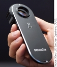
The dermatoscope generates a beam of light that falls on the cutaneous surface at an angle of 20°, allowing visualisation of the dermoscopic characteristics resulting from the presence of melanin and haemoglobin in the different skin layers. The usual magnification provided by the dermatoscope is ten-fold.
PATIENT GROUP AND USE
The use of dermoscopy enables:?
monitoring of pigmented lesions,
diagnosis of melanoma,
the physician to understand naevus morphology beyond what is possible by naked-eye examination alone, and
recognition of different populations of naevi characterised by similar morphological patterns and pigment distribution.
IMPORTANCE
Skin malignancy is an important cause of mortality in the UK, and is rising in incidence every year. An important determinant of outcome is initial recognition and management of the lesion. National Institute for Health and Clinical Excellence (NICE) guidance reports that one-quarter of primary care consultations in England and Wales are related to the diagnosis and management of skin conditions, including skin lesions (1.7%). Cancer research UK data for 2008 reported 2067 deaths due to malignant melanoma.
As with many cancer diagnoses, if melanoma is diagnosed early the survival rates are good; most stage 1 and stage 2 melanomas can be cured. A recent study on the recognition of skin malignancies showed that GPs in the UK missed one-third of malignancies,2 and one systematic review showed the sensitivity for detection of malignant melanoma was as low as 81% in dermatologists and only 41% in primary care physicians.3
PREVIOUS RESEARCH
The performance of dermoscopy has been widely investigated, and two meta-analyses have confirmed its use increases diagnostic accuracy by between 5% and 30% compared with clinical visual inspection.4–6 In one multicentre European trial, GPs were given a 1-day training course in skin cancer detection and dermoscopic evaluation, and randomly assigned to the dermoscopy evaluation arm or naked-eye evaluation arm.7 During a 16-month period, 73 physicians evaluated 2522 patients with skin lesions. All patients were re-evaluated by expert dermatologists. Sensitivity was significantly higher using dermoscopy (79% versus 54%, P<0.01), with identical specificity (71%). Histopathological examination of equivocal lesions revealed 23 malignant skin tumours missed by GPs performing naked-eye observation and only six missed by GPs using dermoscopy (P<0.01). The study concluded that the use of dermoscopy improves the ability of GPs to triage lesions that are suggestive of skin cancer.
In addition, tools such as a three-point checklist to identify melanoma (asymmetry, atypical network, and blue-white structures) and the CASH score (colours, architectural disorder, symmetry, homogeneity/heterogeneity), which can be used in conjunction with dermoscopy, have been developed; and validation studies have shown overall good interobserver reproducibility.8–10
An alternative to dermoscopy called MoleMate™, which uses spectrophotometric intracutaneous analysis (SIAscopy) along with an algorithm specifically developed for primary care, has been evaluated in a multicentre randomised controlled trial in the UK.11 The trial was completed in December 2010; however, the findings are as yet unpublished. Results of this study will be relevant to the diagnosis of melanoma in primary care.
COST-EFFECTIVENESS AND ECONOMIC IMPACT
There is no published evidence on the cost and cost-effectiveness of the use of dermoscopy for the diagnosis of melanoma in primary care. Several researchers have commented that dermoscopy in routine practice may have major implications in large-scale melanoma screening, with a reduction in the dermatological surgery workload of false-positive lesions, leading to cost savings, reduced morbidity, and less scarring.12 It may be cost-effective due to the decreased number of excised benign lesions and the early detection of melanomas.13 In terms of patient-reported outcomes, a study estimating patients’ willingness to pay for handheld dermoscopy, digital dermoscopy, and teledermoscopy was reported to be 40% below a hypothetical method promising 100% accuracy, yet higher than that reported for naked-eye inspection.14
Future research is needed to assess whether the use of dermatoscopes in a primary care setting is cost-effective in terms of early detection of melanomas. 
Acknowledgments
The authors would like to thank Richard Stevens and Nia Roberts for helpful discussions.
Relevant guidelines
NICE clinical guideline. Improving outcomes for people with skin tumours including melanoma (update): the management of low-risk basal cell carcinomas in the community. http://www.nice.org.uk/guidance/index.jsp?action=download&o=48878 (accessed 24 Mar 2011).
Provenance
Freely submitted; not externally peer reviewed.
Funding
The Centre for Monitoring and Diagnosis Oxford (MaDOx) is funded by the National Institute for Health Research, UK programme grant ‘Development and implementation of new diagnostic processes and technologies in primary care’.
Competing interests
The authors have declared no competing interests.
Discuss this article
Contribute and read comments about this article on the Discussion Forum: http://www.rcgp.org.uk/bjgp-discuss
REFERENCES
- 1.Balch CM, Gershenwald JE, Soong SJ, et al. Final version of 2009 AJCC melanoma staging and classification. J Clin Oncol. 2009;27(36):6199–6206. doi: 10.1200/JCO.2009.23.4799. [DOI] [PMC free article] [PubMed] [Google Scholar]
- 2.Pockney P, Primrose J, George S, et al. Recognition of skin malignancy by general practitioners: observational study using data from a population-based randomised controlled trial. Br J Cancer. 2009;100(1):24–27. doi: 10.1038/sj.bjc.6604810. [DOI] [PMC free article] [PubMed] [Google Scholar]
- 3.Chen SC, Bravata DM, Weil E. A comparison of dermatologists’ and primary care physicians’ accuracy in diagnosing melanoma. Arch Dermatol. 2001;137(12):1627–1634. doi: 10.1001/archderm.137.12.1627. [DOI] [PubMed] [Google Scholar]
- 4.Kittler H, Pehamberger H, Wolff K, Binder M. Diagnostic accuracy of dermoscopy. Lancet Oncol. 2002;3(3):159–165. doi: 10.1016/s1470-2045(02)00679-4. [DOI] [PubMed] [Google Scholar]
- 5.Bafounta ML, Beauchet A, Aegerter P, Saiag P. Is dermoscopy (epiluminescence microscopy) useful for the diagnosis of melanoma? Results of a meta-analysis using techniques adapted to the evaluation of diagnostic tests. Arch Dermatol. 2001;137(10):1343–1350. doi: 10.1001/archderm.137.10.1343. [DOI] [PubMed] [Google Scholar]
- 6.Vestergaard ME, Macaskill P, Holt PE, Menzies SW. Dermoscopy compared with naked eye examination for the diagnosis of primary melanoma: a meta-analysis of studies performed in a clinical setting. Br J Dermatol. 2008;159(3):669–676. doi: 10.1111/j.1365-2133.2008.08713.x. [DOI] [PubMed] [Google Scholar]
- 7.Argenziano G, Puig S, Zalaudek I, et al. Dermoscopy improves accuracy of primary care physicians to triage lesions suggestive of skin cancer. J Clin Oncol. 2006;24(12):1877–1882. doi: 10.1200/JCO.2005.05.0864. [DOI] [PubMed] [Google Scholar]
- 8.Henning JS, Dusza SW, Wang SQ, et al. The CASH (color, architecture, symmetry, and homogeneity) algorithm for dermoscopy. J Am Acad Dermatol. 2007;56(1):45–52. doi: 10.1016/j.jaad.2006.09.003. [DOI] [PubMed] [Google Scholar]
- 9.Henning JS, Stein JA, Yeung J, et al. CASH algorithm for dermoscopy revisited. Arch Dermatol. 2008;144(4):554–555. doi: 10.1001/archderm.144.4.554. [DOI] [PubMed] [Google Scholar]
- 10.Zalaudek I, Argenziano G, Soyer HP, et al. Three-point checklist of dermoscopy: an open internet study. Br J Dermatol. 2006;154(3):431–437. doi: 10.1111/j.1365-2133.2005.06983.x. [DOI] [PubMed] [Google Scholar]
- 11.Walter FM, Morris HC, Humphrys E, et al. Protocol for the MoleMate UK Trial: a randomised controlled trial of the MoleMate system in the management of pigmented skin lesions in primary care [ISRCTN 79932379] BMC Fam Pract. 2010;11:36. doi: 10.1186/1471-2296-11-36. [DOI] [PMC free article] [PubMed] [Google Scholar]
- 12.Carli P, De Giorgi V, Crocetti E, et al. Improvement of malignant/benign ratio in excised melanocytic lesions in the ‘dermoscopy era’: a retrospective study 1997–2001. Br J Dermatol. 2004;150(4):687–692. doi: 10.1111/j.0007-0963.2004.05860.x. [DOI] [PubMed] [Google Scholar]
- 13.Massone C, Di Stefani A, Soyer HP. Dermoscopy for skin cancer detection. Curr Opin Oncol. 2005;17(2):147–153. doi: 10.1097/01.cco.0000152627.36243.26. [DOI] [PubMed] [Google Scholar]
- 14.Schiffner R, Schiffner-Rohe J, Landthaler M. Patients’ confidence in dermoscopical methods for detection of malignant melanoma. Dermatol Psychosom. 2002;3:114–118. [Google Scholar]


