
Figure 4.
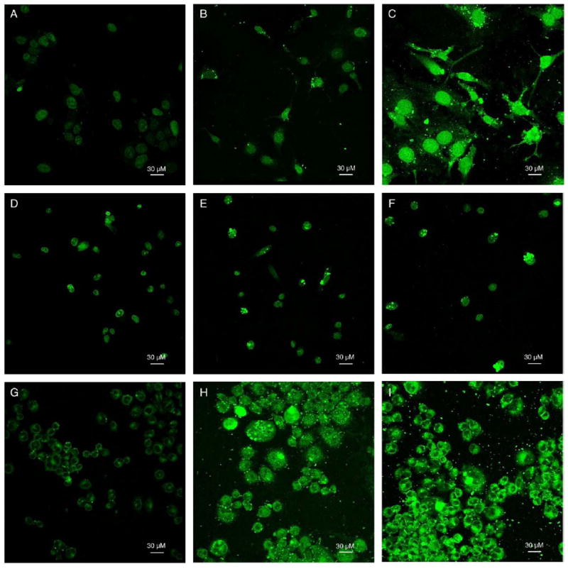
(A–I) Cell images obtained with a Nikon confocal microscope and a 488-nm argon laser at 40× objective magnification. DOX fluorescence images were recorded with software-added green pseudo color. (A–C) 10 μM free DOX, DNPs, or ADNPs were incubated with SKOV-3 cells for 24 hours, respectively. Photomultiplier tube (PMT) was set at gain 7. (D–F) 10 μM free DOX, DNPs, or ADNPs were incubated with MES-SA cells for 24 hours, respectively. PMT was set at gain 6. (G–I) 10 μM free DOX, DNPs, or ADNPs were incubated with Dx5 cells for 24 hours, respectively. PMT was set at gain 7.