Heterotopic pancreatic tissue, otherwise known as pancreatic rest, is pancreatic tissue that lacks anatomic and vascular continuity with the main body of the pancreas. Common locations for this tissue include the stomach, duodenum, jejunum, Meckel diver-ticulum, and ileum.1,2 We report a case of heterotopic pancreatic tissue found in the esophagus, which is an exceedingly rare finding.
Case Report
A 14-year-old girl presented with complaints of peri-umbilical abdominal pain. Episodes of pain tended to occur at least once daily, typically lasting for 1 hour. The patient had no history of constipation, vomiting, fever, melena, or weight loss. Her physical examination revealed a healthy-appearing female patient who was at nearly the 75th percentile for weight. There was no abdominal tenderness or appreciable mass. Complete blood count with differential, chemistries, liver function tests, erythrocyte sedimentation rate, amylase levels, and lipase levels were all within normal limits. Serology tested negative for Helicobacter pylori and celiac disease. Computed tomographic scan of the abdomen and pelvis, along with upper gastrointestinal series with small bowel follow-through, were both unremarkable.
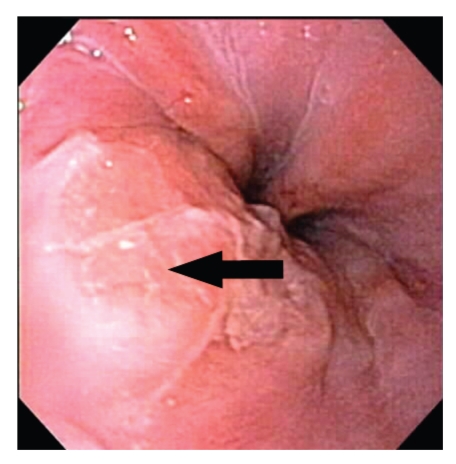
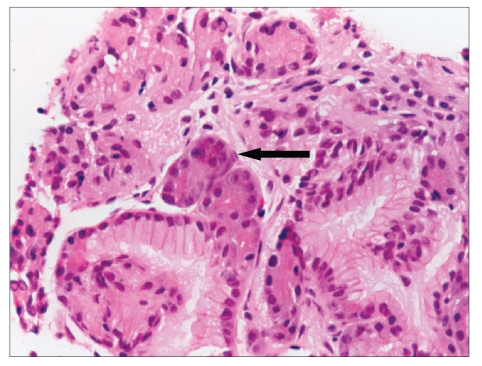
A circular patch of raised shiny yellowish mucosa, approximately 2 cm in diameter, was seen in the distal third of the esophagus during esophagogastroduodenos-copy (Figure 1). Hematoxylin and eosin staining of the biopsies from this area revealed ectopic pancreatic tissue in the submucosa (Figure 2).
Figure 1.
Pancreatic rest of the distal esophagus. Arrow points to pancreatic tissue
Figure 2.
Hematoxylin and eosin stain of esophageal biopsy. Arrow points to pancreatic tissue in the submucosa (original magnification, ×200)
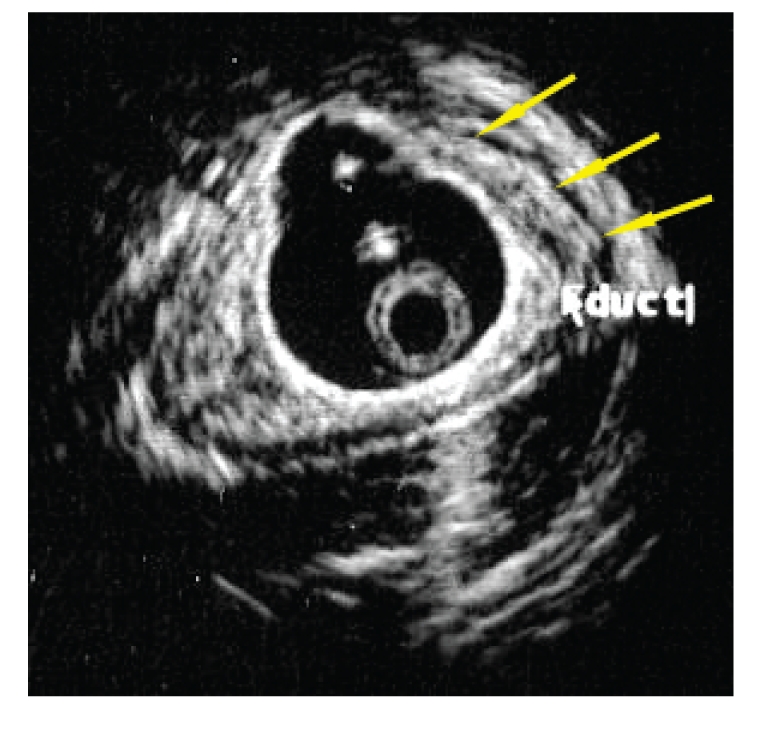
In order to determine the extent of the lesion, endoscopic ultrasound was performed. The pancreatic rest did not appear to extend beyond the submucosa. A small tubular anechoic structure thought to represent a pancreatic ductule was seen within the ectopic tissue (Figure 3). Endoscopic biopsies of the abnormal-appearing esophageal mucosa once again confirmed the presence of pancreatic acinar cells in the submucosa.
Figure 3.

Endoscopic ultrasound image of distal esophagus. Outline of ectopic pancreatic tissue demarcated by thin yellow arrows. Short white arrow points to suspected pancreatic ductule
Discussion
Heterotopic pancreatic tissue, which occurs most commonly in the stomach and duodenum, appears endoscopically as a submucosal lesion that usually contains a central umbilication. Unless the pancreatic rest is large, upper gastrointestinal series is usually normal. His-tologically, the tissue may contain acini, islets, and ducts. Patients do not usually develop symptoms from this entity, which is typically found during an evaluation for unrelated complaints. If symptoms develop, they are most likely secondary to a mass effect. Large lesions may cause obstruction, ulceration, hemorrhage, or intussusception. Other complications include pancreatitis, pseudocyst formation, carcinomas, islet-cell tumors, or inflammatory pseudotumors. Surgical excision is the only cure.1-3
Ectopic pancreatic tissue in the esophagus has been reported in the literature only 10 times. Only 3 of the patients with this lesion were younger than 18 years old. In these children, the ectopic tissue was associated with one or more anatomic abnormalities of the esophagus.2 One patient had a tracheoesophageal fistula, esophageal atresia, and esophageal duplication.4 Another patient had a congenital diverticulum,5 and the third had a congenital cyst.6
Pancreatic rest was found in the distal third of the esophagus in 6 of the 10 patients. In 2 adult patients, the lesion was found to be malignant. Nine patients underwent resection or enucleation of the lesion, and 1 patient was observed.2
The prevalence of malignant transformation of heterotopic pancreatic tissue is unknown. Guillou and associates proposed that a carcinoma should be stated to originate from heterotopic pancreatic tissue only when the following three conditions are met: the tumor is found within or close to the ectopic pancreatic tissue; a direct transition between the pancreatic structures and the carcinoma can be shown (eg, duct cell dysplasia); and the nonneoplastic pancreatic tissue contains fully developed acini and ductal structures.7
Conclusion
The patient described above has no known anatomic abnormalities. Currently, the lesion does not appear to extend beyond the submucosa, and there is no endoscopic or histologic evidence of neoplasia. Routine follow-up of the lesion is planned.
References
- 1.Feldman M, Friedman LS, Brandt LJ, editors. Sleisenger and Fordtran's Gastrointestinal and Liver Disease. 7th ed. Philadelpia, Pa: Saunders; 2002. pp. 883–884. [Google Scholar]
- 2.Temes RT, Menen MJ, Davis MS, Pett SB, Jr, Wernly JA. Heterotopic pancreas of the esophagus masquerading as Boerhaave's syndrome. Ann Thorac Surg. 2000;69:259–261. doi: 10.1016/s0003-4975(99)01223-0. [DOI] [PubMed] [Google Scholar]
- 3.Noffsinger AE, Hyams DM, Fenoglio-Preiser CM. Esophageal heterotopic pancreas presenting as an inflammatory mass. Dig Dis Sci. 1995;40:2373–2379. doi: 10.1007/BF02063240. [DOI] [PubMed] [Google Scholar]
- 4.Ishikawa O, Ishiguro S, Ohhigashi H, Sasaki Y, Yasuda T, et al. Solid and papillary neoplasm arising from an ectopic pancreas in the mesocolon. Am J Gastroenterol. 1990;85:597–601. [PubMed] [Google Scholar]
- 5.Chatterjee PK, Chatterjee SN, Dastidar N, et al. Heterotopic gastric mucosa and pancreatic tissue in congenital diverticulum of oesophagus. Indian J Surg. 1982;44:139–141. [Google Scholar]
- 6.Roshe J, Del Buono E, Domenico D, Colturi TJ. Anaplastic carcinoma arising in ectopic pancreas located in the distal esophagus. J Clin Gastroenterol. 1996;22:242–247. doi: 10.1097/00004836-199604000-00022. [DOI] [PubMed] [Google Scholar]
- 7.Guillou L, Nordback P, Gerber C, Schneider RP. Ductal adenocarcinoma arising in a heterotopic pancreas situated in a hiatal hernia. Arch Pathol Lab Med. 1994;118:568–571. [PubMed] [Google Scholar]


