


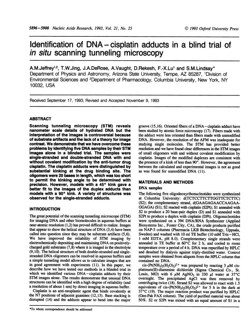
Abstract
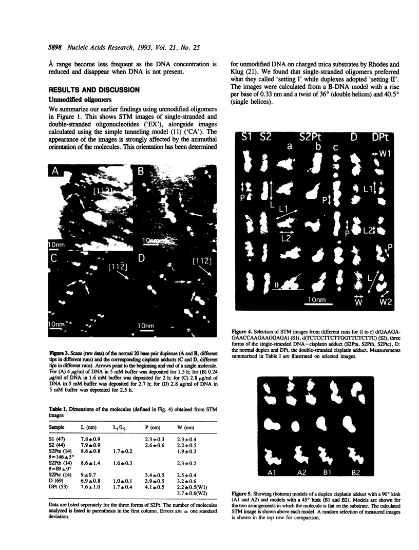
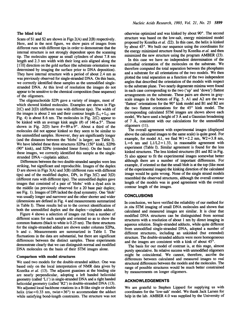
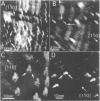
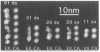
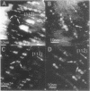
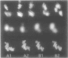
Scanning tunneling microscopy (STM) reveals nanometer scale details of hydrated DNA but the interpretation of the images is controversial because of substrate artifacts and the lack of a theory for image contrast. We demonstrate that we have overcome these problems by identifying five DNA samples by their STM images alone in a blinded trial. The samples were single-stranded and double-stranded DNA with and without covalent modification by the anti-tumor drug cisplatin. The cisplatin adducts were distinguished by substantial kinking at the drug binding site. The oligomers were 20 bases in length, which was too short to permit the kinking angle to be determined with precision. However, models with a 45 degree kink gave a better fit to the images of the duplex adducts than models with a 90 degrees kink. A variety of structures was observed for the single-stranded adducts.
Full text
PDF
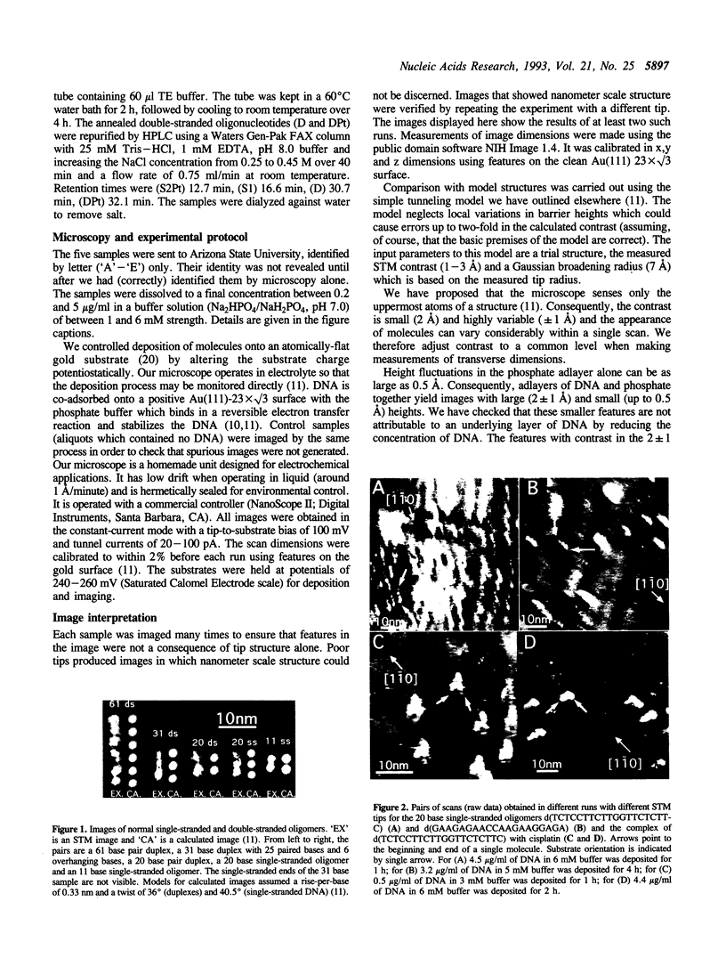
Images in this article
Selected References
These references are in PubMed. This may not be the complete list of references from this article.
- Beebe T. P., Jr, Wilson T. E., Ogletree D. F., Katz J. E., Balhorn R., Salmeron M. B., Siekhaus W. J. Direct observation of native DNA structures with the scanning tunneling microscope. Science. 1989 Jan 20;243(4889):370–372. doi: 10.1126/science.2911747. [DOI] [PubMed] [Google Scholar]
- Brabec V., Reedijk J., Leng M. Sequence-dependent distortions induced in DNA by monofunctional platinum(II) binding. Biochemistry. 1992 Dec 15;31(49):12397–12402. doi: 10.1021/bi00164a014. [DOI] [PubMed] [Google Scholar]
- Clemmer C. R., Beebe T. P., Jr Graphite: a mimic for DNA and other biomolecules in scanning tunneling microscope studies. Science. 1991 Feb 8;251(4994):640–642. doi: 10.1126/science.1992517. [DOI] [PubMed] [Google Scholar]
- Driscoll R. J., Youngquist M. G., Baldeschwieler J. D. Atomic-scale imaging of DNA using scanning tunnelling microscopy. Nature. 1990 Jul 19;346(6281):294–296. doi: 10.1038/346294a0. [DOI] [PubMed] [Google Scholar]
- Heckl W. M., Binnig G. Domain walls on graphite mimic DNA. Ultramicroscopy. 1992 Jul;42-44(Pt B):1073–1078. doi: 10.1016/0304-3991(92)90404-8. [DOI] [PubMed] [Google Scholar]
- Jing T. W., Jeffrey A. M., DeRose J. A., Lyubchenko Y. L., Shlyakhtenko L. S., Harrington R. E., Appella E., Larsen J., Vaught A., Rekesh D. Structure of hydrated oligonucleotides studied by in situ scanning tunneling microscopy. Proc Natl Acad Sci U S A. 1993 Oct 1;90(19):8934–8938. doi: 10.1073/pnas.90.19.8934. [DOI] [PMC free article] [PubMed] [Google Scholar]
- Kozelka J., Archer S., Petsko G. A., Lippard S. J., Quigley G. J. Molecular mechanics modeling of oligonucleotide adducts of the antitumor drug cis-diamminedichloroplatinum(II). Biopolymers. 1987 Aug;26(8):1245–1271. doi: 10.1002/bip.360260804. [DOI] [PubMed] [Google Scholar]
- Kozelka J., Chottard J. C. How does cisplatin alter DNA structure? A molecular mechanics study on double-stranded oligonucleotides. Biophys Chem. 1990 Apr;35(2-3):165–178. doi: 10.1016/0301-4622(90)80006-s. [DOI] [PubMed] [Google Scholar]
- Kozelka J., Fouchet M. H., Chottard J. C. H8 chemical shifts in oligonucleotide cross-linked at a GpG sequence by cis-Pt(NH3)2(2+): a clue to the adduct structure. Eur J Biochem. 1992 May 1;205(3):895–906. doi: 10.1111/j.1432-1033.1992.tb16855.x. [DOI] [PubMed] [Google Scholar]
- Lindsay S. M., Tao N. J., DeRose J. A., Oden P. I., Lyubchenko YuL, Harrington R. E., Shlyakhtenko L. Potentiostatic deposition of DNA for scanning probe microscopy. Biophys J. 1992 Jun;61(6):1570–1584. doi: 10.1016/S0006-3495(92)81961-6. [DOI] [PMC free article] [PubMed] [Google Scholar]
- Lindsay S. M., Thundat T., Nagahara L., Knipping U., Rill R. L. Images of the DNA double helix in water. Science. 1989 Jun 2;244(4908):1063–1064. doi: 10.1126/science.2727694. [DOI] [PubMed] [Google Scholar]
- Rampino N. J. Cisplatin induced alterations in oriented fibers of DNA studied by atomic force microscopy. Biochem Biophys Res Commun. 1992 Jan 15;182(1):201–207. doi: 10.1016/s0006-291x(05)80131-7. [DOI] [PubMed] [Google Scholar]
- Rhodes D., Klug A. Helical periodicity of DNA determined by enzyme digestion. Nature. 1980 Aug 7;286(5773):573–578. doi: 10.1038/286573a0. [DOI] [PubMed] [Google Scholar]
- Rice J. A., Crothers D. M., Pinto A. L., Lippard S. J. The major adduct of the antitumor drug cis-diamminedichloroplatinum(II) with DNA bends the duplex by approximately equal to 40 degrees toward the major groove. Proc Natl Acad Sci U S A. 1988 Jun;85(12):4158–4161. doi: 10.1073/pnas.85.12.4158. [DOI] [PMC free article] [PubMed] [Google Scholar]
- Sonnenfeld R., Hansma P. K. Atomic-resolution microscopy in water. Science. 1986 Apr 11;232(4747):211–213. doi: 10.1126/science.232.4747.211. [DOI] [PubMed] [Google Scholar]