Abstract
Current tuberculosis (TB) vaccine strategies are largely aimed at activating conventional T cell responses to mycobacterial protein antigens. However, the lipid-rich cell wall of Mycobacterium tuberculosis (M. tuberculosis) is essential for pathogenicity and provides targets for unconventional T cell recognition. Group 1 CD1–restricted T cells recognize mycobacterial lipids, but their function in human TB is unclear and their ability to establish memory is unknown. Here, we characterized T cells specific for mycolic acid (MA), the predominant mycobacterial cell wall lipid and key virulence factor, in patients with active TB infection. MA-specific T cells were predominant in TB patients at diagnosis, but were absent in uninfected bacillus Calmette-Guérin–vaccinated (BCG-vaccinated) controls. These T cells were CD1b restricted, detectable in blood and disease sites, produced both IFN-γ and IL-2, and exhibited effector and central memory phenotypes. MA-specific responses contracted markedly with declining pathogen burden and, in patients followed longitudinally, exhibited recall expansion upon antigen reencounter in vitro long after successful treatment, indicative of lipid-specific immunological memory. T cell recognition of MA is therefore a significant component of the acute adaptive and memory immune response in TB, suggesting that mycobacterial lipids may be promising targets for improved TB vaccines.
Introduction
Mycobacterium tuberculosis (M. tuberculosis), the intracellular pathogen that causes tuberculosis (TB), infects around one-third of the human population, causing almost 2 million deaths annually (1, 2). Improved TB vaccines that induce prolonged cellular immunity across whole populations are urgently needed (1–4). Conventional CD4+ and CD8+ T cell responses recognizing peptide antigens presented by polymorphic MHC class II and I molecules, respectively, play key roles in immunity to M. tuberculosis and are targeted by current vaccine and immunodiagnostic strategies (5). However, unconventional T cells that recognize nonpeptidic antigens in an MHC-independent fashion are also implicated in immunity to M. tuberculosis (6–14).
In particular, the M. tuberculosis cell wall, which plays a critical role in its intracellular survival, persistence, and pathogenicity, provides a rich source of diverse lipid antigens for immune recognition (15–18). Moreover, the CD1 family of MHC-like molecules has evolved to sample endocytic compartments, where M. tuberculosis resides, and present such lipid antigens to T cells (19–22), suggesting a potential role in immunity to TB. Furthermore, unlike MHC molecules, CD1 proteins exhibit very limited polymorphism (6), highlighting CD1-restricted M. tuberculosis lipid antigens as attractive candidates for incorporation into vaccine approaches that could induce immunity in genetically diverse human populations.
Current evidence particularly implicates the group 1 CD1 subset comprising CD1a, -b, and -c isoforms (6, 23, 24), which is functionally distinct from the group 2 CD1d isoform recognized by natural killer T cells, in antimycobacterial immunity (25). Remarkably, the diverse array of foreign lipid antigens shown to be presented by group 1 CD1 molecules all derive from the mycobacterial cell wall (20, 23). Secondly, group 1 CD1–restricted T cells have been isolated from Mycobacterium leprae– or M. tuberculosis–infected individuals (8, 9, 12–14, 23, 26, 27); such cells secrete TH1 cytokines, critical for protective immunity to TB (27, 28), and are cytotoxic (13, 25, 28–32). Finally, M. tuberculosis lipid–vaccinated guinea pigs display detectable group 1 CD1–restricted lipid-specific T cell responses and reduced lung pathology after M. tuberculosis challenge (10). However, the role of lipid-specific T cells in human TB infection remains unclear; in particular, almost nothing is known of their relationship to antigen load in vivo, their ability to home to sites of infection, and whether or not they can establish immunological memory.
The predominant lipid component of the M. tuberculosis cell wall is mycolic acid (MA), which was the first CD1-restricted lipid to be identified (7), making it the prototypical microbial lipid antigen. MA is a key virulence factor, protecting M. tuberculosis from dehydration, exposure to drugs, and the hostile environment of the macrophage phagolysosome (33–35). In addition, free MA is a major component of extracellular biofilms formed by M. tuberculosis that harbor drug-tolerant persisting M. tuberculosis populations (36). Although recognition of MA by a T cell clone isolated from a healthy subject established it as the first lipid antigen to be presented by CD1b (7) and MA-specific T cells have been reported in group 1 CD1-transgenic MHC class II–deficient mice (37), ex vivo T cell responses to MA during human M. tuberculosis infection have not been reported. Given its central role in M. tuberculosis virulence and its predominance in the cell wall and extracellularly in biofilm, the interaction of MA with the cellular immune response during M. tuberculosis infection is of considerable interest.
Here, we identified and comprehensively characterized CD1b-restricted T cells specific for MA in TB patients and we investigated their relationship with treatment-induced changes in antigen load in vivo and the induction and persistence of M. tuberculosis lipid–specific adaptive immunity long after curative treatment.
Results
A majority of untreated TB patients have circulating IFN-γ–secreting MA-specific T cells.
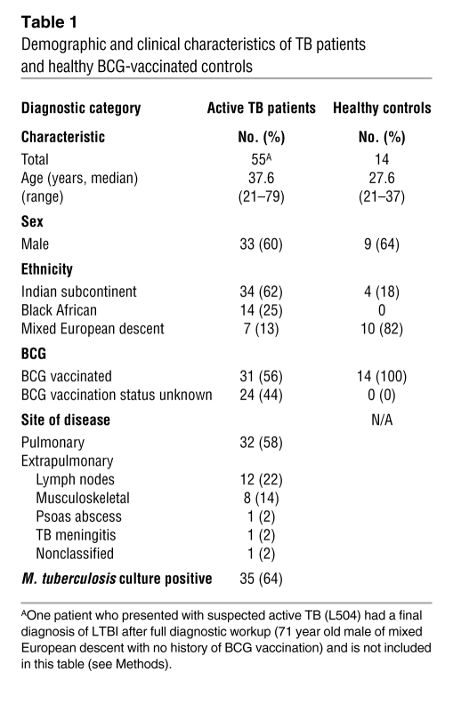
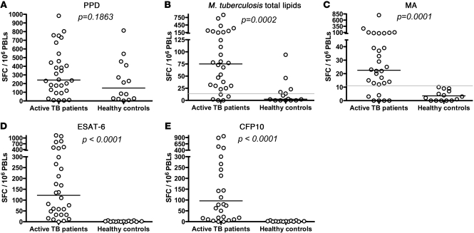
Demographic and clinical characteristics of the TB patients and healthy bacillus Calmette-Guérin–vaccinated (BCG-vaccinated) control subjects are summarized in Table 1. Frequencies of circulating IFN-γ–producing T cells specific for purified protein derivative (PPD), M. tuberculosis total lipids, MA, and the immunodominant M. tuberculosis–specific protein antigens ESAT-6 and CFP10 were assessed by ex vivo IFN-γ ELISpot. Autologous DCs, generated from monocytes in the presence of IL-4 and GM-CSF, were used to present these antigens and phosphatidylinositol (PI), a self lipid used as a negative control (median: 3 spot-forming cells [SFC]/million peripheral blood lymphocytes [PBLs]; interquartile range [IQR]: 1.5–12), to PBLs from active TB patients (n = 30) and healthy BCG-vaccinated controls (n = 14) (Figure 1). Flow cytometry of matured DCs from different individuals confirmed expression of group 1 CD1 molecules on a high proportion of group 1 CD1+ DCs (65%–95%; Supplemental Figure 1; supplemental material available online with this article; doi: 10.1172/JCI46216DS1). Whereas the frequency of PPD-specific T cells was similar in both groups, T cells specific for M. tuberculosis total lipids were preferentially observed in TB, with 25 of 30 (83%) patients displaying detectable responses (median: 75 SFC/million PBLs; IQR: 27.5–181) compared with 5 of 14 (36%) controls (median: 2.5 SFC/million PBLs; IQR: 0–20; P = 0.0002; Figure 1, A and B). Notably, 24 of 30 (80%) TB patients had T cells specific for the single lipid antigen MA (median: 22.5 SFC/million PBLs; IQR: 12.5–67.5; Figure 1C), and these responses were highly specific to TB patients since they were absent in all BCG-vaccinated controls. As expected, responses to the M. tuberculosis–specific immunodominant protein antigens ESAT-6 and CFP10 were detected in most TB patients (90% and 80%, respectively; Figure 1, D and E), but not in BCG-vaccinated uninfected controls. Therefore, MA-specific T cells are observed in most TB patients, and although low in frequency compared with the highly immunodominant protein antigens tested (magnitude hierarchy: PPD > ESAT-6 > CFP10 > M. tuberculosis total lipids > MA), their presence was highly discriminatory for TB, to an extent similar to that of ESAT-6/CFP-10 responses.
Table 1 .
Demographic and clinical characteristics of TB patients and healthy BCG-vaccinated controls
Figure 1. T cell responses to M. tuberculosis peptide and lipid antigens in active TB patients and BCG-vaccinated healthy donors.
PBLs from 30 untreated TB patients and 14 BCG-vaccinated healthy donors were incubated overnight in the presence of autologous blood monocyte–derived DCs pulsed with (A) PPD, (B) M. tuberculosis total lipids, (C) MA, (D) ESAT-6, or (E) CFP10. The production of IFN-γ was measured by IFN-γ ELISpot. Horizontal bars represent the median of each population. The gray lines in B and C represent the limit of detection for this assay and the cut-off for a positive response.
The frequency of MA-specific T cells varied widely between patients but no correlation was observed between the magnitude of the MA-specific IFN-γ response (or the ESAT-6– and CFP10-specific responses) at diagnosis and the site of disease (pulmonary versus extrapulmonary) or acid-fast bacillus smear status (Supplemental Table 1). However, the size of the MA-specific response was significantly larger in patients of black African ethnicity (n = 9) compared with those of Indian subcontinental ethnicity (n = 18) (P = 0.02).
MA and M. tuberculosis total lipid-specific T cell populations contract during successful TB treatment.
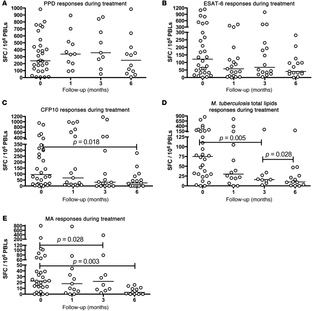
To assess the relationship between MA-specific T cells and mycobacterial antigen load, we compared the frequency of IFN-γ–secreting T cells in TB patients during treatment, which progressively decreases mycobacterial burden in vivo. PPD-specific T cells remained stable during treatment, while the number of ESAT-6– and CFP-10–specific T cells gradually declined (Figure 2, A–C). T cells specific for M. tuberculosis total lipids and MA declined similarly (Figure 2, D and E). Whereas protein-specific T cells remained detectable in most patients after treatment completion (ESAT-6– and CFP10-specific T cells were observed in 87% and 60% of TB patients, respectively), MA-specific T cells were only detectable in 23% of TB patients (P = 0.029 for MA vs. ESAT-6 and P = 0.352 for MA vs. CFP10).
Figure 2. T cell responses to M. tuberculosis antigens in TB patients during and after treatment.
PBLs from active TB patients were incubated overnight in the presence of autologous blood monocyte–derived DCs pulsed with (A) PPD, (B) ESAT-6, or (C) CFP10, (D) M. tuberculosis total lipids, or (E) MA. IFN-γ production in response to these antigens was measured in patients at 4 different stages of treatment: diagnosis and 1, 3, and 6 months after treatment using an IFN-γ ELISpot. Horizontal bars represent the median of each population.
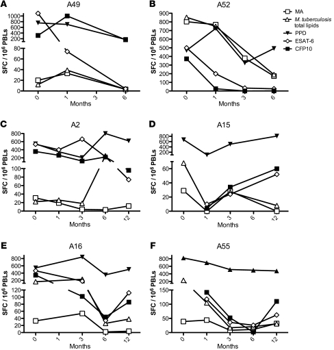
These findings were corroborated by longitudinal tracking of MA- and peptide antigen–specific T cells within a subset of individual TB patients for which pretreatment, 1-month, 3-month, and 6-month samples were available (Figure 3, A–F). The frequency of both peptide-specific and lipid-specific T cells declined over this time period. At the end of treatment (6 months), MA-specific T cells were detectable in only 1 of 5 patients tested, while T cells specific for either ESAT-6 or CFP10 were still present in 4 out of 5. In addition, in 4 patients for whom 12-month samples were available (Figure 3, C–F; patients A2, A15, A16, and A55), all 4 had ESAT-6– and CFP10-specific T cells, whereas only 2 had detectable MA-specific T cells, which were present at very low frequencies. Thus, MA-specific T cells are more closely dynamically related to pathogen burden than ESAT-6/CFP-10–specific T cells and may be a better marker of antigen load than ESAT-6/CFP10–specific IFN-γ responses if confirmed in larger patient cohorts.
Figure 3. Longitudinal study of T cell responses to M. tuberculosis antigens in TB patients during treatment.
IFN-γ production from 6 TB patients in response to MA, M. tuberculosis total lipids, PPD, ESAT-6, and CFP10 was serially monitored from diagnosis up to 6 months after diagnosis for 2 patients (A and B; patients A49 and A52) and for 12 months after diagnosis for 4 patients (C–F; patients A2, A15, A16, and A55) using IFN-γ ELISpot.
Quantification of MA-specific IFN-γ–secreting T cells at site of infection.
To determine whether MA-specific T cells were present at the site of infection, we enumerated frequencies of IFN-γ–secreting T cells specific for MA in 6 active TB patients with paired time-matched blood and bronchoalveolar lavage (BAL) samples at diagnosis (Figure 4A; patients A441, A452, A502, A514, A877, A878). In most cases, MA-specific T cells were detectable in both anatomical compartments. In contrast, for 2 patients with active disease (A877, A878) and 1 (L504) with latent TB infection (LTBI), MA-specific T cells were present in BAL but not in blood. PPD-specific T cells were detectable in both blood and BAL of the 5 active TB patients tested (Figure 4B); for the single LTBI case, PPD-specific T cells were also present in both compartments. Although PPD-specific T cell responses were enhanced in BAL versus blood in 2 patients, the remaining 4 PPD-specific responses were broadly comparable in both compartments, unlike in a previous study that reported lower PPD-specific responses in blood versus BAL samples (38). A likely explanation is that this study did not control for APC numbers in each compartment, instead pulsing PPD onto unfractionated BAL cells or PBMCs. Consequently, the high levels of alveolar macrophages in BAL samples are likely to have preferentially boosted PPD-specific responses within this compartment relative to PBMCs. In contrast, our study used a fixed number of PPD-pulsed APCs (DCs) to compare PPD-specific responses in blood and BAL. Therefore, MA-specific IFN-γ–secreting T cells are present in the lungs during active TB and LTBI, and in some cases preferentially localized to the lung.
Figure 4. MA- and PPD-specific T cell responses from the blood and the BAL of TB patients.
PBLs and BALMCs from 6 active TB patients (A441, A452, A501, A514, A877, A878) and 1 person with LTBI (L504) were incubated overnight in the presence of blood monocyte–derived DCs pulsed with either PI (negative control; data not shown) (A) MA or (B) PPD. IFN-γ production was measured by IFN-γ ELISpot.
MA-specific T cells are CD1b-restricted, include CD4+, CD8+, and CD4–CD8– phenotypes and secrete IFN-γ, IL-2, or both cytokines.
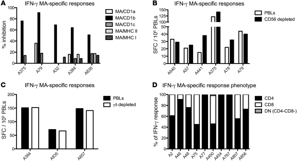
Although previous studies established that recognition of MA by a T cell clone was CD1b restricted (7), we sought to determine whether MA recognition ex vivo by circulating T cells from TB patients is CD1b restricted using CD1/MHC-blocking antibodies. Consistent with presentation of MA by CD1b, MA-specific IFN-γ responses were substantially inhibited by monoclonal antibodies to CD1b (Figure 5A) but not other CD1 isoforms or MHC I/II. To exclude involvement of NK cells or γδ T cells, the effect of depleting CD56+ cells (Figure 5B) and γδ TCR+ T cells (Figure 5C) on the ex vivo MA-specific IFN-γ response was also assessed. As expected, depletion of these cell subsets had no effect on the strength of the IFN-γ response. To further verify the involvement of αβ-TCR+ T cells in the MA-specific response, PBLs from 2 TB patients were stained with an anti–αβ-TCR antibody and their MA-specific IFN-γ and IL-2 responses ascertained by cytokine secretion assay (Supplemental Figure 2). As expected, MA-specific responses derived exclusively from αβ-TCR+ T cells in both patients.
Figure 5. Functional characterization of MA-specific T cells.
(A) PBLs from active TB patients were incubated overnight with MA-pulsed DCs in the presence of an isotype control antibody, anti-CD1a, anti-CD1b, anti-CD1c, anti-HLA ABC, or anti–HLA-DP, -DQ, -DR. IFN-γ response was analyzed using ELISPOT. PBLs from active TB patients were depleted of CD56+ cells (B) and γδ T cells (C) and incubated overnight with MA-pulsed DCs. IFN-γ–secreting T cells were quantified by ex vivo ELISPOT. (D) CD4/CD8 phenotype was assessed for PBLs from 10 active TB patients incubated in the presence of blood monocyte–derived DCs pulsed with MA or PI. IFN-γ–producing T cells were enriched through magnetic separation and analyzed by flow cytometry. Absolute values of IFN-γ–producing cells per 100,000 PBLs after enrichment were as follows for CD4+, CD8+, and double-negative (DN) cells: A2 (24, 15, 0 per hundred thousand [pht]); A46 (140, 14, 0 pht); A48 (56, 18, 0 pht); A75 (7, 9, 0 pht); A77 (9, 6, 0 pht); A454 (226, 0, 0 pht); A450 (463, 522, 20 pht); A787 (78, 0, 0 pht); A807 (72, 3, 0 pht); A856 (19, 6, 0 pht).
Assessment of the CD4/CD8 phenotype of these MA-specific CD1b-restricted T cells in 10 TB patients revealed that circulating MA-specific T cells were often predominantly CD4+ (Figure 5D). Three patients displayed an exclusively CD4+ phenotype among responding T cells, 5 exhibited a dominant CD4+ response with variable CD8+ involvement, and a further 2 patients had broadly equivalent CD8+ and CD4+ responses; in 1 of these (A450), a small response from CD4–CD8– T cells was also detected.
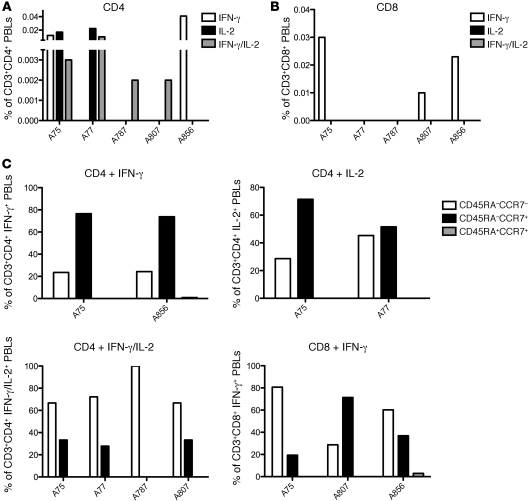
The cytokine profile (Figure 6) and memory phenotype (Figure 6C) of MA-specific T cells was also characterized in 5 untreated TB patients. Four exhibited a CD4+ IFN-γ/IL-2–double-producing MA-specific T cell population (Figure 6A), with responses dominated by T cells with an effector-memory phenotype (CD45RA–CCR7–) (Figure 6C). CD4+ IL-2–only–producing T cells were observed in 2 patients, both associated with a central memory phenotype (Figure 6C). CD4+ IFN-γ–only–producing T cells were observed in 2 patients (Figure 6A), both unexpectedly associated with a dominant central memory phenotype (CD45RA–CCR7+) (Figure 6C). CD8+ T cells were detected in 3 out of 5 patients, and secreted IFN-γ only (Figure 6B), with some variation in the proportion of central and effector memory phenotype populations between different donors (Figure 6C).
Figure 6. Phenotypic characterization of MA-specific T cells.
PBLs from 5 active untreated TB patients were incubated in the presence of blood-derived DCs pulsed with PI or MA. IFN-γ– and IL-2–producing T cells were enriched through magnetic separation and stained with anti-CD3, anti-CD4, anti-CD8, anti-CCR7, anti-CD45RA, and 7-AAD. Stained cells were analyzed by flow cytometry. (A–B) IFN-γ and IL-2 secretion from MA-specific CD4+ T cells (A) and MA-specific CD8+ T cells (B). (C) CD45RA and CCR7 cell-surface marker phenotype of MA-specific T cells of different functional subsets and CD4/CD8 phenotypes according to cytokine production.
Durable memory MA-specific T cells are present more than 1 to 2 years after curative treatment.
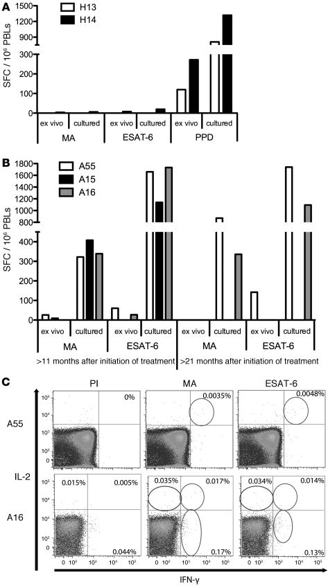
To address whether CD1 group 1–restricted T cells form memory populations that can subsequently reexpand upon antigen reencounter, we cultured PBLs from 3 TB patients (A15, A16, A55) with MA- or ESAT-6–pulsed autologous DCs for 14 days and then quantified MA- or ESAT-6–specific T cells using autologous DCs in IFN-γ ELISpot (Figure 7B). This approach, known as cultured ELISpot, is an established method for determining the presence of MHC-restricted memory T cells in TB (39–41) and non-TB models (refs. 42–44 and Figure 7, A and B). As a negative control, MA-specific T cells were not detectable using the same cultured ELISpot protocol on 2 healthy BCG-vaccinated controls, although PPD-specific T cells were successfully expanded (Figure 7A). In contrast, MA-specific T cells could be expanded from all 3 patients at 27, 21, and 11 months after treatment (for A15, A16, and A55 respectively), with 12- to 16-fold amplification in the frequency of responding T cells relative to direct ex vivo measurement from the same blood samples. As a comparison, ESAT-6–specific T cells were also expanded by cultured ELISpot, with a 27- to 73-fold amplification compared with frequencies measured ex vivo from the same blood samples. Ten months later, MA-specific T cells could still be expanded in A16 and A55 (31 and 21 months after treatment, respectively), both of whom also exhibited weak ex vivo IFN-γ ELISpot responses to MA (25 and 15 SFC/106 PBLs respectively) (Figure 7C). MA-specific T cells in A16 were amplified 15-fold with cultured ELISpot, similar to the previous time point, while the corresponding expansion of ESAT-6–specific T cells was 100-fold. In A55, there was a 59-fold expansion in MA-specific T cells in cultured ELISpot, comparable to the increase of ESAT-6–specific T cells at this time point. For A15, by now 37 months after the end of treatment, no residual responses to MA or ESAT-6 peptides were detectable by ex vivo or cultured ELISpot. These results indicate that long after successful treatment and cure, MA-specific T cells persist at very low frequencies and can expand upon reencounter with cognate lipid antigen.
Figure 7. Expansion of MA-specific T cell responses from the blood of TB patients more than 6 months after curative treatment.
PBLs from (A) 2 healthy donors (H13, H14) and (B) 3 TB patients (A15, A16, and A55 bled 27, 21, and 11 months after initiation of treatment, respectively, and 10 months later) were incubated for 14 days in the presence of blood monocyte–derived DCs pulsed with PI, MA, ESAT-6, or PPD. Cultured T cells were then harvested and restimulated with blood-derived DCs pulsed with their respective antigens, and results analyzed by ELISpot. (C) Direct ex vivo staining of uncultured PBLs from patients A55 and A16 (21 and 37 months after treatment initiation, respectively). PBLs were incubated in the presence of monocyte-derived DCs pulsed with PI, MA, or ESAT-6. IFN-γ– and IL-2–producing T cells were enriched through magnetic separation and stained with anti-CD3, anti-CD4, anti-CD8, anti-CCR7, and anti-CD45RA. Stained cells were analyzed by flow cytometry. Circles represent positive dual IFN-γ/IL-2–secreting T cells (top right quadrants); positive IL-2–only–secreting T cells (top left quadrants); and positive IFN-γ–only–secreting T cells (bottom right quadrants). Percentages of live CD3+ cells are shown in each quadrant in which a response was present.
Ex vivo cytokine signature and phenotype of circulating MA-specific memory T cells.
Finally, we directly assessed the ex vivo cytokine signature and cell-surface phenotype of the rare circulating MA-specific T cells in A16 and A55 at 31 and 21 months after treatment by short-term (5 hours) ex vivo antigen stimulation with MA followed by magnetic enrichment and flow cytometry. These MA-specific T cells produced both IFN-γ and IL-2 and were detectable directly ex vivo by flow cytometry without in vitro culture (Figure 7C), consistent with an effector-memory pool, and were predominantly CD4+ and CD45RA–CCR7– (data not shown). The dual IFN-γ/IL-2–secreting T cell population observed in both donors was accompanied by populations of IL-2–only–secreting and IFN-γ–only–secreting T cells in A16. The cytokine signature and surface marker phenotype of ESAT-6–specific T cells from matching blood samples in each donor were similar to those for the MA-specific T cells (Figure 7C). The cytokine profile and phenotype of these T cells as determined directly ex vivo thus suggests that they contribute to a circulating lipid-specific effector-memory T cell pool.
Discussion
Our results establish for what we believe is the first time that MA is a major target of circulating unconventional T cells in human TB. Focussing on natural mycolate antigens produced by M. tuberculosis rather than synthetic analogs, we show first that responses to MA are highly prevalent in TB, since even in an ethnically diverse patient cohort, a large majority (80%) of untreated TB patients had circulating MA-specific T cells. This prevalence is similar to that of T cells for the highly immunodominant protein antigens ESAT-6 (90%) and CFP10 (80%), establishing MA as a major immune target during active TB. CD1b is the only CD1 isoform capable of presenting lipids with hydrocarbon chains as long as M. tuberculosis–derived MA (7, 14, 26), due to its unique hydrophobic pocket structure relative to other isoforms (45). As anticipated, MA-specific T cells were CD1b restricted, and there was no evidence of NK or γδ T cell involvement, consistent with αβ T cell recognition as also indicated by positive αβ-TCR staining of MA-specific T cells.
The relationship between antigen load and group 1 CD1–restricted T cell responses in TB is poorly understood. However, a quantitative evaluation of this relationship is essential in order to understand the role of such unconventional T cells in host defence and protective immunity and has generated important insights into the role of peptide-specific T cell subsets in TB (46–49). We found that lipid-specific T cells, in common with peptide-specific T cells, have a close relationship with mycobacterial load in vivo. Specifically, cured posttreatment patients had lower frequencies of T cells specific for MA and M. tuberculosis total lipids than untreated patients, and frequencies of both these populations declined progressively with declining pathogen and antigen load during treatment. We believe this is the first study simultaneously to quantify lipid antigen–specific and protein antigen–specific T cells in the same individuals, and the decline in lipid-specific T cells generally mirrored that in IFN-γ–secreting ESAT-6– and CFP-10-peptide–specific T cells, but was somewhat more pronounced and involved lower absolute baseline frequencies of lipid-specific T cells before treatment. This contraction in MA-specific and M. tuberculosis total lipid-specific T cell populations indicates a dynamic relationship between lipid-specific T cells and antigen load characteristic of adaptive immunity.
However, the frequency of MA-specific T cells varied widely among patients, and by comparison, frequencies of peptide-specific T cells for the immunodominant protein antigens PPD, ESAT-6, and CFP-10 were substantially higher. Furthermore MA-specific T cells, unlike those responding to total M. tuberculosis lipids, were not detectable in BCG-vaccinated healthy controls, indicating their presence is a highly specific, as well as sensitive, marker of TB. The substantial and broad immunogenicity of MA specifically in TB, its abundance in the cell wall, and the nonpolymorphic nature of CD1-restricted immune responses highlight MA as a lipid antigen of considerable diagnostic and vaccination potential.
If MA-specific T cells play a significant role in host immunity to M. tuberculosis in vivo, they should localize to anatomical sites of infection, bacillary replication, and tissue pathology. Although CD1b-restricted CD4+ T cells have previously been isolated from skin lesions of leprosy patients (8, 27), the presence of CD1-restricted cells at sites of disease in TB has not been addressed. This is an important issue since responses in peripheral blood may not be representative of those at disease sites. We found that IFN-γ–secreting MA-specific T cells were indeed present in the lungs of TB patients and a latently infected subject. In some of these cases, MA-specific T cells were not detectable in blood. These data establish the presence of lipid-specific T cells at sites of M. tuberculosis infection and demonstrate preferential disease-site localization of these cells in some subjects. Given that MA-specific T cells could contribute to localized TH1 cytokine production in the lung, a more extensive quantitative assessment of the relationship between pulmonary T cell responses and disease pathology versus LTBI would be informative and may help to explain why in vitro responses to the M. tuberculosis lipid antigen glycerol monomycolate (GroMM) were detected in the blood of latently infected individuals but not TB patients (14).
The absence of MA-specific T cells in uninfected BCG-vaccinated controls is interesting and surprising considering that BCG is known to induce substantial CD1-restricted T cell responses (11) and responses to total M. tuberculosis lipids and other defined lipid antigens are detectable in BCG-vaccinated individuals (12, 14). This might be because known slight differences in overall composition between the M. tuberculosis–derived MA used in our study and BCG-derived MA can alter T cell recognition (50). Alternatively, BCG may induce MA-specific responses, as observed in group 1 CD1–transgenic MHC class II–deficient mice (37), but these may be relatively short-lived such that they are no longer detectable more than a decade after vaccination, as in our BCG-vaccinated control subjects. Whatever its mechanism, the unexpectedly high specificity of MA-specific T cell responses for TB may prove useful for future immunodiagnosis.
As described for T cell populations specific for other group 1 CD1–restricted lipid antigens (27, 28, 51), MA-specific T cells were either CD4+ or CD8+ and produced both IFN-γ and IL-2. Although a small IFN-γ response was observed from CD4–CD8– T cells in 1 patient, this does not appear to be a common phenotype among MA-specific T cells. Interestingly, the relative frequency of these populations varied considerably, with CD4+ T cells prevalent in many individuals, CD8+ in others. Furthermore, ex vivo production of IFN-γ and IL-2 by MA-specific T cells is consistent with in vitro group 1 CD1–restricted responses to other lipid antigens, highlighting a TH1 cytokine profile (27, 28). Our findings suggest that MA-specific T cells contribute substantially to TH1 responses in vivo, which are considered critical to immune control of M. tuberculosis infection (52, 53).
We also characterized the memory phenotype of MA-specific T cells during untreated active TB. The dominant effector-memory phenotype displayed by CD4+ IFN-γ/IL-2–producing MA-specific T cells is suggestive of memory CD1b-restricted T cell responses during M. tuberculosis infection and is phenotypically analogous to classical peptide-specific memory T cells (54, 55). Also analogous to peptide-specific T cells, IL-2–only–producing CD4+ T cells predominantly displayed a central memory phenotype (54, 55). However, in contrast to peptide-specific T cells, IFN-γ–only–producing MA-specific CD4+ T cells expressed CCR7, suggesting a dominant central memory phenotype (54, 55). The exclusive presence of IFN-γ–only–producing cells within the CD8+ T cell subset of active TB patients is also observed on their peptide-specific counterparts (56), although the presence of a more dominant effector-memory phenotype was expected (56). These results, based on a limited number of patients, highlight strong similarities and also some differences between lipid-specific and peptide-specific T cells in terms of cytokine production and memory phenotype during active M. tuberculosis infection. Further studies are now required to confirm the generalizability of these findings in larger patient populations.
Collectively, our findings define circulating and pulmonary CD1b-restricted MA-specific T cells as a significant component of the human host response to M. tuberculosis infection and identify MA as an important lipid antigen during TB. MA-specific T cells, as well as group 1 CD1–restricted T cells specific for other lipid antigens, have functional and homing attributes consistent with a role in protective immunity against TB. The fact that M. tuberculosis lipid–based vaccination of guinea pigs confers partial protection against TB (10), coupled with the minimal polymorphism of CD1 molecules, makes CD1-restricted lipid antigens attractive subunit vaccine candidates. While the rationale for lipid antigen–based vaccination is supported by the presence of T cells specific for MA in the majority of the ethnically diverse TB patients we studied, the feasibility of lipid antigen–based vaccination hinges on whether or not CD1-restricted T cells, after priming by natural infection or vaccination, can persist as memory T cells that expand on antigen reencounter. Notably, the mere presence of CD1-restricted responses to lipid antigens during active or latent M. tuberculosis infection (9, 12–14) does not necessarily imply that these T cells go on to form memory populations after antigen clearance. Indeed, there is substantial uncertainty as to whether group 1 CD1–restricted T cells are more akin to CD1d-restricted NKT cells that do not expand upon reencounter with cognate ligand, α-galactosyl-ceramide (57–59), or whether they resemble MHC-restricted T cells of the adaptive immune response in expanding readily on antigen reencounter, as recently suggested by repeat immunization in an M. tuberculosis lipid–immunized MHCII–/– human-CD1–transgenic mouse model (37).
We directly addressed this key question by longitudinally tracking ex vivo IFN-γ–secreting MA-specific and peptide-specific T cells up to 3 years after curative treatment when these cells remained at only very low frequencies in blood or were undetectable. In vitro stimulation of PBMCs from these time points with MA resulted in robust expansions of MA-specific T cells, suggesting that long after completion of curative treatment and pathogen clearance, they contribute to a lipid-specific memory T cell pool that efficiently expands upon antigen reencounter. Corresponding expansions of peptide-specific T cells from the same patients at the same time points were 2- to 5-fold higher. Furthermore, this proliferative potential of the lipid-specific memory T cell pool is consistent with the predominantly dual IFN-γ/IL-2–secreting profile and CD4+ CD45RA–CCR7– effector memory phenotype measured directly ex vivo in these circulating T cells. These cytokine profiles and accompanying memory signature were identical to those observed for ESAT-6 peptide–specific T cells in the same cured patients. Thus, our data suggest that MA-specific T cells function more as part of the adaptive immune system than the innate immune system and are qualitatively similar to conventional MHC-restricted peptide-specific T cells but distinct from CD1d-restricted NKT cells (59). Our results therefore highlight the potential for harnessing CD1-restricted T cells by vaccination in humans, making broadly immunogenic antigens such as MA potentially important new components of future subunit vaccines against TB.
Methods
Participants.
55 consenting adult patients with suspected active TB were prospectively recruited at Birmingham Heartlands Hospital and St. Mary’s Hospital; of these, 54 had a final diagnosis of active TB and 1 had a final diagnosis of LTBI on the basis of positive tuberculin skin test (17 mm) and ESAT-6/CFP-10 IFN-γ ELISpot results and continued absence of pathology and symptoms over 12-month follow-up. Blood samples were collected before or within the first week of antituberculous treatment and at subsequent time points as indicated. Fourteen healthy BCG-vaccinated individuals with no history of TB exposure and uniformly negative ESAT-6/CFP-10 IFN-γ ELISpot assays were recruited as uninfected controls. In 35 TB patients, the diagnoses were bacteriologically confirmed on culture; the remaining 20 cases were diagnosed on the basis of previously validated criteria including strongly suggestive clinical and radiological findings and a good response to antituberculous therapy (60); 11 also had supportive histology on tissue biopsies. All active TB patients were treated in accordance with British Thoracic Society guidelines. Ethical approval was granted by the NHS National Research Ethics Service, St. Mary’s Research Ethics Committee (reference 07/H0712/85). Consenting adult patients were prospectively recruited from St. Mary’s Hospital, Imperial College Healthcare NHS Trust. Written informed consent was given in all cases.
Preparation of lymphocytes and DCs from participants.
Blood was collected from TB patients upon diagnosis and from healthy control subjects at day 0. PBMCs were harvested by density gradient centrifugation using Lymphoprep (AXIS-SHIELD UK Limited) and monocytes then separated using plastic adherence (PBLs were frozen for subsequent use). Monocytes were then incubated overnight in RPMI 1640 (Sigma-Aldrich) with 5% human serum (HS) (Sigma-Aldrich) and GMCSF (1000 U/ml) (Miltenyi Biotec); on day 1, they were cultured in 5% HS RPMI in the presence of IL-4 (1000 U/ml) (Miltenyi Biotec) and GMCSF (1000 U/ml) (Miltenyi Biotec), and on day 3, they were fed with 5% HS RPMI 1640 (Sigma-Aldrich) with GMCSF (1000 U/ml) (Miltenyi Biotec). On day 6, the immature DCs were pulsed with PI, a self lipid (Avanti Polar Lipids Inc.), MA, and M. tuberculosis total lipids at a concentration of 0.5 μg/ml, or 20 μg/ml tuberculin PPD (States Serum Institut). On day 7, the DCs were harvested. To assess the kinetics of the T cell response, these steps were repeated at 1 month, 3 months, 6 months, and 12 months after diagnosis. Mononuclear cells from the BAL (BALMCs) were harvested by mixing the BAL samples 1:1 with RPMI 1640 and filtering them through a 70-μm nylon cell strainer (BD Falcon). The filtered BAL samples were then centrifuged, and the resulting BALMCs were frozen down for subsequent use.
Preparation of mycobacterial antigens.
For the keto-MA sample, methyl keto-mycolate was initially purified from a methyl-mycolate preparation of M. tuberculosis (containing α, methoxy, and keto esters) by silica gel chromatography as described by Minnikin and Polgar (61, 62). The purified keto-mycolate was then base saponified to completion (63) to regenerate the keto-MA. The keto-MA used in this study is from the original stringent purification carried out by Minnikin and Polgar (61) and was extensively characterized by nuclear magnetic resonance and mass spectrometry methods after column chromatographic steps, making contamination by peptide, lipopeptide, protein, or lipoprotein extremely unlikely.
Total M. tuberculosis lipids were prepared by extracting freeze-dried mycobacteria M. tuberculosis H37Rv using chloroform-methanol (2:1 by volume) at 50°C. Dried extracts were redissolved in a mixture of chloroform-methanol-water (8:4:2 by volume) and the contents from the lower organic phase recovered and dried to yield the washed total-lipid extract (63).
Analysis of keto-MA and total lipid for purity by SDS-PAGE and silver staining procedures as previously described (12) revealed no detectable protein bands, excluding any contamination greater than 0.01% by weight for any single protein constituent.
ESAT-6 and CFP10 peptide antigens comprised 15-mer peptides overlapping by 10 amino acids (10 μg/ml final concentration of each peptide; Louisiana State University Health Science Center Core Laboratories, New Orleans, Louisiana, USA) spanning the length of ESAT-6 (17 peptides) or CFP-10 (18 peptides) (60).
Ex vivo ELISpot assays.
These were carried out using precoated 96-well IFN-γ ELISpot plates (Mabtech), which were preincubated with RPMI containing 10% HS for one hour at 37°C. For lipid ELISpot assays, DCs cultured from TB patients and controls pulsed with the appropriate lipid antigen were harvested and 1 × 10⁵ added to wells of the ELISpot plate. 5 × 10⁵ autologous previously frozen mononuclear cells (from blood or BAL) were then thawed, washed, and added to the DCs. Plates were then incubated overnight at 37°C, coated with biotinylated anti–IFN-γ mAbs. and incubated for 90 minutes at room temperature. Blocking assays used anti-CD1a (OKT-6), anti-CD1b (BCD1b.3), and anti-CD1c (F10/2/A3.1) nonclonal blocking antibodies (all gifts from Branch Moody, Division of Rheumatology, Immunology and Allergy, Brigham and Women’s Hospital, Harvard Medical School, Boston, Massachusetts, USA) and the anti–HLA-ABC (clone W6/32; BD Biosciences), anti–HLA-DR, -DP, and -DQ (BD Biosciences — Pharmingen) at 10 μg/ml. ELISpot assays were then developed by addition of avidin-conjugated peroxidase substrate and incubation at room temperature in the dark. Peptide ELISpot assays were carried out by addition of 10% FCS media to IFN-γ ELISpot plate wells (Mabtech), incubation at 37°C for 60 minutes, and then addition of PBMCs at 2.5 × 10⁶/ml along with the appropriate peptide pools at a final concentration of 10 μg/ml for ESAT-6 and CFP10 or 20 μg/ml for PPD (States Serum Institut), as previously described (60). Plates were then incubated overnight at 37°C and developed as previously described for lipid ELISpot assays. All peptide and lipid ELISpot plates were then analyzed using an AID ELISpot reader (AID-Diagnostika) with the same predefined settings for all assays.
Cultured ELISpot assays.
PBLs from TB patients or BCG-vaccinated controls were cultured with autologous DCs presenting relevant antigen (PI, MA, or ESAT-6 for patients; PPD for controls) at a 5:1 ratio in flat-bottom 96-well plates (BD Falcon) for 14 days in RPMI 1640, 10% HS, and 100 U/ml penicillin-streptomycin, as described (64). Cells were fed at day 3 and 6 with media containing IL-2 (10 ng/ml) and on day 10 with media alone. At day 14, cells were harvested and restimulated with autologous DCs pulsed with appropriate antigen and cultured overnight in a 96-well IFN-γ ELISpot plate (Mabtech). Plates were developed as previously described.
Cell depletion experiments.
Previously frozen PBLs from TB patients were stained with either anti-CD56 (anti-CD56 Microbead Kit; Miltenyi Biotec) or anti-TCRγδ (anti-TCRγδ Microbead Kit: Miltenyi Biotec) and passed through a magnetic MS column (Miltenyi Biotec). Unbound cells were collected and incubated overnight at 37°C in the presence of autologous DCs pulsed with either PI or MA in a 96-well IFN-γ ELISpot plate (Mabtech). Plates were developed as previously described.
Ex vivo cytokine secretion and capture assay and magnetic enrichment.
Previously frozen PBMCs from TB patients were incubated in the presence of autologous DCs pulsed with either MA or PPD at a ratio of 5:1 for 4 hours at 37°C. PBMCs were then harvested and IFN-γ capture assay was carried out using the human IFN-γ secretion assay detection kit (PE) (Miltenyi Biotec). Cells were then stained with CD4 FITC (BD), CD8 APC (BD), and 7-ADD (BD) before magnetic enrichment using anti-PE magnetic beads and MS columns (Miltenyi Biotec). Cells were analyzed through flow cytometry on a Cyan7 (Dako). For analysis of cytokine secretion and memory phenotype, cells were treated as above but the human IL-2 secretion assay detection kit (APC) (Miltenyi Biotec) was used with the IFN-γ secretion assay detection kit (FITC) (Miltenyi Biotec). The cells were stained with CD3 Pacific orange (Invitrogen), CD4 Pacific blue (BD), CD8 APC-Cy7 (BD), CCR7 PE-Cy7 (BD), CD45RA Qdot 605 (Invitrogen), and 7-ADD (BD); for 2 patients (Supplemental Figure 2), cells were also stained with anti–αβ-TCR PE (BD). After magnetic enrichment for IFN-γ–secreting and IL-2–secreting T cells using anti-FITC and anti-APC beads, respectively, and MS columns (Miltenyi Biotec), cells were analyzed through flow cytometry on a BD LSR II (BD).
Statistics.
All statistical analysis was undertaken using GraphPad Prism version 4 software. We performed analysis for statistically significant differences between groups using a 2-tailed Mann-Whitney U test. P < 0.05 was considered significant.
Supplementary Material
Acknowledgments
This work was supported by Wellcome Trust Project Grant funding to B.E. Willcox, A. Lalvani, and G.S. Besra, supporting D.J. Montamat-Sicotte as a postdoctoral fellow. A. Lalvani is a Wellcome Trust Senior Research Fellow in Clinical Science and a National Institute for Health Research (NIHR) Senior Investigator. D.E. Minnikin was a Leverhulme Trust Emeritus Fellow. G.S. Besra acknowledges support in the form of a Personal Research Chair from James Badrick, a Royal Society Wolfson Research Merit Award, as a former Lister Institute-Jenner Research Fellow, and from the Medical Research Council and the Wellcome Trust. We would like to thank Saranya Sridhar for statistical advice.
Footnotes
Conflict of interest: Damien J. Montamat-Sicotte, Kerry A. Millington, Gurdyal S. Besra, Benjamin E. Willcox, and Ajit Lalvani hold patents in the field of T cell–based diagnosis. Ajit Lalvani holds minority equity in Oxford Immunotec Ltd. and has royalty entitlements.
Citation for this article: J Clin Invest. 2011;121(6):2493–2503. doi:10.1172/JCI46216.
References
- 1. WHO. Global tuberculosis control; epidemiology, strategy, financing. In:World Health Report 2009 . Geneva, Switzerland: World Health Organization; 2009:6–33. [Google Scholar]
- 2.Kaufmann SH, McMichael AJ. Annulling a dangerous liaison: vaccination strategies against AIDS and tuberculosis. Nat Med. 2005;11(4 suppl):S33–S44. doi: 10.1038/nm1221. [DOI] [PMC free article] [PubMed] [Google Scholar]
- 3.Dye C. Doomsday postponed? Preventing and reversing epidemics of drug-resistant tuberculosis. Nat Rev Microbiol. 2009;7(1):81–87. doi: 10.1038/nrmicro2048. [DOI] [PubMed] [Google Scholar]
- 4.Colditz GA, et al. Efficacy of BCG vaccine in the prevention of tuberculosis. Meta-analysis of the published literature. Jama. 1994;271(9):698–702. doi: 10.1001/jama.271.9.698. [DOI] [PubMed] [Google Scholar]
- 5.Kaufmann SH, Baumann S, Nasser Eddine A. Exploiting immunology and molecular genetics for rational vaccine design against tuberculosis. Int J Tuberc Lung Dis. 2006;10(10):1068–1079. [PubMed] [Google Scholar]
- 6.Brigl M, Brenner MB. CD1: antigen presentation and T cell function. Annu Rev Immunol. 2004;22:817–890. doi: 10.1146/annurev.immunol.22.012703.104608. [DOI] [PubMed] [Google Scholar]
- 7.Beckman EM, Porcelli SA, Morita CT, Behar SM, Furlong ST, Brenner MB. Recognition of a lipid antigen by CD1-restricted alpha beta+ T cells. Nature. 1994;372(6507):691–694. doi: 10.1038/372691a0. [DOI] [PubMed] [Google Scholar]
- 8.Sieling PA, et al. CD1-restricted T cell recognition of microbial lipoglycan antigens. Science. 1995;269(5221):227–230. doi: 10.1126/science.7542404. [DOI] [PubMed] [Google Scholar]
- 9.Moody DB, et al. CD1c-mediated T-cell recognition of isoprenoid glycolipids in Mycobacterium tuberculosis infection. Nature. 2000;404(6780):884–888. doi: 10.1038/35009119. [DOI] [PubMed] [Google Scholar]
- 10.Dascher CC, et al. Immunization with a mycobacterial lipid vaccine improves pulmonary pathology in the guinea pig model of tuberculosis. Int Immunol. 2003;15(8):915–925. doi: 10.1093/intimm/dxg091. [DOI] [PubMed] [Google Scholar]
- 11.Kawashima T, et al. Cutting edge: major CD8 T cell response to live bacillus Calmette-Guerin is mediated by CD1 molecules. J Immunol. 2003;170(11):5345–5348. doi: 10.4049/jimmunol.170.11.5345. [DOI] [PubMed] [Google Scholar]
- 12.Ulrichs T, Moody DB, Grant E, Kaufmann SH, Porcelli SA. T-cell responses to CD1-presented lipid antigens in humans with Mycobacterium tuberculosis infection. Infect Immun. 2003;71(6):3076–3087. doi: 10.1128/IAI.71.6.3076-3087.2003. [DOI] [PMC free article] [PubMed] [Google Scholar]
- 13.Gilleron M, et al. Diacylated sulfoglycolipids are novel mycobacterial antigens stimulating CD1-restricted T cells during infection with Mycobacterium tuberculosis. J Exp Med. 2004;199(5):649–659. doi: 10.1084/jem.20031097. [DOI] [PMC free article] [PubMed] [Google Scholar]
- 14.Layre E, et al. Mycolic acids constitute a scaffold for mycobacterial lipid antigens stimulating CD1-restricted T cells. Chem Biol. 2009;16(1):82–92. doi: 10.1016/j.chembiol.2008.11.008. [DOI] [PubMed] [Google Scholar]
- 15.Schaible UE, Kaufmann SH. CD1 and CD1-restricted T cells in infections with intracellular bacteria. Trends Microbiol. 2000;8(9):419–425. doi: 10.1016/S0966-842X(00)01829-1. [DOI] [PubMed] [Google Scholar]
- 16.Brennan PJ, Nikaido H. The envelope of mycobacteria. Annu Rev Biochem. 1995;64:29–63. doi: 10.1146/annurev.bi.64.070195.000333. [DOI] [PubMed] [Google Scholar]
- 17.Nikaido H. Prevention of drug access to bacterial targets: permeability barriers and active efflux. Science. 1994;264(5157):382–388. doi: 10.1126/science.8153625. [DOI] [PubMed] [Google Scholar]
- 18.Barry CE, 3rd, Mdluli K. Drug sensitivity and environmental adaptation of mycobacterial cell wall components. Trends Microbiol. 1996;4(7):275–281. doi: 10.1016/0966-842X(96)10031-7. [DOI] [PubMed] [Google Scholar]
- 19.Moody DB, Porcelli SA. Intracellular pathways of CD1 antigen presentation. Nat Rev Immunol. 2003;3(1):11–22. doi: 10.1038/nri979. [DOI] [PubMed] [Google Scholar]
- 20.Cohen NR, Garg S, Brenner MB. Antigen presentation by CD1 lipids, T cells, and NKT cells in microbial immunity. Adv Immunol. 2009;102:1–94. doi: 10.1016/S0065-2776(09)01201-2. [DOI] [PubMed] [Google Scholar]
- 21.Jackman RM, et al. The tyrosine-containing cytoplasmic tail of CD1b is essential for its efficient presentation of bacterial lipid antigens. Immunity. 1998;8(3):341–351. doi: 10.1016/S1074-7613(00)80539-7. [DOI] [PubMed] [Google Scholar]
- 22.Sugita M, et al. Cytoplasmic tail-dependent localization of CD1b antigen-presenting molecules to MIICs. Science. 1996;273(5273):349–352. doi: 10.1126/science.273.5273.349. [DOI] [PubMed] [Google Scholar]
- 23.Willcox BE, Willcox CR, Dover LG, Besra G. Structures and functions of microbial lipid antigens presented by CD1. Curr Top Microbiol Immunol. 2007;314:73–110. doi: 10.1007/978-3-540-69511-0_4. [DOI] [PubMed] [Google Scholar]
- 24.Behar SM, Porcelli SA. CD1-restricted T cells in host defense to infectious diseases. Curr Top Microbiol Immunol. 2007;314:215–250. doi: 10.1007/978-3-540-69511-0_9. [DOI] [PubMed] [Google Scholar]
- 25.Stenger S, Niazi KR, Modlin RL. Down-regulation of CD1 on antigen-presenting cells by infection with Mycobacterium tuberculosis. J Immunol. 1998;161(7):3582–3588. [PubMed] [Google Scholar]
- 26.Moody DB, et al. Structural requirements for glycolipid antigen recognition by CD1b-restricted T cells. Science. 1997;278(5336):283–286. doi: 10.1126/science.278.5336.283. [DOI] [PubMed] [Google Scholar]
- 27.Sieling PA, et al. Evidence for human CD4+ T cells in the CD1-restricted repertoire: derivation of mycobacteria-reactive T cells from leprosy lesions. . J Immunol. 2000;164(9):4790–4796. doi: 10.4049/jimmunol.164.9.4790. [DOI] [PubMed] [Google Scholar]
- 28.Rosat JP, et al. CD1-restricted microbial lipid antigen-specific recognition found in the CD8+ alpha beta T cell pool. . J Immunol. 1999;162(1):366–371. [PubMed] [Google Scholar]
- 29.Ochoa MT, et al. T-cell release of granulysin contributes to host defense in leprosy. Nat Med. 2001;7(2):174–179. doi: 10.1038/84620. [DOI] [PubMed] [Google Scholar]
- 30.Stenger S, et al. Differential effects of cytolytic T cell subsets on intracellular infection. Science. 1997;276(5319):1684–1687. doi: 10.1126/science.276.5319.1684. [DOI] [PubMed] [Google Scholar]
- 31.Stenger S, et al. An antimicrobial activity of cytolytic T cells mediated by granulysin. Science. 1998;282(5386):121–125. doi: 10.1126/science.282.5386.121. [DOI] [PubMed] [Google Scholar]
- 32.De Libero G, Mori L. Recognition of lipid antigens by T cells. Nat Rev Immunol. 2005;5(6):485–496. doi: 10.1038/nri1631. [DOI] [PubMed] [Google Scholar]
- 33.Takayama K, Wang C, Besra GS. Pathway to synthesis and processing of mycolic acids in Mycobacterium tuberculosis. Clin Microbiol Rev. 2005;18(1):81–101. doi: 10.1128/CMR.18.1.81-101.2005. [DOI] [PMC free article] [PubMed] [Google Scholar]
- 34.Barry CE 3rd, et al. Mycolic acids: structure, biosynthesis and physiological functions. Prog Lipid Res. 1998;37(2–3):143–179. doi: 10.1016/S0163-7827(98)00008-3. [DOI] [PubMed] [Google Scholar]
- 35.Dubnau E, et al. Oxygenated mycolic acids are necessary for virulence of Mycobacterium tuberculosis in mice. Mol Microbiol. 2000;36(3):630–637. doi: 10.1046/j.1365-2958.2000.01882.x. [DOI] [PubMed] [Google Scholar]
- 36.Ojha AK, et al. Growth of Mycobacterium tuberculosis biofilms containing free mycolic acids and harbouring drug-tolerant bacteria. Mol Microbiol. 2008;69(1):164–174. doi: 10.1111/j.1365-2958.2008.06274.x. [DOI] [PMC free article] [PubMed] [Google Scholar]
- 37.Felio K, et al. CD1-restricted adaptive immune responses to Mycobacteria in human group 1 CD1 transgenic mice. J Exp Med. 2009;206(11):2497–2509. doi: 10.1084/jem.20090898. [DOI] [PMC free article] [PubMed] [Google Scholar]
- 38.Barry SM, Lipman MC, Bannister B, Johnson MA, Janossy G. Purified protein derivative-activated type 1 cytokine-producing CD4+ T lymphocytes in the lung: a characteristic feature of active pulmonary and nonpulmonary tuberculosis. . J Infect Dis. 2003;187(2):243–250. doi: 10.1086/346112. [DOI] [PubMed] [Google Scholar]
- 39.Goletti D, Butera O, Bizzoni F, Casetti R, Girardi E, Poccia F. Region of difference 1 antigen-specific CD4+ memory T cells correlate with a favorable outcome of tuberculosis. . J Infect Dis. 2006;194(7):984–992. doi: 10.1086/507427. [DOI] [PubMed] [Google Scholar]
- 40.Pathan AA, et al. Direct ex vivo analysis of antigen-specific IFN-gamma-secreting CD4 T cells in Mycobacterium tuberculosis-infected individuals: associations with clinical disease state and effect of treatment. J Immunol. 2001;167(9):5217–5225. doi: 10.4049/jimmunol.167.9.5217. [DOI] [PubMed] [Google Scholar]
- 41.McShane H, et al. Recombinant modified vaccinia virus Ankara expressing antigen 85A boosts BCG-primed and naturally acquired antimycobacterial immunity in humans. Nat Med. 2004;10(11):1240–1244. doi: 10.1038/nm1128. [DOI] [PubMed] [Google Scholar]
- 42.Lalvani A, et al. Potent induction of focused Th1-type cellular and humoral immune responses by RTS,S/SBAS2, a recombinant Plasmodium falciparum malaria vaccine. J Infect Dis. 1999;180(5):1656–1664. doi: 10.1086/315074. [DOI] [PubMed] [Google Scholar]
- 43.Lalvani A, Brookes R, Hambleton S, Britton WJ, Hill AV, McMichael AJ. Rapid effector function in CD8+ memory T cells. . J Exp Med. 1997;186(6):859–865. doi: 10.1084/jem.186.6.859. [DOI] [PMC free article] [PubMed] [Google Scholar]
- 44.Godkin AJ, Thomas HC, Openshaw PJ. Evolution of epitope-specific memory CD4(+) T cells after clearance of hepatitis C virus. J Immunol. 2002;169(4):2210–2214. doi: 10.4049/jimmunol.169.4.2210. [DOI] [PubMed] [Google Scholar]
- 45.Gadola SD, et al. Structure of human CD1b with bound ligands at 2.3 A, a maze for alkyl chains. Nat Immunol. 2002;3(8):721–726. doi: 10.1038/ni821. [DOI] [PubMed] [Google Scholar]
- 46.Casey R, et al. Enumeration of functional T-cell subsets by fluorescence-immunospot defines signatures of pathogen burden in tuberculosis. PLoS One. 2010;5(12):e15619. doi: 10.1371/journal.pone.0015619. [DOI] [PMC free article] [PubMed] [Google Scholar]
- 47.Millington KA, et al. Dynamic relationship between IFN-gamma and IL-2 profile of Mycobacterium tuberculosis-specific T cells and antigen load. . J Immunol. 2007;178(8):5217–5226. doi: 10.4049/jimmunol.178.8.5217. [DOI] [PMC free article] [PubMed] [Google Scholar]
- 48.Lalvani A. Diagnosing tuberculosis infection in the 21st century: new tools to tackle an old enemy. Chest. 2007;131(6):1898–1906. doi: 10.1378/chest.06-2471. [DOI] [PubMed] [Google Scholar]
- 49.Lalvani A. Counting antigen-specific T cells: a new approach for monitoring response to tuberculosis treatment? Clin Infect Dis. 2004;38(5):757–759. doi: 10.1086/381763. [DOI] [PubMed] [Google Scholar]
- 50.Grant EP, et al. Fine specificity of TCR complementarity-determining region residues and lipid antigen hydrophilic moieties in the recognition of a CD1-lipid complex. J Immunol. 2002;168(8):3933–3940. doi: 10.4049/jimmunol.168.8.3933. [DOI] [PubMed] [Google Scholar]
- 51.Porcelli S, Morita CT, Brenner MB. CD1b restricts the response of human CD4–8- T lymphocytes to a microbial antigen. . Nature. 1992;360(6404):593–597. doi: 10.1038/360593a0. [DOI] [PubMed] [Google Scholar]
- 52.North RJ, Jung YJ. Immunity to tuberculosis. Annu Rev Immunol. 2004;22:599–623. doi: 10.1146/annurev.immunol.22.012703.104635. [DOI] [PubMed] [Google Scholar]
- 53.Casanova JL, Abel L. Genetic dissection of immunity to mycobacteria: the human model. Annu Rev Immunol. 2002;20:581–620. doi: 10.1146/annurev.immunol.20.081501.125851. [DOI] [PubMed] [Google Scholar]
- 54.Sallusto F, Lenig D, Forster R, Lipp M, Lanzavecchia A. Two subsets of memory T lymphocytes with distinct homing potentials and effector functions. Nature. 1999;401(6754):708–712. doi: 10.1038/44385. [DOI] [PubMed] [Google Scholar]
- 55.Harari A, Vallelian F, Meylan PR, Pantaleo G. Functional heterogeneity of memory CD4 T cell responses in different conditions of antigen exposure and persistence. J Immunol. 2005;174(2):1037–1045. doi: 10.4049/jimmunol.174.2.1037. [DOI] [PubMed] [Google Scholar]
- 56.Caccamo N, et al. Analysis of Mycobacterium tuberculosis-specific CD8 T-cells in patients with active tuberculosis and in individuals with latent infection. PLoS One. 2009;4(5):e5528. doi: 10.1371/journal.pone.0005528. [DOI] [PMC free article] [PubMed] [Google Scholar] [Retracted]
- 57.Uldrich AP, et al. NKT cell stimulation with glycolipid antigen in vivo: costimulation-dependent expansion, Bim-dependent contraction, and hyporesponsiveness to further antigenic challenge. . J Immunol. 2005;175(5):3092–3101. doi: 10.4049/jimmunol.175.5.3092. [DOI] [PMC free article] [PubMed] [Google Scholar]
- 58.Parekh VV, et al. Glycolipid antigen induces long-term natural killer T cell anergy in mice. J Clin Invest. 2005;115(9):2572–2583. doi: 10.1172/JCI24762. [DOI] [PMC free article] [PubMed] [Google Scholar]
- 59.Chiba A, Dascher CC, Besra GS, Brenner MB. Rapid NKT cell responses are self-terminating during the course of microbial infection. J Immunol. 2008;181(4):2292–2302. doi: 10.4049/jimmunol.181.4.2292. [DOI] [PubMed] [Google Scholar]
- 60.Dosanjh DP, et al. Improved diagnostic evaluation of suspected tuberculosis. Ann Intern Med. 2008;148(5):325–336. doi: 10.7326/0003-4819-148-5-200803040-00003. [DOI] [PMC free article] [PubMed] [Google Scholar]
- 61.Minnikin DE, Polgar N. The methoxymycolic and ketomycolic acids from human tubercle bacilli. Chem Commun (London). 1967;22:1172–1174. [Google Scholar]
- 62.Minnikin DE, Polgar N. The mycolic acids from human and avian tubercle bacilli. Chem Commun (London). 1967;18:916–918. [Google Scholar]
- 63.Besra GS. Preparation of cell-wall fractions from mycobacteria. Methods Mol Biol. 1998;101:91–107. doi: 10.1385/0-89603-471-2:91. [DOI] [PubMed] [Google Scholar]
- 64.Vuola JM, et al. Differential immunogenicity of various heterologous prime-boost vaccine regimens using DNA and viral vectors in healthy volunteers. J Immunol. 2005;174(1):449–455. doi: 10.4049/jimmunol.174.1.449. [DOI] [PubMed] [Google Scholar]
Associated Data
This section collects any data citations, data availability statements, or supplementary materials included in this article.


