Abstract
Mass mortality events are increasing dramatically in all coastal marine environments. Determining the underlying causes of mass mortality events has proven difficult in the past because of the lack of prior quantitative data on populations and environmental variables. Four-year surveys of two shallow-water sponge species, Ircinia fasciculata and Sarcotragus spinosulum, were carried out in the western Mediterranean Sea. These surveys provided evidence of two severe sponge die-offs (total mortality ranging from 80 to 95% of specimens) occurring in the summers of 2008 and 2009. These events primarily affected I. fasciculata, which hosts both phototrophic and heterotrophic microsymbionts, while they did not affect S. spinosulum, which harbors only heterotrophic bacteria. We observed a significant positive correlation between the percentage of injured I. fasciculata specimens and exposure time to elevated temperature conditions in all populations, suggesting a key role of temperature in triggering mortality events. A comparative ultrastructural study of injured and healthy I. fasciculata specimens showed that cyanobacteria disappeared from injured specimens, which suggests that cyanobacterial decay could be involved in I. fasciculata mortality. A laboratory experiment confirmed that the cyanobacteria harbored by I. fasciculata displayed a significant reduction in photosynthetic efficiency in the highest temperature treatment. The sponge disease reported here led to a severe decrease in the abundance of the surveyed populations. It represents one of the most dramatic mass mortality events to date in the Mediterranean Sea.
Introduction
Global warming is an emerging threat to ecosystems worldwide [1]. Marine coastal environments, especially in the tropics, are suffering dramatic increases in mass mortality events and diseases associated to ocean warming [2], [3]. In the Caribbean alone, 28 major epidemics have been documented in a wide variety of taxa since 1980 [2]. Information on temperate seas is far less abundant, although reports point to a rising trend in major epidemics [2], [3].
In the temperate western Mediterranean Sea, two major mass mortality events of sessile epibenthic invertebrates occurred in 1999 and 2003 [4]. They involved a wide range of species (approximately 30 species from 5 phyla) and covered an extensive geographical area (1000 km of coastline) [4]–[6]. In addition to these two major events, other mass mortality events that affected fewer species or were geographically restricted have been recorded in the Mediterranean Sea during the last decades [7]–[11]. In general, these events were concomitant with anomalous high temperature conditions [4]. The occurrence of these mortality events has raised concern among scientists and managers about the potential effects of global warming on Mediterranean biodiversity [4], [12].
To date, the effects of mass mortality events have only been quantified at the population level in gorgonian species (e.g., [4]–[5], [13]–[15]). The lack of baseline data hinders quantification of the extent of mortality on other dramatically affected organisms, such as sponges. In addition, few studies have examined the biological mechanisms causing mass mortality events in the Mediterranean (but see [11], [16]–[17]). The study of sponge diseases is particularly challenging because sponges host a high density and diversity of microorganisms, including heterotrophic bacteria, cyanobacteria, unicellular algae, diatoms, viruses, fungi and archaea [18]; reviewed in [19].
In late summers of 2008 and 2009, extensive die-offs of shallow water sponge populations were observed in Cabrera National Park and Scandola Marine Reserve. The species with the highest proportion of die-offs was Ircinia fasciculata, which hosts cyanobacteria and heterotrophic bacteria. Sarcotragus spinosulum, another keratose sponge that harbors only heterotrophic bacteria, was hardly affected. Because the mortality outbreak was concomitant with high temperature conditions, the working hypothesis was that high temperatures may induce a breakdown of the cyanobacteria-sponge symbiosis, causing I. fasciculata mortality.
The aims of the present study were (i) for the first time, to provide accurate quantitative data (before and after the events) on Mediterranean sponge die-offs and comparatively evaluate their impact on two keratose sponges with and without symbiotic cyanobacteria (I. fasciculata and S. spinosulum, respectively) in Cabrera and Scandola; (ii) to assess the relationship between seawater temperature, sponge habitat and sponge mortality; and (iii) to experimentally determine the effect of elevated temperatures on the photosynthetic efficiency of symbiotic cyanobacteria hosted by I. fasciculata.
Materials and Methods
Study sites
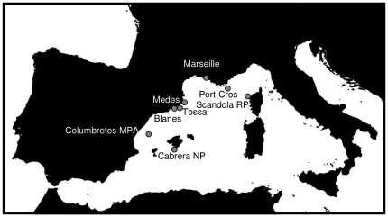
Quantitative surveys on the sponge mortality were carried out in two Marine Protected Areas in the western Mediterranean Sea: Cabrera National Park (Balearic Islands, Spain, hereafter Cabrera NP) and Reserve Naturelle de Scandola (Corsica, France, hereafter Scandola RN) (Fig. 1). Quantitative assessments of sponge population's status started in 2007 in Cabrera NP and were extended in 2008 to Scandola RN till late summer 2010. After the first observation of mortality signs in October 2008, qualitative surveys (presence/absence) of mortality (presence of partial or total necrosis) were carried out in six other zones encompassing the Spanish and French coasts (Table 1; Fig. 1). Finally, sponge mortality recorded by other research teams in several areas of western Mediterranean was also compiled to assess the geographical extent of the sponge die-offs (Table 1).
Figure 1. The western Mediterranean Sea, showing the surveyed localities of Ircinia fasciculata.
Table 1. Localities and geographical position where the presence (+) or absence (−) of mass mortality was assessed.
| Locality | coordinates | I. fasciculate die-off |
| *Blanes (2 sites) | 41.674N ; 2.74E | − |
| *Tossa de Mar (1 site) | 41.720N ; 2.93E | − |
| *Medes Islands MPA (3 sites) | 42.047N ; 3.22E | − |
| *Cabrera NP (3 sites) | 39.140N ; 2.94E | + |
| *Scandola RP (1 site) | 42.356N ; 8.56E | + |
| *Marseille (4 sites) | 43.297N ; 5.38E | − |
| *Port-Cros (3 sites) | 43.009N ; 6.38E | − |
| *Columbretes MPA (6 sites) | 39.055N ; 0.44E | − |
| **Granada Coast | 36.728N ; 3.44W | + |
| **Chafarinas Islands | 35.005N ; 2.25W | + |
Present study
Maldonado et al. 2010.
Target species
The species selected for the survey were Ircinia fasciculata (Esper 1794) and Sarcotragus spinosulum (Schmidt 1862). Both species are abundant in shallow waters of the western Mediterranean [20], are phylogenetically close (Sarcotragus was considered a subgenus of Ircinia until 2002 [21]), and belong to the category of “bacteriosponges” because they harbor a high proportion of symbiotic microbes relative to sponge biomass [22]. I. fasciculata harbors both cyanobacteria and heterotrophic bacteria, while S. spinosulum hosts only heterotrophic bacteria. Both species inhabit the photophilic assemblages and are usually restricted to the first 20 m of depth.
Sponge survey and mortality assessment
Several populations of I. fasciculata and S. spinosulum were monitored for sponge densities and injury incidence in July and October (before and after the period of warmest temperatures) by placing 20 quadrats of 2500 cm2 haphazardly between 10 and 15 m depth at each surveyed site. We counted the total number of specimens and those suffering partial or total mortality (when specimens were totally dead, the skeletons remained in situ for ca. 1 month after the sponge detached from the substratum [23], [24]). The surveys were carried out in three sites in Cabrera NP from July 2007 to October 2010 and in one site in Scandola RN from October 2008 to October 2010. In the remaining surveyed areas we annually surveyed only for the presence or absence of mortality after summer (Table 1).
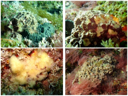
As affected sponges we considered those individuals displaying small whitish spots on the sponge surface characteristics of the early sponge disease; as well as those specimens displaying one or more necrotic zones, where the skeleton (spongin network) was free of cellular material (see Results) characteristic of the later disease stages (see Results Fig. 2).
Figure 2. Aspects of Ircinia fasciculata mortality.
a) First stage of the affectation on I. fasciculata (arrow head) compared with healthy S. spinosulum (arrow), b) small whitish spots on the I. fasciculata surface, c) completely dead I. fasciculata with the skeleton (spongin network) free of cellular material, (d) white bacterial veil on unhealthy Sarcotragus spinosulum surface.
The density and percentage of injured sponges in Cabrera NP was analyzed for both species by two-way ANOVA with time (fixed) and site (random) as factors. Differences in the density and percentage of injured sponges in Scandola RN across time were analyzed by one-way ANOVA. Tukey tests were used for post-hoc comparisons. Prior to analysis, the homogeneity of variances was verified with Cochran's test. The analyses were carried out by using the Statistica 8.0 software.
Ultrastructural study
Healthy and injured tissues from the same specimen of I. fasciculata were examined by transmission electron microscopy (TEM) to assess the main ultracellular changes in sponge cells, cyanobacteria, and heterotrophic bacteria, associated with the sponge disease. For TEM observation, small samples (2–3 mm in diameter) were fixed in a solution consisting of 1% OsO4 and 2% glutaraldehyde in 0.4 M sodium cacodylate buffer (pH 6.4) with 10% sucrose for 2 h at 4°C [25]. The samples were then rinsed in the buffer solution, dehydrated in a graded ethanol series to 70% and embedded in Spurr's resin. Semi-thin sections were stained with uranyl acetate and lead citrate and observed through a Hitachi H-600 transmission electron microscope (Microscopy Unit of the Scientific and Technical Services at the University of Barcelona).
Experimental approach to assess the effects of temperature on the photochemical efficiency of I. fasciculata cyanobacteria symbionts
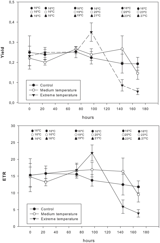
Variation on the cyanobacteria photochemical efficiency under several temperatures was measured in a set of laboratory experiments. Eighteen specimens (ca. 25 cm in diameter) of I. fasciculata were randomly collected in Tossa de Mar, Girona, Spain, at a depth of 10 m in January 2010, when the water temperature was 14°C. The samples were transported in aerated seawater under low-light conditions at 14°C to the CEAB aquarium facilities, where the experiments were conducted. The specimens of I. fasciculata were randomly transferred to three experimental tanks (50 L): one control and two temperature treatments. The experiment temperature treatments were: “control” (16°C), “normal summer T” (23°C) and “extreme summer T” (27°C) according to the T values recorded in the NW Mediterranean [26]. Light was provided by several metal halide lamps (Philips, HPIT 40 W) at an intensity of 400 µmol photon m−2 s−1 to simulate the irradiance conditions of the sponge habitat [27]. Irradiance was measured using a spherical Li-Cor underwater quantum sensor (LI-193SA). Sponges were maintained for a 48 h adaptation period under low (16°C) temperature conditions in the three tanks. After the adaptation period, we progressively increased the temperature (1°C every 12 h) in the treatment tanks for 8 days until the “medium” (23°C) and for 10 days until the “extreme” (27°C) temperatures were reached, using aquarium heaters connected to electronic controllers (±0.1°C). Temperatures were subsequently maintained at 16°C, 23°C and 27°C for 48 h. The effect of temperature on cyanobacteria was estimated by measuring the symbionts' photochemical efficiency by means of chlorophyll fluorescence parameters (effective quantum yield: ΦPSII quantum yield and photosynthetic electron transfer: ETR = ΦPSII *PAR*0.5*AF, where ΦPSII is the effective quantum yield of Photosystem II and PAR is the photosynthetically available irradiance reaching the sample) using a PAM fluorometer (DIVING-PAM, Walz, Germany) [28].
One-way ANOVA on ETR and ΦPSII values obtained during the first day of the experiment was carried out to verify that sponges undergoing different treatments started at similar photosynthetic activity conditions. To assess the effects of increasing temperatures on cyanobacteria photosynthetic activity, one-way ANOVA was used to test for differences in ETR and ΦPSII values obtained for each treatment at the end of the experiment. Prior to analysis, the homogeneity of variances was verified by Cochran's test. These analyses were carried out with the Statistica 8.0 program.
Temperature conditions
High-resolution (hourly records) temperature series were obtained at Cabrera NP and Scandola RN at 15 m depth since 2007 and 2008 respectively. Temperature was recorded in situ by Stowaway Tidbits, autonomous sensors (0.2 °C precision, 0.15 °C resolution), and recovered annually by divers (see [26] for more details). For the data analysis we considered the period from August 1 to September 30 as the summer period. The average mean and maximum temperatures for each summer period were calculated. Moreover, given the short time interval between measurements (1 h), the data set allowed the calculation of the percentage of time spent above the following temperature thresholds: 23, 24, 25 and 26°C. We used these different descriptors of summer temperature conditions to explore the relationship between the mortality on sponge populations and temperature.
Relationship between mortality and temperature conditions
A regression analysis between the percentage of time above the maximum temperature threshold during the summer period and the percentage of injured I. fasciculata specimens was performed to explore the potential role of the high temperatures in the I. fasciculata die-off. As thermal regimes are particular for each study area, thresholds selected were those showing the highest differences between mortality and non mortality years in Cabrera NP (>26°C) and Scandola RN (25°C) (see results). The analysis was performed with Statistica 8.0.
Results
Morphological and structural features of the sponge disease
The disease in I. fasciculata specimens was initially characterized by small yellowish spots that soon transformed into small zones of necrotic tissue, and then enlarged and fused to form, rounded areas of necrotic ectosome in a few days (Fig. 2a, b). Soon after, the necrotic, rounded areas interconnected, forming large areas of exposed skeleton deprived of tissue. Some partially affected specimens still surviving at the end of the summer were able to heal these wounds in few weeks, while others progressively amassed necrotic tissue and completely died (Fig. 2c). Skeletons of completely dead specimens were eventually detached by storms, leaving a conspicuous mark of naked substrata in the rock that lasted several months.
In contrast, the few injured S. spinosulum never displayed the above described pattern, but showed a typical bacterial veil covering the sponge ectosome (Fig 2d), as frequently reported in rotten surfaces of many other sponges (e.g., [29], [30]).
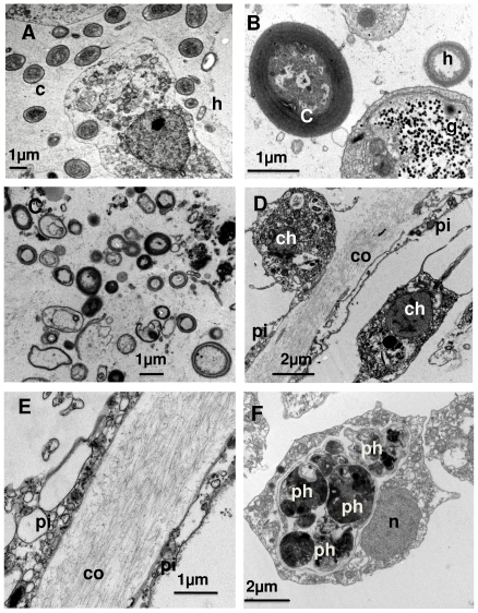
TEM observation of spotted (death) areas, zones in close vicinity to the spots, and healthy zones of I. fasciculata showed remarkable differences at an ultrastructural level. Healthy zones contained healthy cyanobacteria (Fig. 3a, b), heterotrophic “symbiotic” bacteria (Fig 3c), abundant collagen bundles (Fig. 3d, e) and choanocytes with huge amounts of phagosomes (Fig. 3f). Copepod exoskeletons, semi-digested heterotrophic bacteria, and muscle tissue, were among the identifiable materials within choanocyte phagosomes.
Figure 3. Several ultrastructural aspects of a healthy Ircinia fasciculata.
A) Archeocyte of a subectosomal zone, surrounded by numerous cyanobacteria (c) and some heterotrophic bacteria (h); B) Detail of a healthy cyanobacteria, close to an archeocyte plenty of glycogen rosettes (g); C) “symbiotic” bacterial community, typically found in the sponge; D) collagen layer (co) between endopinacocytes (pi) separating two choanocyte chambers (ch: choanocytes); E) collagen bundles, surrounded by endopinacocytes (pi); F) basal section of a choanocyte, full of heterogeneous phagosomes (ph), which is characteristic of the species (n: nucleus).
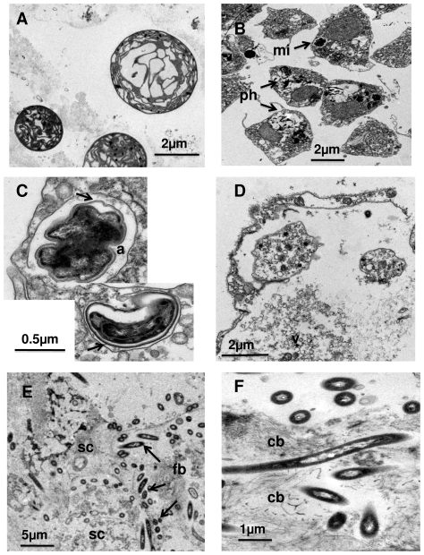
The sponge tissue in close vicinity to the necrotic zones contained both choanocytes and archeocytes, plenty of phagocytosed cell debris, and empty vesicles. The cell nuclei were irregular in shape, with a poorly defined nuclear membrane. Cyanobacteria were always degraded, showing a cell volume that was twice to three times the volume of normal cyanobacteria (Fig. 4 a, b). Collagen fibrils were equally abundant in affected and healthy tissue although showed a poorer organization in the former (Fig. 4f). A non-identified microorganism, with a multilayered membrane was quite frequent in cellular vacuoles of disorganized choanocytes, where it divided (Fig. 4c). However, it could not be related unambiguously to the disease.
Figure 4. Several ultrastructural aspects of unhealthy specimens of Ircinia fasciculata.
A, B, C) Zone in the vicinity of a pustule (whitish spot): A) cyanobacteria under several degradation stages; B) section of a degenerating choanocyte chamber with multiple phagosomes (ph) and an unknown microorganism (mi); C) closer view of the “rare” microorganism, which divides within cell vacuoles (a) and shows a membrane complex (arrows) and an irregular, dark inner zone; D, E, F). Zone corresponding to a pustule (died whitish spot): D) completely degenerated cells with multiple released vesicles (v); E) Abundant foreign bacteria (fb), similar to the morphotype reported to consume on the skeleton of dead sponges (Vacelet et al. 1994), among completely degenerated sponge cells (sc); F) Close view of this particular bacterium, which is always associated to the collagen bundles (cb).
The necrotic zones (whitish spots) showed total tissue destruction, absence of cyanobacteria, numerous vesicles, broken membranes, and collagen remains (Fig. 4d). A characteristic bacterium invaded the collagen areas (Fig. 4e, f). This bacteria was similar to that described tunneling spongin fibers in free skeletons of dead keratose, species (e.g. [31]), and have also been observed perforating the spongin basal plate of several healthy encrusting species such as Crambe crambe (authors obs.).
No specific pathogen involved in the disease could be identified in the ultrastructural study. Cell destruction as indicated by broken membranes and cell disorganization, degeneration and then absence of cyanobacteria and the presence of heterotrophic bacteria similar in ultrastructure to those present in healthy tissue were the main features of the affected sponge tissue.
Impact of the sponge disease
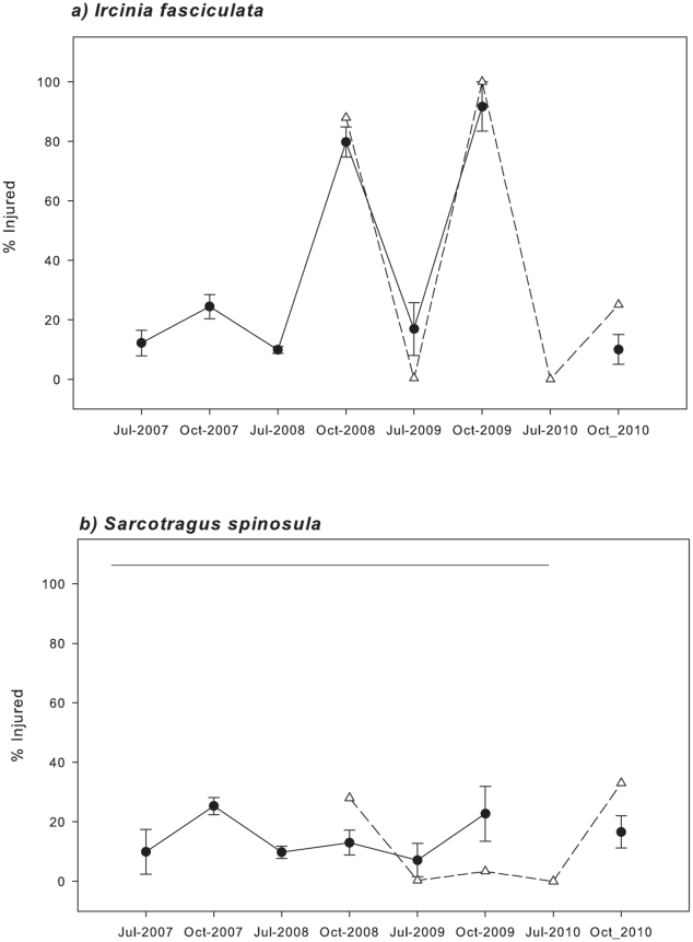
The percentage of injured I. fasciculata specimens peaked up to 80–100% of the surveyed individuals for both areas, Cabrera NP and Scandola RN, in October 2008 and again in October 2009 (Fig. 5a, Table 2). In contrast, during the rest of the study period, injured individuals never exceeded 30% (Fig. 5a).
Figure 5. Mean percentages of injured Ircinia fasciculata (a) and Sarcotragus spinosulum (b) specimens recorded in Cabrera NP (circles) and Scandola RN (triangles) during the monitoring period.
Bars represent standard errors. Mean concentrations, which were not significantly different in a Tukey test, are joined by horizontal lines.
Table 2. Two-way ANOVA results for Cabrera NP Ircinia fasciculata and Sarcotragus spinosulum densities and % of injured specimens, with site (random) and time (fixed) factors.
| Cabrera NP | |||
| I. fasciculata density | |||
| MS | F | Post-hoc (Tukey-test) | |
| Site | 0.307 | 3.01 | |
| Time | 3.071 | 30.16** | J07 = O07 = J08 = O08>J09 = O09 = O10 |
| Site*Time | 0.67 | 0.65 | |
| S. spinosulum density | |||
| MS | F | Post-hoc (Tukey-test) | |
| Site | 0.218 | 2.851 | |
| Time | 0.522 | 6.823* | (O07 = J08)>O09 |
| Site*Time | 0.577 | 0.744 | |
| % I. fasciculata Injured | |||
| MS | F | Post-hoc (Tukey-test) | |
| Site | 17.00 | 0.09 | |
| Time | 7672 | 42.05** | J07 = O07 = J08 = J09<O08 = O09 = O10 |
| Site*Time | 237 | 1.30 | |
| % S. spinosulum Injured | |||
| MS | F | Post-hoc (Tukey-test) | |
| Site | 295.9 | 2.868 | |
| Time | 315.6 | 3.059 | |
| Site*Time | 137.7 | 1.334 |
ANOVA results for Scandola RP Ircinia fasciculata and Sarcotragus spinosulum densities and % of injured specimens across time.
For S. spinosulum, the percentage of injured specimens varied between 10% and 30% in both areas from 2007 to 2010, but differences over time were not significant in any area (Fig. 5b, Table 2).
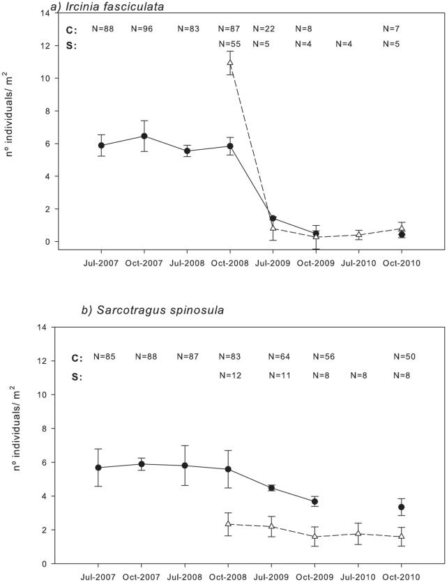
In both sites, densities of I. fasciculata decreased dramatically from around 10 (in Scandola RN) and 7 (in Cabrera NP) specimens·m−2 before the mortality event (summer 2008) to less than 1 specimens·m−2 at the end of autumn 2010 (Fig. 6a). The ANOVA analyses showed that time had a significant effect on I. fasciculata densities in Cabrera NP and Scandola RN, which were significantly lower in July and October 2009 and July 2010, after the first outbreak (Fig. 6a, Table 2). In Cabrera NP, where three sites were monitored, no significant differences in I. fasciculata densities were observed between sites (populations); neither was the interaction between locality and time significant (Table 2), indicating that the pattern of density losses was similar everywhere (Table 2).
Figure 6. Mean densities of Ircinia fasciculata (a) and Sarcotragus spinosulum (b) recorded in Cabrera NP (circles, C) and Scandola RN (triangles, S) during the monitoring period.
Bars represent standard errors. Mean concentrations, which were not significantly different in a Tukey test, are joined by horizontal lines. N = number of specimens sampled.
S. spinosulum densities in Cabrera NP ranged from 6.8 to 3.35 specimens·m−2 from July 2008 to October 2010. Time had a significant effect on S. spinosulum densities; the densities in October 2009 and July 2010 were lower than those recorded in October 2007 and July 2008 (Fig. 6b, Table 2). In Cabrera NP, no significant differences in S. spinosulum densities were observed between sites, and there was no interaction between locality and time, which indicated a similar density pattern in the three localities (Table 2). Conversely, in Scandola RN, time had no significant effect, even though densities decreased from 2.33 to 1.60 specimens·m−2 between October 2008 and October 2010 (Fig. 6b, Table 2).
Surveys along the Catalan and Provence coasts and in the Balearic Islands indicated that no appreciable mortality occurred after the summer period, from 2007 to 2010. In contrast, mortality records from other western Mediterranean areas by other research teams indicated that I. fasciculata was also injured in the southern coast of Spain (Table 1).
Temperature conditions during the sponge die-off
The mean (± s.e.) and maximum temperatures during summer periods are summarized in Table 3. Summer mean and maximum temperatures at 15 m observed in 2008 and 2009 were among the highest values recorded in both areas during the 2007–2010 period (Table 3). Mean temperatures ranged between 22.45 and 24.4°C in Scandola RN and between 25.4 to 26.1°C in Cabrera NP; while maximum temperatures at 15 m ranged between 24.5 and 26.9°C in Scandola RN and between 26.7 and 27.7°C in Cabrera NP (Table 3).
Table 3. Temperature records (from 1 August to 30 September) at 15 m depth obtained in Scandola RN and Cabrera NP.
| Scandola RN -15 m | |||
| TX | s.e. | Tmax | |
| 2007 | 22.45 | 0.1500 | 24.53 |
| 2008 | 23.55 | 0. 353 | 26.27 |
| 2009 | 24.40 | 0.100 | 26.90 |
| 2010 | 22.49 | 0.0437 | 25.30 |
TX and T max refer to the average mean and maximum temperatures, respectively, obtained for the available period in each study area.
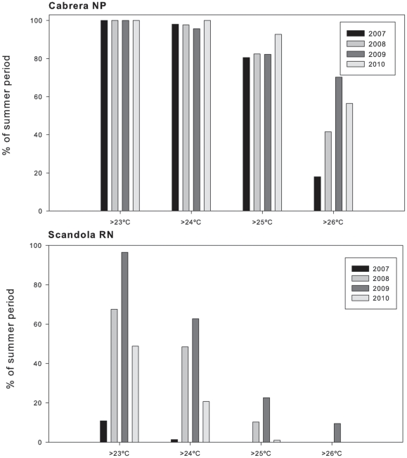
Regarding temperature thresholds, in Cabrera NP the years 2008 and 2009 displayed higher percentages of time of exposition than in 2007 regardless the temperature threshold considered, although the percentage of time above 26°C showed the largest differences. In 2010, values were similar to those of the mortality years; however by then the density of populations of I. fasciculata were really low (see Impact of sponge disease section) preventing the assessment of sponge mortality (Figure 7a).
Figure 7. Percentage of the summer period with temperatures above 23°C, 24°C, 25°C and 26°C in Cabrera NP (a) and above 23°C, 24°C and 25°C in Scandola RN (b).
Data from SOMLIT (temperature sensors) data series.
In contrast, temperatures exceeded 25°C and 26°C in Scandola RN only in the mortality years (Table 3, Fig. 7b). Considering the percentages of time above 24 and 25°C, differences among years with and without sponge mortality were evident: in 2008 and 2009 the percentage of time with water temperatures above 25°C were at least two-fold than that in 2007 and 2010 (Fig. 7b). Overall, summer temperatures conditions at 15 m were much warmer, in Cabrera NP than in Scandola RN (Table 3).
Relationship between sponge mortality and temperature
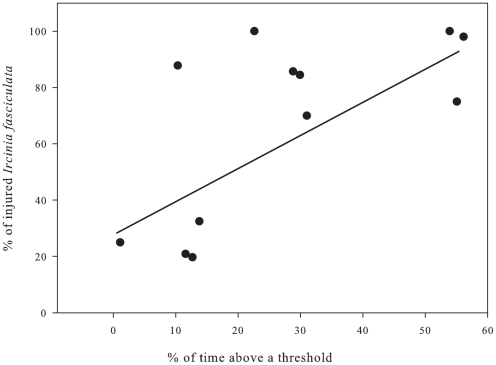
The percentage of time above 26°C for Cabrera populations and above 25°C for the Scandola population was positively correlated to the percentage of affected individuals of I. fasciculata for each site (Fig. 8, R = 0.672, F = 8.15, p = 0.0171). The greater percentage of summer time above a maximum threshold, the higher the impact on sponge populations (Fig. 8).
Figure 8. Relationship between the percentage of time above a temperature threshold (26°C) from August 1 to September 30 and the percentage of affected I. fasciculata colonies in Cabrera NP.
Experimental outcome of increased temperature on the photosynthetic microsymbionts
The effective quantum yield (ΦPSII) and the photosynthetic electron transfer (ETR) were similar for all specimens at the beginning of the experiment (ANOVA F-test; p>0.1; Fig. 9). At the end of the experiment, photosynthetic parameters were significantly lower in I. fasciculata specimens submitted to extreme temperatures, compared to specimens submitted to low (control) and medium temperatures, which did not differ between them (Table 4, Fig. 9). Photosynthesis was inhibited in specimens submitted to extreme temperatures because both ΦPSII and ETR significantly decreased to nearly zero at the end of the experiment (Table 4, Fig. 9).
Figure 9. The effective quantum yield (ΦPSII) and photosynthetic electron transfer (ETR) during the experiment for the "control,” “medium” and “extreme” treatments.
Bars represent standard errors.
Table 4. 1-Results of one-way ANOVA comparing initial and final fluorescence variables (Yield, ETR) for the Control, Medium and Extreme temperature treatments, and 2- results of one-way ANOVA comparing different treatments fluorescence variables (Yield, ETR) for the initial and final time point of the experiment.
| Yield | ETR | ||||||
| MS | F | MS | F | ||||
| 1 | Control | 0.009 | 0.340 | n.s. | 32.76 | 0.367 | n.s. |
| 1 | Medium temperature | 0.018 | 2.591 | n.s. | 91.81 | 2.267 | n.s. |
| 1 | High temperature | 0.075 | 7.924 | ** | 307.70 | 6.198 | ** |
| 2 | Initial time (among treatments) | 0.001 | 0.066 | n.s. | 0.454 | 0.005 | n.s. |
| 2 | Final time (among treatments) | 0.021 | 4.566 | * | 81.71 | 4.140 | * |
*show significant differences p<0.05 and
**p<0.01.
Discussion
Sponge mass mortality events occurred during the late summers of 2008 and 2009, affecting I. fasciculata populations thriving in shallow sublittoral habitats in central and southern parts of the western Mediterranean Sea. These mortality events affected 80 to 100% of I. fasciculata individuals in the studied populations. During the rest of the study period, basal mortality of I. fasciculata was also observed. In contrast, we did not detect significant mortality rates for S. spinosulum during the study period. Populations of other keratose sponge species involved in previous mortality events (e.g., Spongia agaricina, Hippospongia communis) [4]–[6] did not show any signs of mortality.
Our surveys detected the same mortality pattern for all populations studied, even though they were carried out in two Mediterranean areas hundreds of kilometers apart (Cabrera NP and Scandola RN). Almost all of the I. fasciculata individuals showed partial or total mortality. Conversely, even though S. spinosulum shared habitat with I. fasciculata in both localities, it only occasionally displayed small necrotic areas covered by a white bacterial veil.
The disease extended along the coasts of Corsica, the Balearic Islands, and the southern Spanish littoral (Granada and Chafarinas). We did not find signs of mortality in any of the other surveyed northwest Mediterranean areas (Table 1) nor in Columbretes MPA (authors obs., Diego K. Kersting, pers. comm.).
The geographical distribution of the disease strongly suggests that only the warmest areas of the western Mediterranean were affected. Mortality was observed only in areas with mean summer temperatures at least 1 to 2°C above those previously recorded on the northern Mediterranean coast [26], [31]. This result strongly contradicts the hypothesis of [32] suggesting the disease originated in the northwestern Mediterranean and only recently reached southern areas. Although significant sponge mortalities have been reported several times in the Mediterranean [5]–[6], [8], [33]–[34], this new disease episode exclusively affects I. fasciculata, pointing to a possible role of symbiotic cyanobacteria in the disease. Current disease features differ from those previously reported (before 2008) for other Mediterranean sponges, which start with grey or whitish zones and a bacterial veil on the sponge surface [6], [34]. In contrast, in the current mortality event, the affected sponges were characterized by small round pustules that developed into large, rounded areas of dead tissue, which led to the complete death of most specimens. The external symptoms of the current disease were more akin to those reported for Spongia officinalis, Hippospongia communis communis or Ircinia variabilis, which also started with the formation of white spots on the sponge surface [5], [8], [35]–[37].
Our study reports, for the first time, population descriptors (mortality incidence and densities) of sponge species before and after mortality outbreaks, allowing an accurate assessment of the impact of mortality on the species. Previous studies were carried out when mortality had already affected many individuals in the population, causing the density decline to not be properly evaluated. Shallow water populations of I. fasciculata were clearly decimated after the 2008 and 2009 outbreaks in both studied areas, with mortality rates similar to those reported for the most dramatic examples of sponge mortality (Spongia spp.) in the Caribbean Sea (70 to 95%; [38]). In contrast, in southeastern Spain (Granada coast; [32]), the impact was at least three-fold lower than in Cabrera NP and Scandola RN (present study). Bearing in mind that affected sponges fall apart and disappear about one month after displaying the first signs of injury ([23]–[24]; authors obs.), this inter-regional difference could be attributed to mortality underestimation in the Granada area, where surveys were carried out two months after the 2008 outbreak [32].
Recovery of the impacted populations is uncertain. It has been reported that when a small proportion of the sponges are affected, populations are able to recover [8]. In contrast, when mortality rates are high (similar to the values reported here) or if other mortality sources are acting synergistically (e.g., overfishing), sponge populations may disappear [39]. As a result, we may predict a dramatic extinction of the populations studied. However, only continued monitoring of the affected and unaffected populations will allow us to elucidate the long-term consequences of these events, which may not only harm sponge populations but also alter benthic assemblages.
Previous massive sponge mortality events in the Mediterranean have been related to high temperatures [4]–[6], [33]. Our temperature records also indicate that the I. fasciculata outbreak coincided with particularly high seawater temperatures recorded in 2008 and 2009 in Cabrera NP and in 2008 in Scandola RN (Table 3 and Fig. 5). We found a significant positive relationship between the mortality rate and the percentage of time that sponge populations experienced temperatures above a certain threshold (Fig. 7). In contrast, Maldonado et al. (2010) studied the same mortality outbreak in I. fasciculata populations but did not find any relationship between temperature and mortality incidence. However, in the later study, the data used to search for a temperature-disease relationship were sea surface temperature (SST) from satellite retrievals, which likely are not representative of the temperatures experienced by sponge populations in situ [26]. Interestingly, northern unaffected areas (Marseilles, Port Cros, Medes Islands, Blanes; www.t-mednet.org and data available from the authors) also reached warm-temperature conditions at 15 m depth in 2008 and in 2009, but temperatures never surpassed 24°C, which commonly occurred in the affected zones. This observation points to 24°C as the threshold for triggering I. fasciculata disease. However, I. fasciculata populations from Cabrera NP were often subjected to temperatures above 24°C in other years (Figure 7a), and only when temperatures exceed the 26°C threshold are clear-cut inter-annual differences observed. Meanwhile, in Scandola RN, temperatures above 26°C were rarely reached (Figure 7b and [26]), suggesting a differential temperature threshold for triggering mortality in the Cabrera NP and Scandola RN populations. It has been already demonstrated that different populations of the same species subjected to contrasting temperature regimes can show differential resilience to global warming [40]. The acquisition of a long-term series of biological and temperature data will allow us to better establish the linkage of mortality events with future temperature conditions.
The other surveyed species, S. spinosulum, was not significantly affected in 2008 and 2009 or other more recent mass mortality events [4]. Individuals of this species have larger sizes and higher abundances in the warmer waters of the central and eastern Mediterranean than in the colder western Mediterranean [20], while individuals of I. fasciculata are of similar size in colder and warmer Mediterranean regions. This positive relationship between maximum sponge size and water temperature suggests that S. spinosulum is better adapted to withstand higher temperatures than I. fasciculata.
Although current and previous outbreaks of I. fasciculata are positively related to high temperatures, the primary cause of the disease remains controversial. Exposure to high temperatures may cause partial or total death of specimens as a result of physiological stress [41]–[44], a decrease in the efficacy of defense mechanisms [45], [46], and/or the development of pathogens [17], [47]–[48]. However, sponges more frequently affected by periodic diseases belong to the so-called “bacteriosponge” category because they harbor abundant a priori symbiotic bacteria; future research should focus on changes in the composition of the symbiotic bacterial community under different temperature conditions. In fact, [49] reported shifts in the composition of the microsymbiotic community in tropical sponges subjected to elevated temperatures, and they attributed sponge mortality to the loss of symbionts and the consequent establishment of alien microbial populations, including potential pathogens.
I. fasciculata and S. spinosulum populations showed significantly different responses to the mass mortality events analyzed in this study. To explain this differential impact, the working hypothesis was that high temperatures might induce a breakdown of the cyanobacteria-sponge symbiosis present in I. fasciculata, but not in S. spinosulum. The decay of cyanobacteria might play a key role in sponge mortality because these symbionts seem to greatly contribute to the carbon requirements of the sponge [50]–[54].
However, the sponge-cyanobacteria symbiosis also implies biochemical reactions with harmful consequences for the sponge. Elevated temperatures may lead to an increase in reactive oxygen species (ROS) [55] and to a lower antioxidant efficiency [56]. This ROS increase has been related to the presence of tissue necrosis (white patches on the ectosome) in the Mediterranean sponge, Petrosia ficiformis, after a period of calm, warm water [57], which may be a plausible hypothesis to explain the present I. fasciculata disease.
Our experiment demonstrated that high temperatures result in a significant reduction in photosynthetic efficiency (Fv/Fm) and ETR of the symbiotic cyanobacteria, with consequent inhibition of photosynthesis (Fig. 9). The TEM observations revealed degeneration or even total absence of cyanobacteria on the affected sponge tissues, while heterotrophic bacteria were present in both affected and unaffected tissues. A particular bacterial type that was only present in the affected tissues is ultrastructurally similar to that reported for tunneling spongin plates and dead skeletal fibers of both healthy sponges and exposed spongin fibers. Thus, it seems to be an opportunistic microorganism that consumes skeletal material of dead sponges, as suggested by [8] and [58], rather than the primary cause of infection, as proposed in [32]. Although a dysfunction in cyanobacterial functioning cannot be considered the only cause of sponge mortality, impairment of sponge fitness due to increasing temperatures might trigger the virulence of pathogens that would otherwise have remained inactive or few in numbers. Furthermore, symbiotic relationships have evolved under constant environmental conditions, and thus, slight changes in environmental variables may prompt their imbalance. In this sense, invertebrates that harbor symbionts, such as corals and sponges, seem to be particularly threatened by warming [59]–[61], which causes these organisms to become more susceptible to pathogenic agents when exposed to a warming climate [2], owing to basic modifications in their biological properties.
In the present study, we were able to quantify devastating mortalities in the sponge I. fasciculata, leading to quasi-local extinctions of the studied populations. The current mortality event is the most severe reported to date in the Mediterranean.
Because future climatic projections for the Mediterranean region clearly indicate warming, with increases in extreme temperatures and heat waves [62]–[64], new mass mortality events are extremely likely to occur during the next several decades. Sponges have been demonstrated to be very sensitive to global change perturbations (e.g. [34], [49], [65]–[67]). In particular, the so-called bacteriosponges are especially vulnerable to environmental changes that may cause imbalances in their delicate symbiotic relationships with microbes. These massive sponges play a structural role in benthic assemblages, and their alteration may threaten rich Mediterranean biodiversity.
Acknowledgments
We would like to thank the director and staff of the Archipelago of Cabrera National Park, Scandola Natural Reserve and Columbretes MPA for permissions and facilities.
Footnotes
Competing Interests: The authors have declared that no competing interests exist.
Funding: Financial support was provided by the Fundación Biodiversidad, an EU Reintegration grant (ERG-2009-248252), the BioRocK project from the Spanish Ministry of Science and Innovation (CTM2009-08045), and the GRACCIE project C5D2007-00067 from the Spanish Ministry of Science and Innovation. EC was funded by a Juan de la Cierva Postdoctoral Fellowship. The funders had no role in study design, data collection and analysis, decision to publish, or preparation of the manuscript.
References
- 1.Salomon S, Qin D, Manning M, Maquis M, Averys K, et al. Fourth's assessment report:The Physical Science Basis. Geneve: 2007. Intergovernmental Panel Climate Change. p. 114 p. [Google Scholar]
- 2.Harvell CD, Kim K, Burkholder JM, Colwell RR, Epstein PR, et al. Emerging marine diseases —climate links and anthropogenic factors. Science. 1999;285:1505–1510. doi: 10.1126/science.285.5433.1505. [DOI] [PubMed] [Google Scholar]
- 3.Harvell CD, Mitchell JR, Ward S, Altizer A, Dobson RS, et al. Climate warming and disease risks for terrestrial and marine biota. Science. 2002;296:2158–2162. doi: 10.1126/science.1063699. [DOI] [PubMed] [Google Scholar]
- 4.Garrabou J, Coma R, Bally M, Bensoussan N, Chevaldonné P, et al. Mass mortality in Northwestern Mediterranean rocky benthic communities: effects of the 2003 heat wave. Glob Change Biol. 2009;15:1090–1103. [Google Scholar]
- 5.Cerrano C, Bavestrello G, Bianchi N, Cattaneo-Vietti R, Bava S, et al. A catastrophic mass-mortality episode of gorgonians and other organisms in the Ligurian Sea (NW Mediterranean), summer 1999. Ecol Let. 2000;3:284–293. [Google Scholar]
- 6.Perez T, Garrabou J, Sartoretto S, Harmelin JG, Francour P, et al. Mortalité massive d'invertébrés marins: Un événement sans précédent en Méditerranée nord-occidentale. Compt Ren Acad Sci Paris III. 2000;323:853–865. doi: 10.1016/s0764-4469(00)01237-3. [DOI] [PubMed] [Google Scholar]
- 7.Gaino E, Pronzato R. Ultrastructural evidence of bacterial damage to Spongia officinalis fibres (Porifera, Demospongiae). Dis Aquat Organ. 1989;6:67–74. [Google Scholar]
- 8.Gaino E, Pronzato R, Corriero G, Buffa P. Mortality of commercial sponges: Incidence in two Mediterranean areas. Bollet Zool. 1992;59:79–85. [Google Scholar]
- 9.Bavestrello G, Bertone S, Cattaneo-Vietti R, Cerrano C, Gaino E, et al. Mass mortality of Paramuricea clavata (Anthozoa, Cnidaria) on Portofino Promontory cliffs, Ligurian Sea, Mediterranean Sea. Marine Life. 1994;4:15–19. [Google Scholar]
- 10.Rodolfo-Metalpa R, Bianchi CN, Peirano A, Morri C. Coral mortality in NW Mediterranean. Coral Reefs. 2000;19:24. [Google Scholar]
- 11.Vezzulli L, Pruzzo C, Huq A, Colwell R. Environmental reservoirs of Vibrio cholerae and their role in cholera. Environ Microbiol Rep. 2010;2:27–33. doi: 10.1111/j.1758-2229.2009.00128.x. [DOI] [PubMed] [Google Scholar]
- 12.Bianchi N. Biodiversity issues for the forthcoming tropical Mediterranean Sea. Hydrobiologia. 2007;580:7–21. [Google Scholar]
- 13.Garrabou J, Perez T, Sartoretto S, Harmelin JG. Mass mortality event in red coral (Corallium rubrum, Cnidaria, Anthozoa, Octocorallia) populations in Provence region (France, NW Mediterranean). Mar Ecol Prog Ser. 2001;217:263–272. [Google Scholar]
- 14.Linares C, Coma R, Diaz D, Zabala M, Hereu B, et al. Immediate and delayed effects of a mass mortality event on gorgonian population dynamics and benthic community structure in the NW Mediterranean Sea. Mar Ecol Prog Ser. 2005;305:127–137. [Google Scholar]
- 15.Coma R, Linares C, Ribes M, Diaz D, Garrabou J, et al. Consequences of a mass mortality in populations of Eunicella singularis (Cnidaria: Octocorallia) in Menorca (NW Mediterranean). Mar Ecol Prog Ser. 2006;327:51–60. [Google Scholar]
- 16.Kushmaro A, Bani E, Loya Y, Stackebrandt E, Rosenberg E. Vibrio shiloi sp. nov. the causative agent of bleaching of the coral Oculina patagonica. Int J Syst Evol Micr. 2001;51:1383–1388. doi: 10.1099/00207713-51-4-1383. [DOI] [PubMed] [Google Scholar]
- 17.Bally M, Garrabou J. Thermodependent bacterial pathogens and mass mortalities in temperate benthic communities: a new case of emerging disease linked to climate change. Glob Change Biol. 2007;13:2078–2088. [Google Scholar]
- 18.Hentschel U, Usher KM, Taylor MW. Marine sponges as microbial fermenters. FEMS Microbiol Ecol. 2006;55:167–177. doi: 10.1111/j.1574-6941.2005.00046.x. [DOI] [PubMed] [Google Scholar]
- 19.Taylor MW, Radax R, Steger D, Wagner M. Sponge-associated microorganisms: evolution, ecology, and biotechnological potential. Microbiol Mol Biol Rev. 2007;71:295–347. doi: 10.1128/MMBR.00040-06. [DOI] [PMC free article] [PubMed] [Google Scholar]
- 20.Uriz MJ, Rosell D, Martin D. The sponge population of the Cabrera archipelago (Balearic Islands): characteristics, distribution and abundance of the most representative species. PSZNI Mar Ecol. 1992;113:101–117. [Google Scholar]
- 21.Cook SC, Bergquist PR. Family Irciniidae. In: Hooper JNA, Van Soest RWM, editors. Systema Porifera: A guide of the classification of the sponges. New York: Kluwer Academic; 2002. pp. 1021–1050. [Google Scholar]
- 22.Simpson TL. New York, Berlin, , Heidelberg, Tokyo: Springer Verlag; 1984. The Cell Biology of Sponges. p. 662 p. [Google Scholar]
- 23.Smith FGW. Sponge disease in British Honduras, and its transmission by water currents. Ecology. 1941;22:415–421. [Google Scholar]
- 24.Wulff J. Rapid diversity and abundance decline in a Caribbean coral reef sponge community. Biol Conserv. 2006;127:167–176. [Google Scholar]
- 25.Leys SP, Reiswig HM. Transport pathways in the neotropical sponge Aplysina. Biol Bull. 1998;195:30–42. doi: 10.2307/1542773. [DOI] [PubMed] [Google Scholar]
- 26.Bensoussan N, Romano JC, Harmelin JG, Garrabou J. High resolution characterization of northwest Mediterranean coastal waters thermal regimes: to better understand responses of benthic communities to climate change. Estuar Coast Shelf Sci. 2010;87:431–441. [Google Scholar]
- 27.Ballesteros E. Els vegetals i la zonació litoral: espècies, comunitats i factors que influeixen en la seva distribució. Arx Sec Ciènc IEC. 1992;101:1–616. [Google Scholar]
- 28.Schreiber U, Schliwa U, Bilger W. Continuous recording of photochemical and non-photochemical chlorophyll fluorescence quenching with a new type of modulation fluorimeter. Photosynth Res. 1986;10:51–62. doi: 10.1007/BF00024185. [DOI] [PubMed] [Google Scholar]
- 29.Perez T, Vacelet J, Rebouillon P. In situ comparative study of several Mediterranean sponges as potential biomonitors of heavy metals. In: Pansini M, Pronzato R, Bavestrello G, Manconi R, editors. Sponge Science in the New Millennium. Genova: Officine Grafiche Canessa Rapallo; 2004. pp. 517–525. [Google Scholar]
- 30.De Caralt S, Uriz MJ, Wijffels RH. Grazing, differential size-class dynamics and survival of the Mediterranean sponge Corticium candelabrum. Mar Ecol Prog Ser. 2008;360:97–106. [Google Scholar]
- 31.Antonov J, Levitus S, Boyer TP, Conkright M, O'Brien T, et al. World Ocean Atlas 1998 Vol. 1. In: Temperature of the Atlantic Ocean. NOAA Atlas NESDIS, 1998;27:166 p. [Google Scholar]
- 32.Maldonado M, Sánchez-Tocino L, Navarro C. Recurrent disease outbreaks in corneous demosponges of the genus Ircinia: epidemic incidence and defense mechanisms. Mar Biol. 2010;157:1577–1590. [Google Scholar]
- 33.Vacelet J. Control of the severe sponge epidemic - Near east and Europe: Algeria, Cyprus, Egypt, Lebanon, Malta, Morocco, Syria, Tunisia, Turkey, Yugoslavia. Technical Report:The struggle against the epidemic which is decimating Mediterranean sponges, FI:TCP/RAB/8853, FAO, Rome. 1994:39 p. [Google Scholar]
- 34.Webster NS. Sponge disease: a global threat? Environ Microbiol. 2007;9:1363–1375. doi: 10.1111/j.1462-2920.2007.01303.x. [DOI] [PubMed] [Google Scholar]
- 35.Allemande-Martin A. Unpublished PhD thesis, Université Lyon, Lyon; 1906. Étude de physiologie appliqué sur la spongiculture sur les côtes de Tunisie. p. 195 p. [Google Scholar]
- 36.Pansini M, Pronzato R. Observations on the dynamics of a Mediterranean sponge community. In: Rützler K, editor. New perspectives in sponge biology. Washington DC: Smithsonian Institution; 1990. pp. 404–415. [Google Scholar]
- 37.Pronzato R, Gaino E. La malattia delle spugne commerciali: considerazioni storico-economiche. Bollettino Museo Instituto Biologia Universitá Genova. 1991;55:17–25. [Google Scholar]
- 38.Galstoff PS. Wasting diseases causing mortality of sponges in the West Indies and Gulf of Mexico. Proceedings 8th American Scientific Congress. 1942;3:411–421. [Google Scholar]
- 39.Pronzato R. Sponge-fishing, disease and farming in the Mediterranean Sea. Aquat Conserv. 1999;9:485–493. [Google Scholar]
- 40.Hughes TP, Baird AH, Bellwood DR, Card M, Connolly SR, et al. Climate Change, Human Impacts, and the Resilience of Coral Reefs. Science. 2003;301:929–933. doi: 10.1126/science.1085046. [DOI] [PubMed] [Google Scholar]
- 41.Coma R, Ribes M, Gili JM, Zabala M. Seasonality of in situ respiration rate in three temperate benthic suspension feeders. Limnol Oceanogr. 2002;47:324–331. [Google Scholar]
- 42.Torrents O, Tambutté E, Caminiti N, Garrabou J. Upper thermal thresholds of shallow vs. deep populations of the precious Mediterranean red coral Corallium rubrum (L.): Assessing the potential effects of warming in the NW Mediterranean. J Exp Mar Biol Ecol. 2008;357:7–19. [Google Scholar]
- 43.Rodolfo-Metalpa R, Richard C, Allemand D, Bianchi CN, Morri C, et al. Response of zooxanthellae in symbiosis with the Mediterranean corals Cladocora caespitosa and Oculina patagonica to elevated temperatures. Mar Biol. 2006;150:45–55. doi: 10.1242/jeb.02550. [DOI] [PubMed] [Google Scholar]
- 44.Ferrier-Pagès C, Tambutté E, Zamoum T, Segonds N, Merle PL, et al. Physiological response of the symbiotic gorgonian Eunicella singularis to a long-term temperature increase. J Exp Biol. 2009;212:3007–3015. doi: 10.1242/jeb.031823. [DOI] [PubMed] [Google Scholar]
- 45.Cossins AR, Bowler K. London: Chapman and Hall; 1987. Temperature Biology of Animals. p. 339 p. [Google Scholar]
- 46.Coma R, Ribes M. Seasonal energetic constraints in Mediterranean benthic suspension feeders: effects at different levels of ecological organization. Oikos. 2003;101:205–215. [Google Scholar]
- 47.Toren A, Landau L, Kushmaro A, Loya Y, Rosenberg E. Effect of temperature on adhesion of Vibrio strain AK- 1 to Oculina patagonica and on coral bleaching. Appl Environ Microbiol. 1998;64:1379–1384. doi: 10.1128/aem.64.4.1379-1384.1998. [DOI] [PMC free article] [PubMed] [Google Scholar]
- 48.Banin E, Israely T, Fine M, Loya Y, Rosenberg E. Role of endosymbiotic zooxanthellae and coral mucus in the adhesion of the coral-bleaching pathogen Vibrio shiloi to its host. FEMS Microbiol Let. 2001;199:33–37. doi: 10.1111/j.1574-6968.2001.tb10647.x. [DOI] [PubMed] [Google Scholar]
- 49.Webster NS, Cobb RE, Negri AP. Temperature thresholds for bacterial symbiosis with a sponge. ISME J. 2008;2:830–842. doi: 10.1038/ismej.2008.42. [DOI] [PubMed] [Google Scholar]
- 50.Wilkinson CR. Productivity and abundance of large sponge populations on Flinders Reef flats, Coral Sea. Coral Reefs. 1987;5:183–188. [Google Scholar]
- 51.Wilkinson CR, Fay P. Nitrogen fixation in coral reef sponges with symbiotic cyanobacteria. Nature. 1979;279:527–529. [Google Scholar]
- 52.Rützler K. Mangrove sponge disease induced by cyanobacterial symbionts: failure of a primitive immune system? Dis Aquat Organ. 1988;5:143–149. [Google Scholar]
- 53.Arillo A, Bavestrello G, Burlando B, Sarà M. Metabolic integration between symbiotic cyanobacteria and sponges: a possible mechanism. Mar Biol. 1993;117:159–162. [Google Scholar]
- 54.Lemloh ML, Fromont J, Brümmer F, Usher KM. Diversity and abundance of photosynthetic sponges in temperate Western Australia. BMC Ecol. 2009;9:1–13. doi: 10.1186/1472-6785-9-4. [DOI] [PMC free article] [PubMed] [Google Scholar]
- 55.Regoli F, Cerrano C, Chierici E, Chiantore E, Bavestrello F. Seasonal variability of prooxidant pressure and antioxidant adaptation to symbiosis in the Mediterranean demosponge Petrosia ficiformis. Mar Ecol Prog Ser. 2004;275:129–137. [Google Scholar]
- 56.Zocchi E, Carpaneto A, Cerrano C, Bavestrello G, Giovine M, et al. The temperature-signaling cascade in sponges involves a heat-gated cation channel, abscisic acid, and cyclic ADP-ribose. Proc Nat Acad Sci USA. 2001;98:14859–14864. doi: 10.1073/pnas.261448698. [DOI] [PMC free article] [PubMed] [Google Scholar]
- 57.Cerrano C, Magnino G, Sarà A, Bavestrello G, Gaino E. Necrosis in a population of Petrosia ficiformis (Porifera, Demospongiae) in relations with environmental stress. Ital J Zool. 2001;68:131–136. [Google Scholar]
- 58.Vacelet J, Vacelet E, Gaino E, Gallissian MF. Bacterial attack of spongin skeleton during the 1986–1990 Mediterranean sponge disease. In: van Soest RWM, van Kempen TMG, Braekman JC, editors. Sponges in time and space. Rotterdam: Balkema; 1994. pp. 355–362. [Google Scholar]
- 59.Hoegh-Guldberg O. Climate change, coral bleaching and the future of the world's coral reefs. Mar Freshwater Res. 1999;50:839–866. [Google Scholar]
- 60.Porter JW, Dustan P, Jaap WC, Patterson KL, Kosmynin V, et al. Patterns of spread of coral disease in the Florida Keys. Hydrobiologia. 2001;460:1–24. [Google Scholar]
- 61.Brand ME. The effect of species and colony size on the bleaching response of reef-building corals in the Florida Keys during the 2005 mass bleaching event. Coral Reefs. 2009;28:911–924. [Google Scholar]
- 62.Déqué M. Frequency of precipitation and temperature extremes over France in an anthropogenic scenario: model results and statistical correction according to observed values. Glob Planet Change. 2007;57:16–26. [Google Scholar]
- 63.Somot S, Sevault F, Déqué M. Crepon M. 21st century climate change scenario for the Mediterranean using a coupled atmosphere-ocean regional climate model. Global Planet Change. 2008;63:112–126. [Google Scholar]
- 64.Fischer EM, Schär C. Future changes in daily temperature variability: driving processes and role for temperature extremes. Clim Dynam. 2009;33:917–935. [Google Scholar]
- 65.Cebrian E, Martí R, Agell G, Uriz MJ. Response to heavy metal pollution of the Mediterranean sponge Chondrosia reniformis Nardo. Environ Pollut. 2006;141:452–458. doi: 10.1016/j.envpol.2005.08.070. [DOI] [PubMed] [Google Scholar]
- 66.Cebrian E, Uriz MJ, Martí R, Turon X. Sublethal effects of contamination on the Mediterranean sponge Crambe crambe: copper accumulation and biological responses. Mar Pollut Bull. 2003;46:1273–1284. doi: 10.1016/S0025-326X(03)00190-5. [DOI] [PubMed] [Google Scholar]
- 67.Cebrian E, Uriz MJ, Turon X. Sponges as bioaccumulators of heavy metals: spatial and temporal variations. Environ Toxicol Chem. 2007;26:2430–2439. doi: 10.1897/07-292.1. [DOI] [PubMed] [Google Scholar]


