Abstract
The endocannabinoid system consists of G-protein coupled cannabinoid receptors that can be activated by cannabis-derived drugs and small lipids called endocannabinoids, plus associated biochemical machinery (precursors, synthetic and degradative enzymes, transporters). The endocannabinoid system in the brain primarily influences neuronal synaptic communication, and affects biological – functions including eating, anxiety, learning and memory, growth and development – via an array of actions throughout the nervous system. While many aspects of synaptic regulation by endocannabinoids are becoming clear, details of the subcellular organization and regulation of the endocannabinoid system are less well understood. This review focuses on recent investigations that illuminate fundamental issues of endocannabinoid storage, release, and functional roles.
Endocannabinoids as retrograde messengers for synaptic plasticity
In the central nervous system, the active agent in cannabis preparations (marijuana, hashish, etc), Δ9-tetrahydrocannabinol (THC) [1], mainly activates the type 1 cannabinoid receptor (CB1R), a G-protein-coupled receptor (GPCR) often found in high density on certain presynaptic nerve terminals. Two fatty acid derivatives, N-arachidonoyl-ethanolamide (anandamide) and 2-arachidonoyl glycerol (2-AG), are the major endogenous ligands for CB1R. Endocannabinoids have numerous functions, and it is useful to distinguish between intercellular “signaling”, “growth-related or metabolic”, and “housekeeping” [2] roles. At synapses, endocannabinoids generally decrease neurotransmitter release via transient retrograde actions (Box 1) that are mainly detected electrophysiologically. Growth-related or metabolic actions take place over longer time periods and are detected with morphological or behavioral methods. In housekeeping roles, endocannabinoids are precursors or products of chemical processes not directly related to CB1R activation. Growth-regulation and housekeeping are considered “non-signaling” functions.
Box 1. Retrograde signaling with endocannabinoids.
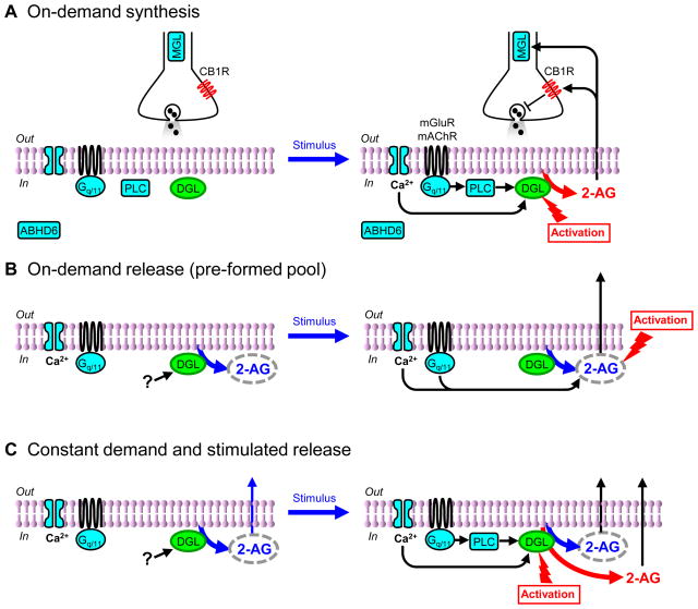
Endocannabinoids are generally considered to be “retrograde” signals, despite some examples of “autocrine” action [21], because they move across the synaptic cleft in the reverse direction from that of normal synaptic transmission; i.e., they are produced in the postsynaptic cell, go “backwards” across the cleft, and activate CB1Rs on presynaptic nerve terminals (Fig. 1A). This model is based on an extensive body of evidence, much of it electrophysiological, because endocannabinoids in the synaptic cleft cannot be collected and measured with biochemical methods. CB1R-mediated suppression of GABAergic or glutamatergic synaptic transmission is often used to bioassay endocannabinoid actions. Key observations (see [5][7] for reviews): include 1) Quantal analyses showing that endocannabinoids reduce synaptic responses by decreasing the quantity of transmitter released from the presynaptic terminal. 2) Triggering or blocking endocannabinoid signaling by manipulations that are confined to the postsynaptic cell (e.g., increasing or decreasing [Ca2+]i). 3) Lack of alteration in postsynaptic receptors for the conventional neurotransmitter. 4) Localization of the CB1Rs on presynaptic nerve terminals, and localization of enzymes necessary for endocannabinoid synthesis, e.g., DGLα, in postsynaptic cells. 5) Endocannabinoid-mediated reduction in presynaptic Ca2+ influx and transmitter vesicle fusion.
In some regions, e.g., hippocampus, the highest densities of CB1R are on axon terminals of interneurons that co-express GABA and cholecystokinin (CCK) [3][4]. In other regions, such as in the cerebellum, CB1Rs are more equally distributed on both excitatory and inhibitory terminals. Comprehensive reviews of the endocannabinoid system [2][5][6][7] are recommended for additional background. Glia express CB1Rs [8] and respond to CB1R agonists by releasing glutamate and influencing synaptic transmission [9]; neuronal and glial contributions cannot be distinguished in the work covered in this review.
Endocannabinoids are directly synthesized from membrane phospholipids in response to an increase in postsynaptic intracellular calcium ([Ca2+]i) alone, or combined with activation of postsynaptic GPCRs, such as group I metabotropic glutamate receptors (mGluRs) [10][11], or M1/M3 muscarinic acetylcholine receptors (mAChRs) [12][13] (Figure 1A). Short-term forms of CB1R-mediated suppression of synaptic transmission are typically triggered by increases in postsynaptic [Ca2+]i, and are called depolarization-induced suppression of inhibition (DSI) [14][15] or excitation (DSE)[16]. DSI and DSE are mediated by brief (~secs) stimulation of CB1Rs, which prevents transmitter release primarily by inhibiting voltage-gated Ca2+ channels [16][17][18][19], or sometimes by increasing K+ conductance [20]. At the less-than-physiological temperatures usually used experimentally, DSI and DSE last for 10’s of sec. CB1Rs can be activated as autoreceptors by endocannabinoids produced within certain cells, and cause a long-lasting, slow self-inhibition (SSI) mediated by a Ca2+-dependent K+ conductance [21][22][23]. SSI is produced by repetitive action potential trains induced over several min, and lasts for ~20 min. CB1R-mediated, long-term synaptic depression (eCB-LTD) can be triggered by mGluRs activated pharmacologically or by synaptic stimulation lasting from a few sec [24][25] to >10 min [26] and requires prolonged (~mins) CB1R stimulation [26][24][25] [26], but lasts for >30 min. Endocannabinoid synthesis and release (“mobilization”) begins within ~150 ms of starting cell stimulation [27], hence the reasons for the different temporal requirements for endocannabinoid-mediated effects are not clear, but may relate to events downstream of CB1R as well as to different modes of endocannabinoid mobilization. (In most experiments endocannabinoid synthesis cannot be distinguished from its release, therefore “mobilization” [28] is used to refer to the occurrence of a CB1R-dependent response following cellular stimulation). CB1R-mediated responses differ widely in their susceptibility to perturbations of the endocannabinoid system, supporting the hypothesis that several different mechanisms are involved. This review focuses on the identity of the signaling endocannabinoid, the molecular mechanisms of, and coupling between, endocannabinoid synthesis and release.
Figure 1. Hypothetical models of three different modes of 2-AG signaling.
A. In a conventional on-demand synthesis model, 2-AG synthesis is tightly linked to demand, which is triggered by neuronal activation. Stimulation causes [Ca2+]i elevation and/or G- protein (Gq/11) activation, leading to activation of the synthetic enzyme for 2-AG, DGL. Once released into the synaptic cleft, 2-AG binds to presynaptic CB1Rs and suppresses synaptic transmission. Presynaptic MGL is a major degradative enzyme for 2-AG, while another degradative enzyme, ABHD6 is located postsynaptically. B. An on-demand release model postulates de-coupling between synthesis and release of 2-AG. 2-AG can be constitutively synthesized in, but not immediately released from, unstimulated neurons. It is proposed to be stored in biochemically undefined pre-formed pools. Activation of DGL in resting neurons is sensitive to the basal level of [Ca2+]i, constitutive G protein activation, or unknown mechanisms. Stimulation-induced signals mediated by Ca2+ and/or G proteins trigger the release of 2-AG. C. In the combined model, both constitutive release from unstimulated cells and stimulation-induced 2-AG mobilization occur. In unstimulated neurons, 2-AG is synthesized as in B, but is also constitutively released into the extracellular space. Neuronal stimulation increases synthesis and induces stimulated release of additional 2-AG.
Supply side
As lipids, endocannabinoids cannot be stored in vesicles and yet their quantities increase with stimulation, leading to the concept that endocannabinoids are mobilized as needed (“on-demand”) [2][29] (Figure 1A). Strategies for manipulating the system for the treatment of human disease will require a thorough understanding of the roles of different endocannabinoids, their sources, and the stimuli that mobilize them. Globally activating the system, e.g., via “medical marijuana” or pharmacological agonists of CB1Rs (to treat multiple sclerosis or chronic pain), or conversely, inhibiting CB1Rs (to treat obesity or tobacco addiction), often cause unacceptable side effects [30] [31] [32]. Alternative approaches would avoid these effects by, e.g., decreasing endocannabinoid degradation, thus locally increasing their concentration precisely where they are naturally generated [33].
Anandamide and 2-AG have different in vivo effects, though both act on CB1Rs [34][35]. Blocking anandamide degradation reduces pain [36][37], inflammation [38], depression [39], and anxiety [40], but does not cause hypothermia, movement disorders or weight gain [40], whereas blocking 2-AG degradation induces hypothermia and hypomotility and analgesia [37]. Simultaneously blocking degradation of both mimics THC in drug discrimination tests, but blocking degradation of only one does not [34]. It will be important to know whether all stimuli that mobilize endocannabinoids are equally effective in altering basal and stimulated release, whether endocannabinoids can remain within a cell without being released, how the intracellular storage of endocannabinoids (if any) could be maintained, and whether all stimuli generate the same endocannabinoids (Figure 1).
General demand and specific supply for endocannabinoids
Physiologically, the involvement of an endocannabinoid in a particular phenomenon is inferred from the ability of a CB1R antagonist to block it, or the absence of the phenomenon in CB1R−/− animals. Of the known candidate endocannabinoids, attention has converged on anandamide [41] and 2-AG [42][43][44], but technical limitations make determining which endocannabinoid is active at a given synapse quite difficult. Endocannabinoids are labile, released in minute quantities, and rapidly taken up and degraded. Biochemical techniques can detect tiny amounts of 2-AG and anandamide in bulk tissue samples, but lack temporal and spatial resolution [45]. Single-cell electrophysiological analysis of synaptic transmission offers sensitive, highly localized, real-time readout of endocannabinoid actions, but by itself cannot identify an endocannabinoid. A combination of biochemical, molecular and electrophysiological methods constitutes the most powerful approach for investigating the endocannabinoid system.
Both 2-AG and anandamide are produced from ubiquitous lipids via several biosynthetic pathways [2][46]. 2-AG can be formed when Ca2+ stimulates phospholipase C (PLC) which then transforms membrane phosphoinositides into a diacylglycerol (DAG), from which 2-AG is liberated by DAG lipase (DGL) [45]. Alternatively, DAG can be produced from phosphatidic acid, a reaction catalyzed by either phospholipase-A2 or -D [47]. 2-AG is metabolized mainly by monoglyceride lipase (MGL)[48] with a lesser contribution from α/β hydrolase 6 (ABHD6)[35][49]. There is no consensus as to which of the multiple pathways of anandamide synthesis [2][46] is physiologically most relevant. Anandamide is degraded by fatty-acid amide hydrolase (FAAH) [50]. 2-AG and anandamide are taken up by an endocannabinoid transport mechanism [51][52] that is incompletely characterized.
While 2-AG and anandamide both induce physiological effects in vivo, thus far anandamide appears to mediate CB1R-dependent retrograde signaling only in limited situations [53] [54], and the majority of the evidence suggests that 2-AG mediates most forms of CB1R-mediated retrograde regulation of synaptic transmission:: DSI and DSE are enhanced by blockade of the degradative enzyme for 2-AG, but are unaffected by changes to FAAH [55][56]. Neuronal stimulation in some brain regions selectively increases 2-AG levels [44][57][58]. DGLα is often located in postsynaptic spines directly across from excitatory afferent terminals, though it is rarely found near CB1R-expressing perisomatic inhibitory terminals [59][60] at which DSI occurs [14][15]. A striking exception to this rule has recently been reported at an unusual “invaginating synapse” in the basal amygdala, where the concave postsynaptic pocket into which the presynaptic nerve terminal fits is densely lined with DGLα, while CCK, MGL and CB1Rs are all highly expressed in the inhibitory nerve terminal [61]. Again, aggregation of DGLα was not observed either at similarly invaginating inhibitory synapses in the dentate gyrus, or at “flat” inhibitory synapses in the lateral amygdala, confirming that the basal amygdala is a special case. Whether the failure to detect DGLα at other inhibitory synapses reflects limitations of detection methods or differences in 2-AG synthesis mechanisms is not known.
Two isoforms of DGL have been cloned: DGLα and DGLβ [62]. Over-expression of DGLα enhances 2-AG levels, and short hairpin RNA (shRNA) knock-down of DGLα diminishes 2-AG in a neuronal cell line [63]. Construction of a 2-AG-generating system in a model cell requires heterologous expression of only mGluR5, DGLα, and the structural protein Homer 2b [64]. In DGLα−/− mice, all forms of endocannabinoid signaling that were tested are abolished, whereas they are normal in DGLβ−/− mice [65][66]. In the cerebellum, hippocampus, and striatum of DGLα−/− mice, DSE and DSI, and three different forms of GPCR-induced endocannabinoid release are eliminated, although the basic machinery related to endocannabinoid actions – Ca2+ influx, mGluRs, mAChRs, PLC [66], MGL, FAAH, CB1Rs and CB2Rs [65] – were unaffected. DGLα−/− and DGLβ−/− mice are fertile and behaviorally normal, with the DGLα−/− animals having a reduced mean body weight [65], consistent with the prominent endocannabinoid involvement in energy metabolism [67]. These data suggest that DGLα, and by extension 2-AG, are necessary and sufficient for most endocannabinoid signaling (but see discussion of DGLβ below). It will be interesting to investigate the excitatory synapses in the auditory system of DGL−/− mice, as they are suppressed by retrograde endocannabinoid actions, and yet DGLβ, not DGLα, is densely present in the dendritic spines [68].
Supply from separate pools of 2-AG
In a simple on-demand model, endocannabinoid supply and demand are tightly coupled because the endocannabinoid concentration gradient created by synthesis drives release. Yet high basal 2-AG levels [44], together with its multiple roles in lipid metabolism [45], have led to speculation that some 2-AG is not immediately released, and may function as a messenger for intercellular signaling [2][49][69]. Recent pharmacological and genetic studies suggest that the synthesis and release of 2-AG are not tightly coupled, although the evidence is indirect and alternative interpretations of the data are possible.
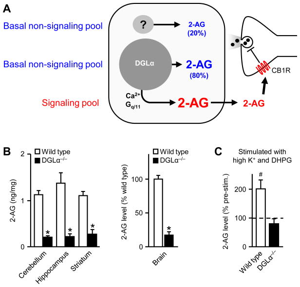
Absolute concentrations of labile products such as endocannabinoids are difficult to measure, and levels vary according to the precise experimental conditions present, rising quickly after death, for example [70]. Nevertheless, under given measurement conditions, large relative differences in endocannabinoid levels should be significant. Comparison of the basal level of 2-AG in DGLα−/− and wild-type mice shows it is much lower in the former [65][66], supporting the idea that a substantial amount of 2-AG is present in unstimulated brains. Although 2-AG-mediated signaling is absent in DGLα−/− animals [65][66], basal 2-AG is only ~80% reduced, suggesting that ~20% of the basal 2-AG pool emerges from another source and plays no role in signaling (Figure 2), in agreement with the finding of significant quantities of 2-AG in COS cells untransfected with DGL [62]. The relatively large size of the basal DGLα-sensitive 2-AG pool –~ 4-fold greater than the insensitive pool – also raises the question of whether DGLα is intrinsically active, or whether low levels of spontaneous neuronal activity are sufficient to stimulate it significantly. A comparative quiescence of neuronal activity could exaggerate the significance of on-demand endocannabinoid synthesis in vitro, whereas in vivo, where spontaneous activity is high, an on-going constitutive process might be more significant. Alternatively, DGLβ might contribute to non-signaling related 2-AG [65] (Box 2). Taken together, the data appear to argue for the existence of 2-AG in apparently resting neurons.
Figure 2. Functional distinctions among intracellular 2-AG pools.
A. Measured differences in basal amounts of 2-AG in DGLα−/− and wild type mice [59][60] supports the postulation of three functionally distinct pools of 2-AG in neurons: 1) basal 2-AG produced independently of DGLα, possibly by DGLβ, although the source is unknown (“?”), 2) basal 2-AG synthesized by DGLα, and, 3) 2-AG that is produced and released upon stimulation such as an increase in [Ca2+]i or activation of Gq/11 proteins. The basal pools are present in “unstimulated” neurons and might not participate in constitutive retrograde signaling onto presynaptic terminals, whereas the stimulus-induced 2-AG mediates retrograde signaling (thus is considered a “signaling pool”). B. Ablation of DGLα (in two independent mouse models [59, 60]) does not eliminate basal 2-AG, as about 20% remains; this appears to constitute a basal, non-signaling pool. C. Stimulation of neurons with high K+ and DHPG increases the 2-AG levels to twice the total basal amount in wild-type mice (the “signaling” pool in panel A), whereas it fails to change 2-AG levels in DGLα−/− tissue, consistent with the idea that DGLα-independent 2-AG (mediated by “?” in panel A) constitutes a basal, non-signaling pool. Modified, with permission from [60] (panels C and B, left) and [65] (panel B, right).
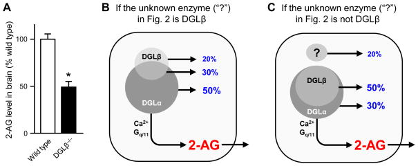
Box 2. Role of DGLβ in generating basal 2-AG.
A decrease of ~50% in basal 2-AG was observed in brain tissue from DGLβ−/− animals compared to wild-type mice [65] (Fig. IA). 2-AG produced by DGLβ might be considered a “non-signaling” pool because endocannabinoid-mediated retrograde signaling to presynaptic CB1Rs is abolished in DGLα−/− mice [65][66]. However, DGLβ contributes 2-AG to the regulation of adult neurogenesis, a growth-related process in which 2-AG from DGLα also participates [65]. Estimates of the overlap between DGLβ- and DGLα-synthesized 2-AG pools depend on the identity of the unknown enzyme (“?” in Figure 2) that produces the 20% of total basal 2-AG remaining in DGLα−/−. If this enzyme is DGLβ, then it is solely responsible for 20% of basal 2-AG; and overlapping contributions from DGLβ and DGLα produce 30% of basal 2-AG for a pool that serves growth-related or metabolic functions [65](see main text) (Fig.IB). This model can explain the observed 50% decrease in basal 2-AG in DGLβ−/− and 80% decrease in DGLα−/− brains (the overlap makes the sum >100%). Alternatively, if the unknown enzyme is neither DGLβ nor DGLα, then 50% of the total basal 2-AG would be produced by the overlapping combination of DGLα and DGLβ in a pool that might not directly serve signaling (Fig. IC). Again, this model can explain the observed decreases in basal 2-AG in each knock-out. Conceptually, therefore, the basal 2-AG pools could be subdivided according to the identity of synthetic enzymes. Further refinement of these estimates require unequivocal determination of the contributions of DGLβ to basal 2-AG pools (cf [66]). Panel A, modified from [65] with permission.
Figure I.
Provisional acceptance of this idea leads to the question as to whether basal 2-AG would be stored in cells or continuously released into the extracellular space. If all DGLα-generated, basal 2-AG were released, constitutive CB1R-mediated suppression of synaptic transmission should be widespread. In hypothalamic proopiomelanocortin [71] and oxytocin-expressing [72] neurons, constitutive CB1R activation suppresses GABAergic transmission, although 2-AG is not known to be responsible. Constitutive CB1R activation in hippocampal slices tonically suppresses GABA release from some interneurons [53] [73][74][53][75], but again, 2-AG involvement in this action appears minimal [53]. Vigorous basal activity of 2-AG degradative enzymes could prevent 2-AG from tonically activating CB1Rs [56]. However, if 2-AG is released and then quickly degraded as soon as it is synthesized, the apparently large basal quantities of 2-AG in unstimulated tissue [44] would be difficult to explain. Indeed, potent and selective MGL and ABHD6 inhibitors, which profoundly enhance stimulated tissue levels of 2-AG, do not affect basal levels [34][35]. Hence, the subcellular sites of production of basal and signaling 2-AG could be separated, and basal 2-AG may not be involved in constitutive CB1R activation. Taken together, the data suggest that a significant amount of DGLα-sensitive 2-AG may not be immediately released from cells (Box 3). T constitutive endocannabinoid actions in hippocampus are probably mediated by anandamide [53] (discussed below).
Box 3. Outstanding unresolved questions in endocannabinoid signaling.
How do endocannabinoids cross membranes? It is not known how the lipophilic endocannabinoids leave the membranes where they are synthesized and travel across the synaptic cleft to reach trans-synaptic CB1Rs. Also, the molecular identity of the “endocannabinoid transporter” remains obscure. Until it is isolated and cloned, some questions surrounding endocannabinoid uptake mechanisms, regulation of the spatial spread of endocannabinoids, and termination of endocannabinoid actions, will persist.
How can trans-synaptic movements of endocannabinoids be visualized? Molecular tools are needed to understand why the functional spread of endocannabinoids is so limited, and how endocannabinoids actually get to their targets.
What maintains the prolonged activation of CB1Rs that is required for eCB-LTD? Stimulation lasting a few seconds gives rise to CB1R activation lasting minutes. Does this reflect prolonged release of 2-AG from the stimulated cell, or the operation of presently unknown mechanisms?
Can DGLα activity be quickly halted (ie. via photolytically caged inhibitors, or inducible DGLα knockout mice)? This would allow comparisons of persistent 2-AG release and DGLα activity, perhaps allowing the discovery of alternative 2-AG supply pathways, as well as the kinetics of 2-AG changes in tissue to be measured.
Are the stimulation-induced, biochemically-measured increases in 2-AG indicative of the signaling or the non-signaling pools, or both? If they exist, can non-signaling and signaling pools communicate with each other? If not, how are they kept separate?
Where is the DGLα responsible for generating 2-AG at most somatic inhibitory synapses? If 2-AG synthesized in the dendrites is able to trans-locate to perisomatic regions, how does it do so?
What biochemical pathway(s) are responsible for the PLC-independent synthesis of 2-AG in responses such as DSI and DSE? Such responses seem to require DAG, since they depend on DGL, yet the other sources of DAG have not been identified.
How do endocannabinoids participate in physiological circuit behaviors? Numerous GPCR-coupled neurotransmitters (eg. mGluRs, mAChRs, dopamine D2Rs) have central roles in circuit interactions such as oscillations and also generate eCBs, yet there is little evidence that eCBs normally influence circuit activity.
Do other retrograde messengers, such as other lipids, gases (including nitric oxide), neuropeptides and growth factors [125], interact with the biochemical pathways of the endocannabinoid system?
Does anandamide mediate tonic activation of CB1Rs everywhere in the brain?
What are the functional implications of the interactions between anandamide and 2-AG that have been revealed by biochemical [81] and genetic [65] studies?
Neuro genesis in the adult hippocampus is important for high order cognitive functions, including pattern separation and integration, and learning and memory [76]. Pharmacological studies implicated DGL activation in adult neurogenesis in the subventricular zone (SVZ) and dentate gyrus [77]. Ependymal and proliferating cells in the SVZ express DGL, and proliferation is inhibited by blocking CB2R. Deleting one or both copies of DGLα markedly reduces the number of proliferating cells in the SVZ, and deleting both has a similar effect in dentate gyrus [65]. DGLβ deficiency does not alter SVZ neurogenesis, but diminishes the numbers of proliferating cells in the dentate gyrus. Thus, while apparently not involved in generating 2-AG for retrograde signaling, DGLβ can participate in adult neurogenesis. Given that DGLα also affects neurogenesis, it is reasonable to propose that DGLα can also contributes to the same non-signaling-related 2-AG pool that DGLβ does. This would be compatible with the existence of three distinguishable 2-AG pools (Box 2). Although this is a plausible hypothesis, direct evidence for the existence of separate pools is needed.
How could distinct 2-AG pools be kept separate?
With the foregoing caveats in mind, it is interesting to consider how the postulated functionally distinct endocannabinoid pools might be established physically. Identifying mechanisms that could segregate groups of lipids would be a major advance in testing the pools hypothesis, but itself poses formidable technical challenges [45]. Nevertheless, three major classes of possibilities exist: 1) compartmentalization of endocannabinoid-system-related proteins, 2) heterogeneity of the lipid matrix, and 3) endocannabinoid sequestration by lipid-binding proteins.
Separate pools of 2-AG could reflect spatially distinct groupings of synthetic or degradative machinery maintained by either cellular compartmentalization or cytoskeletal structures. Generation of 2-AG by mGluRs and mAChRs requires PLCβ1 [78] or PLCβ4 [58]. In contrast, neither DSI nor DSE are affected by the pharmacological inhibition or genetic deletion of PLC [25, 78–80], and DGL inhibitors do not always alter CB1R-dependent synaptic plasticity (see below). Differential distribution of the degradative enzymes MGL (presynaptic, mainly soluble) and ABHD6 (postsynaptic, membrane bound) could enable them to establish different 2-AG pools [35]. Anandamide can control DGL by activating transient receptor potential vanilloid type 1 (TRPV1) channels [81], thus, selective arrangements of TRPV1 channels and DGL could create compartments for 2-AG production. DGLα is found in dendritic spines apposed to excitatory axon terminals [45][59][60]. 2-AG produced in spines should affect local glutamate release only, because the lateral spread of endocannabinoids along dendrites is only ~10 μm [82]. This restricted sphere of action makes dendritic spines an unlikely source for the 2-AG that suppresses somatic inhibitory terminals 50–100 μm away; perhaps another DGLα pool remains undetected.
At excitatory synapses, the structural proteins called Homers could tie together elements of the endocannabinoid system. Long forms of Homer have a protein-binding motif at one end and a coiled-coil (CC) domain at the other; CC-domain interconnections create cytoskeletal scaffolding [83]. Long Homers attenuate CB1R-independent mGluR actions [64], and facilitate CB1R-dependent mGluR actions [64][84] by tethering DGLα to the plasma membrane [63]. A short form, Homer 1a, lacks the CC tail and disrupts Homer scaffolding. When elevated in cultured hippocampal neurons, Homer 1a depresses mGluR-induced endocannabinoid responses, while simultaneously enhancing DSE [84]. Long-form Homers might inversely link mGluR-dependent and Ca2+-dependent endocannabinoid mobilization. Interestingly, in a mouse model of Fragile X syndrome (Fmr1−/−), the functional coupling between group I mGluRs and endocannabinoids is enhanced in hippocampus [85] and striatum [86], resulting in marked CB1R-dependent depression of GABAergic transmission following modest activation of mGluRs. DGL and MGL activities are both increased in Fmr1−/− [86], and the enhanced mGluR-endocannabinoid coupling is associated with heightened susceptibility to long-term plasticity [85]. The mechanism of enhanced coupling is not known, but regulation of Homers is a possibility [87]. Regional localization or regulation of the mGluR-Homer apparatus might establish discrete 2-AG pools.
The heterogeneous composition of the membrane lipid bilayer could cause variability in 2-AG production in different parts of a cell [45]. Lateral partitioning of the bilayer into distinct domains, possibly corresponding to “lipid rafts”, can concentrate protein components of the endocannabinoid system [88], including DGLα [89], into small regions that do not communicate freely with the entire cell. Lipid-based co-localization could also facilitate protein-protein interactions. In addition, the different shapes of individual lipid molecules can cause deformations in the bilayer that directly alter protein function [45]; conceivably, the “membrane bending” properties of lipids could alter the operation of endocannabinoid-synthesizing or degrading enzymes in restricted regions. Finally, proteins such as serum albumin and lipid transfer proteins [90] serve as carriers for lipids, including anandamide [91]. Under appropriate conditions it might be possible to liberate bound 2-AG from these intracellular proteins. Determining the physical basis of functionally distinguishable sources of 2-AG would be an important advance towards testing of the multiple pools hypothesis.
Balancing supplies of endocannabinoids with demand
The concept of 2-AG pools prompts a number of new questions: how large would the signaling pool be? How might the signaling, stimulated, and basal pools be related? A high concentration of the mGluR agonist, DHPG, plus high [K+]o, applied to cerebellar tissue, increased 2-AG levels by ~2-fold above unstimulated levels [66], similar to previous reports [44][92](Figure 2). The treatment did not increase 2-AG in DGLα−/− tissue, suggesting that DGLβ was not activated. As strong activation of DGLα doubles the amount of 2-AG already present, the total stimulated increase of 2-AG appears comparable to the amount of the DGLα-sensitive basal 2-AG (Figure 2). Furthermore, the stimulated 2-AG might not all be releasable. Only 20% of the 2-AG produced in stimulated neuroblastoma cells is released into the incubation medium [93]. In nucleus accumbens, the amounts of 2-AG detected by microdialysis in the extracellular space following stimulation are much lower than the resting quantities [57], again implying that much of the basal 2-AG is not readily released.
Probing these issues with pharmacological DGL inhibitors (tetrahydrolipstatin, THL; RHC 80267 or OMDM-188) has produced complex results. Some variance correlates with the response type. eCB-LTD is generally abolished by DGL inhibitors, as in hippocampus [25][79], cerebellum [94] and prefrontal cortex [95]. In striatum, adenosine A2 receptors potentiate 2-AG release and eCB-LTD in the indirect, but not the direct, medium spiny neurons (MSNs); and a DGL inhibitor blocks A2-initiated LTD initiated with moderate frequency (20 Hz) stimulation. In the cases of mouse amygdala [96] or developmentally-regulated striatal eCB-LTD that is induced by high frequency stimulation (100 Hz) [97][98], the CB1R-dependent plasticity is insensitive to DGL inhibition because it is mediated by anandamide rather than 2-AG.
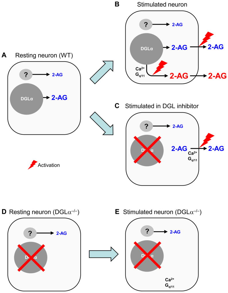
Short-term 2-AG-mediated responses are more variable. Striatal DSE and DSI are both blocked by a DGL inhibitor [99], whereas in cerebellum, DSE is unaffected [94] while DSI is blocked by a DGL inhibitor [80]. Depending on experimental conditions, hippocampus DSI may [25] [51] [100] or may not [25] [79] [80] [101] be inhibited. In the negative cases, the ineffectiveness of DGL inhibitors appears to contradict the molecular evidence for 2-AG involvement [65][66]. Variable penetration of the inhibitors into brain slices is not an obvious explanation when they prevent only one of two 2-AG mediated responses in the same cell [25][90]. An alternative hypothesis could be that some of the signaling 2-AG is released from a pre-formed pool [79][102][102] [103] basal DGLα-sensitive pool, which would not be immediately depleted when DGL is blocked (Figure 3A–C). Constitutively deleting DGLα would preclude the formation of all 2-AG accessible for signaling (Figure 3D–E).
Figure 3. Hypothetical mobilization of 2-AG.
The hypothesis of distinct 2-AG pools may further suggest that differences in the mechanisms of 2-AG mobilization can help reconcile apparent discrepancies between observations produced by pharmacological and genetic silencing of DGL. A. In a resting neuron in wild type animals, basal 2-AG is produced by either DGLα-dependent or -independent mechanisms, and most of it remains in the cell and does not substantially activate presynaptic CB1Rs constitutively. B. In stimulated neurons, Ca2+ and/or G proteins activate DGLα-dependent synthesis of 2-AG, which is then released and mediates retrograde signaling. Simultaneously, neuronal activation may trigger 2-AG release from a basal pre-formed pool. C. DGL inhibitors are sometimes ineffective in blocking Ca2+- or G protein-dependent 2-AG signaling (see text for examples). This could be explained if Ca2+ or G proteins facilitate the release process per se of 2-AG from a pre-formed basal 2-AG pool. Acute blockade of DGLα would inhibit 2-AG synthesis, but would not immediately deplete the pre-formed pool. D. In a DGLα−/− neuron, the DGLα-dependent portion of basal 2-AG pool is eliminated (see also Fig. 2B). E. Stimulation of DGLα−/− neurons will fail to mobilize any 2-AG, because the formation of all releasable 2-AG has been abolished (also Fig. 2C).
The pre-formed pool hypothesis makes a number of testable predictions: 1) Pharmacological DGL inhibitors might not block all 2-AG-mediated responses, or decrease 2-AG tissue levels uniformly, as pool size or accessibility might vary. 2) DGL-inhibitor-induced reduction of 2-AG responses and tissue levels would be use-dependent, and proceed faster if the system was repetitively stimulated. Other tests would rely on the ability to halt DGL activity quickly (e.g., inducible knock-outs). 3) 2-AG responses mediated by release from a pre-formed pool would continue after DGLα inactivation until the pool was depleted; and 4) stimulation should reduce tissue levels of 2-AG, once DGLα was inactivated.
Some of these predictions were tested in single CA1 pyramidal cells filled with OMDM-188 or THL [103]. In each cell, both DSI and mGluR-initiated endocannabinoid responses (eCBmGluR) were measured and DSI was found to be more readily suppressed than eCBmGluR. While the possibility of non-specific pharmacological effects always exists, the data revealed differences in the relationship of DGL to these two forms of endocannabinoid mobilization. Although eCBmGluR was much less sensitive to OMDM-188, if evoked repeatedly with brief DHPG applications, eCBmGluR declined significantly owing to a use-dependent rundown in eCBmGluR, not slow infusion of OMDM-188 into the cells. If the abolition of DSI implies that DGLα activity was blocked, the continued occurrence of eCBmGluR supports the concept that 2-AG sourcing is not necessarily tightly coupled to DGLα. This could reflect either release of 2-AG from a pre-formed pool, or its temporary generation by some other mechanism (discussed below).
Release from a pre-formed pool could also explain why GPCR-dependent endocannabinoid production is abolished in PLCβ1−/− or [78] or PLCβ4−/− [58] mice, while a potent PLC inhibitor, U73122, failed to block the same short-term endocannabinoid-dependent responses in the hippocampus [25][79]. U73122 also prevents 2-AG synthesis in cultured neurons [44] [92]. Regional or tissue-specific variability may exist, because U73122 is partially effective in the cerebellum [104] and ventral tegmentum [105], while it completely prevents short-term GPCR-induced endocannabinoid effects in other cases [22] [106] [107] [108]. Interestingly, U73122 abolishes hippocampal [25][79] and neocortical [95] eCB-LTD, despite its variable effects on short-term responses, underscoring the possibility of distinct long-term and short-term endocannabinoid mobilization mechanisms.
Alternatives to physically separate pools of 2-AG
Despite the appeal of the separate pools idea, alternative models are possible. For instance, 2-AG could arise from multiple synthetic pathways instead of physically distinct pools. 2-AG is a component of numerous metabolic pathways apart from endocannabinoid synthesis. It is not clear what form the basal 2-AG is in prior to its measurement with biochemical methods, and its absolute levels are highly labile [70]. Basal 2-AG might be rapidly turning over in ongoing biochemical reactions normally unrelated to endocannabinoid production. If DGLα-dependent 2-AG production is halted, mGluR activation might temporarily tap into an alternative 2-AG stream. In this case, DGLα would have to interact directly or indirectly with the alternative stream, since eCBmGluR is abolished in the DGLα−/− mouse [66], and is eventually reduced by DGLα inhibitors [103].
Another alternative to intracellular pools of 2-AG would be complexity in the regulation of 2-AG release. Neither bulk tissue measurements nor physiological assays distinguish directly between endocannabinoid synthesis and release. If these two processes can be de-coupled, then stimulation could conceivably trigger 2-AG efflux separately from its synthesis. In this case, apparently different pools of 2-AG might reflect different forms of stimulus-induced release. There is evidence that endocannabinoid release from the postsynaptic cell might not occur solely by diffusion. The presence of an endocannabinoid experimentally loaded into a cell via a whole-cell recording pipette leads to suppression of CB1R-sensitive synaptic inputs to the cell [109], indicating that the applied endocannabinoid reaches presynaptic CB1Rs. Release of the loaded endocannabinoids is influenced by afferent fiber stimulation [110][111]. Importantly, including an endocannabinoid transporter inhibitor in the electrode prevents a loaded endocannabinoid from leaving the cell. Possibly the naturally synthesized 2-AG can also be transported to the extracellular space. A dedicated export mechanism could account for the prolonged release of endocannabinoids that is required for eCB-LTD and that is produced by brief stimulation [25] [109]. Intracellular application of a transporter blocker prevents the induction of CB1R-mediated, neocortical timing-dependent LTD [112], and inhibitory eCB-LTD in hippocampus [28][109] and striatum [111]. Reversed transport is not mandatory for endocannabinoid release however, because DSI is not affected by a transport inhibitor [28][109].
In summary, some recent work seems to pose challenges to the long-held concept of “on-demand” mobilization of endocannabinoids. While the competing hypotheses are in need of rigorous testing, data are beginning to hint that endocannabinoid efflux may not always be tightly coupled to its production, that some endocannabinoid release may occur from pre-formed pools or novel 2-AG metabolic pathways, and even that release per se, separate from synthesis, might be triggered by stimulation.
Supply and demand for anandamide
Although anandamide was the first endocannabinoid discovered [41], evidence from DGL−/− mice suggests that 2-AG mediates all forms of endocannabinoid signaling tested thus far. However, in DGLα−/− animals, the basal levels of anandamide are also lowered by ~50% [65][66], pointing to an unsuspected convergence between the synthesis pathways of anandamide and 2-AG, and leaving open the possibility that anandamide might be involved in some DGL-regulated signaling. Evidence arguing against the latter interpretation is that strong stimulation did not increase anandamide in wild type animals [34][44][66]. Yet stimulation can increase anandamide levels [113], and anandamide evidently mediates some forms of eCB-LTD [96] [98]. The decrease in neural proliferation found in DGLα−/− mice [65] could reflect the decrease in anandamide. FAAH−/− mice have a higher rate of neuroprogenitor proliferation than do wild-type mice [114], and inhibition of FAAH induces proliferation [77], but does not degrade 2-AG in intact cells [48]. Whether inhibiting 2-AG degradation by MGL [48] or ABHD6 [35][49] is equally effective in the proliferation assay is not known, and should be tested before the effects observed in the DGL−/− mice could be attributed to decreases in anandamide.
Anandamide is implicated in chronic behavioral states such as pain [36][37], anxiety [40], depression [39], and analgesia [115][116], as well as constitutive activation of CB1Rs [53]. “Synaptic homeostasis” refers to the maintenance of global neuronal network stability despite persistent changes in strengths of synapses within the network [117]. Uncompensated, non-uniform alterations in synaptic strengths can lead to network instabilities. Endocannabinoids are sensitive to neuronal excitability and are ideal candidates for mediating synaptic homeostatic plasticity. Silencing of neuronal activity for several days with tetrodotoxin strengthened synapses between CB1R-positive interneurons and pyramidal cells in organotypic hippocampal cultures [53]. Synaptic strengthening resulted from a decrease in the tonic inhibitory action of endocannabinoids, and not a change in CB1Rs. Pharmacological tests showed that the endocannabinoid involved is anandamide, and that anandamide uptake and degradation, rather than its synthesis or release were increased by prolonged inactivity. Tonic anandamide release depended on a low constant [Ca2+]i in the postsynaptic pyramidal cell, consistent with the dependence of anandamide synthesis on Ca2+ [7]. The data revealed a new signaling role for anandamide, and illustrated a novel mechanism whereby variable endocannabinoid demand is met by down-regulation of endocannabinoid removal and degradation, rather than by up-regulation in supply. Finally, the study showed that ongoing background activity in a neural network can regulate endocannabinoids without there being any well-defined demand for them. Given the partial dependence of anandamide synthesis on DGL, this phenomenon should be investigated in DGL−/− tissue [65][66]. Moreover, it will be important to determine if anandamide is the tonically released endocannabinoid in other cells where constitutive CB1R-mediated effects have been recorded.
Anandamide and TRPV1
In this review, responses induced by endocannabinoids have been assumed to be mediated by CB1R However, recent studies show that anandamide, well-known pharmacologically as a full agonist at TRPV1 channels [118], in fact does have physiological actions that are mediated by TRPV1. In the striatum, anandamide inhibits the production of 2-AG in some cells by activating TRPV1 channels, thereby decreasing glutathione levels and suppressing DGL [81]. In the indirect pathway MSNs of the nucleus accumbens [54] and in the dentate gyrus [119], activation of mGluR5 triggers the production of anandamide, which in turn activates TRPV1 present on those cells. TRPV1 activation then induces postsynaptic LTD by internalizing AMPA receptors. In the indirect pathway MSNs, anandamide is also released as a retrograde transmitter that activates presynaptic CB1Rs and induces LTD of glutamate release [54]. TRPV1 is abundant in the brain and, as TRPV1 rather than CB1R becomes more widely used as a bioassay, a wider range of anandamide actions will undoubtedly emerge. A major synthetic enzyme for anandamide, N-acylphosphatidylethanolamine-specific phospholipase D (NAPE-PLD) [120] is found in some glutamatergic nerve terminals, but not postsynaptic pyramidal cell bodies [121][122], arguing against its being the source of anandamide in either TRPV1-mediated LTD [119] [54], or CB1R-mediated homeostatic plasticity [53]. The association of NAPE-PLD with intracellular, perhaps Ca2+-containing cisternae [121], might explain the recent finding that anandamide can induce quantal glutamate release via a CB1R- and TRPV1-independent mechanism [123]. Working out the biosynthetic mechanisms [124] of anandamide production and its modes of action are pressing tasks for the future.
Concluding remarks
Endocannabinoid synthesis, release, and perhaps even storage, have multiple features that suggest a complex underlying organization. DGLα provides all of the signaling-related 2-AG assayed thus far, although DGLβ may contribute in other ways. Both DGLα and DGLβ participate in regulating adult neurogenesis, an important observation that could lead to selective therapeutic interventions. Different pathways upstream of DGLα generate Ca2+-dependent and GPCR-dependent 2-AG; the Homer proteins may be positively coupled to the GPCR-PLC pathway. DSI and DSE require DGLα but are independent of PLC. Taken together, the current evidence suggests that functionally distinct pools of endocannabinoids may exist, although other hypotheses are not ruled out, and there is little information on the mechanisms underlying the apparent pools. Probably not all of the DGLα-dependent 2-AG is involved in signaling, though the signaling and non-signaling pools cannot be distinguished biochemically. Resolution of this issue is crucial for the physiological interpretation of measured tissue endocannabinoid levels. An unexpected finding is that anandamide levels are significantly decreased by DGLα elimination, thus anandamide could be involved in certain DGL-dependent responses. Anandamide acting at CB1Rs seems to be responsible for chronic or constitutive endocannabinoid-mediated regulation of phenomena such as pain, anxiety and analgesia, adult neurogenesis, as well as a form of homeostatic synaptic plasticity in vitro. The actions of anandamide at TRPV1 channels, its ability to influence 2-AG, and regulate synaptic plasticity on its own will undoubtedly become a rich field for future investigations, among many other outstanding questions to be addressed (Box 3).. Future work will undoubtedly lead to nuanced modifications of the present on-demand model of endocannabinoid synthesis and release.
Acknowledgments
Research in the authors’ laboratories was supported by NIH grants R01 DA014625 and R01 MH077277 to B.E.A. and RO1 AG036794 to J.K.
Footnotes
Publisher's Disclaimer: This is a PDF file of an unedited manuscript that has been accepted for publication. As a service to our customers we are providing this early version of the manuscript. The manuscript will undergo copyediting, typesetting, and review of the resulting proof before it is published in its final citable form. Please note that during the production process errors may be discovered which could affect the content, and all legal disclaimers that apply to the journal pertain.
Reference List
- 1.Gaoni Y, Mechoulam R. Isolation, structure and partial synthesis of an active constituent of hashish. J Am Chem Soc. 1964;86:1646–1647. [Google Scholar]
- 2.Piomelli D. The molecular logic of endocannabinoid signaling. Nat Rev Neurosci. 2003;4:873–884. doi: 10.1038/nrn1247. [DOI] [PubMed] [Google Scholar]
- 3.Marsicano G, Lutz B. Expression of the cannabinoid receptor CB1 in distinct neuronal subpopulations in the adult mouse forebrain. Eur J Neurosci. 1999;11:4213–4225. doi: 10.1046/j.1460-9568.1999.00847.x. [DOI] [PubMed] [Google Scholar]
- 4.Katona I, et al. Presynaptically located CB1 cannabinoid receptors regulate GABA release from axon terminals of specific hippocampal interneurons. J Neurosci. 1999;19:4544–4558. doi: 10.1523/JNEUROSCI.19-11-04544.1999. [DOI] [PMC free article] [PubMed] [Google Scholar]
- 5.Alger BE. Retrograde signaling in the regulation of synaptic transmission: focus on endocannabinoids. Prog Neurobiol. 2002;68:247–286. doi: 10.1016/s0301-0082(02)00080-1. [DOI] [PubMed] [Google Scholar]
- 6.Freund TF, et al. Role of endogenous cannabinoids in synaptic signaling. Physiol Rev. 2003;83:1017–1066. doi: 10.1152/physrev.00004.2003. [DOI] [PubMed] [Google Scholar]
- 7.Kano M, et al. Endocannabinoid-Mediated Control of Synaptic Transmission. Physiol Rev. 2009;89:309–380. doi: 10.1152/physrev.00019.2008. [DOI] [PubMed] [Google Scholar]
- 8.Stella N. Cannabinoid signaling in glial cells. Glia. 2004;48:267–277. doi: 10.1002/glia.20084. [DOI] [PubMed] [Google Scholar]
- 9.Navarrete M, Araque A. Endocannabinoids potentiate synaptic transmission through stimulation of astrocytes. Neuron. 2010;68:113–126. doi: 10.1016/j.neuron.2010.08.043. [DOI] [PubMed] [Google Scholar]
- 10.Maejima T, et al. Presynaptic inhibition caused by retrograde signal from metabotropic glutamate to cannabinoid receptors. Neuron. 2001;31:463–475. doi: 10.1016/s0896-6273(01)00375-0. [DOI] [PubMed] [Google Scholar]
- 11.Varma N, et al. Metabotropic glutamate receptors drive the endocannabinoid system in hippocampus. J Neurosci. 2001;21:RC188, 1–5. doi: 10.1523/JNEUROSCI.21-24-j0003.2001. [DOI] [PMC free article] [PubMed]
- 12.Kim J, et al. Activation of muscarinic acetylcholine receptors enhances the release of endogenous cannabinoids in the hippocampus. J Neurosci. 2002;22:10182–10191. doi: 10.1523/JNEUROSCI.22-23-10182.2002. [DOI] [PMC free article] [PubMed] [Google Scholar]
- 13.Ohno-Shosaku T, et al. Postsynaptic M1 and M3 receptors are responsible for the muscarinic enhancement of retrograde endocannabinoid signalling in the hippocampus. Eur J Neurosci. 2003;18:109–116. doi: 10.1046/j.1460-9568.2003.02732.x. [DOI] [PubMed] [Google Scholar]
- 14.Wilson RI, Nicoll RA. Endogenous cannabinoids mediate retrograde signalling at hippocampal synapses. Nature. 2001;410:588–592. doi: 10.1038/35069076. [DOI] [PubMed] [Google Scholar]
- 15.Ohno-Shosaku T, et al. Endogenous cannabinoids mediate retrograde signals from depolarized postsynaptic neurons to presynaptic terminals. Neuron. 2001;29:729–738. doi: 10.1016/s0896-6273(01)00247-1. [DOI] [PubMed] [Google Scholar]
- 16.Kreitzer AC, Regehr WG. Retrograde inhibition of presynaptic calcium influx by endogenous cannabinoids at excitatory synapses onto Purkinje cells. Neuron. 2001;29:717–727. doi: 10.1016/s0896-6273(01)00246-x. [DOI] [PubMed] [Google Scholar]
- 17.Wilson RI, et al. Presynaptic specificity of endocannabinoid signaling in the hippocampus. Neuron. 2001;31:453–462. doi: 10.1016/s0896-6273(01)00372-5. [DOI] [PubMed] [Google Scholar]
- 18.Diana MA, et al. Short-term retrograde inhibition of GABAergic synaptic currents in rat Purkinje cells is mediated by endogenous cannabinoids. J Neurosci. 2002;22:200–208. doi: 10.1523/JNEUROSCI.22-01-00200.2002. [DOI] [PMC free article] [PubMed] [Google Scholar]
- 19.Hoffman AF, Lupica CR. Mechanisms of cannabinoid inhibition of GABAA synaptic transmission in the hippocampus. J Neurosci. 2000;20:2470–2479. doi: 10.1523/JNEUROSCI.20-07-02470.2000. [DOI] [PMC free article] [PubMed] [Google Scholar]
- 20.Kreitzer AC, et al. Inhibition of interneuron firing extends the spread of endocannabinoid signaling in the cerebellum. Neuron. 2002;34:787–796. doi: 10.1016/s0896-6273(02)00695-5. [DOI] [PubMed] [Google Scholar]
- 21.Bacci A, et al. Long-lasting self-inhibition of neocortical interneurons mediated by endocannabinoids. Nature. 2004;431:312–316. doi: 10.1038/nature02913. [DOI] [PubMed] [Google Scholar]
- 22.Marinelli S, et al. The endocannabinoid 2-arachidonoylglycerol is responsible for the slow self-inhibition in neocortical interneurons. J Neurosci. 2008;28:13532–13541. doi: 10.1523/JNEUROSCI.0847-08.2008. [DOI] [PMC free article] [PubMed] [Google Scholar]
- 23.Marinelli S, et al. Self-modulation of neocortical pyramidal neurons by endocannabinoids. Nat Neurosci. 2009;12:1488–1490. doi: 10.1038/nn.2430. [DOI] [PubMed] [Google Scholar]
- 24.Gerdeman GL, et al. Postsynaptic endocannabinoid release is critical to long-term depression in the striatum. Nature Neurosci. 2002;5:446–451. doi: 10.1038/nn832. [DOI] [PubMed] [Google Scholar]
- 25.Chevaleyre V, Castillo PE. Heterosynaptic LTD of hippocampal GABAergic synapses. A novel role of endocannabinoids in regulating excitability. Neuron. 2003;38:461–472. doi: 10.1016/s0896-6273(03)00235-6. [DOI] [PubMed] [Google Scholar]
- 26.Robbe D, et al. Endogenous cannabinoids mediate long-term synaptic depression in the nucleus accumbens. Proc Natl Acad Sci USA. 2002;99:8384–8388. doi: 10.1073/pnas.122149199. [DOI] [PMC free article] [PubMed] [Google Scholar]
- 27.Heinbockel T, et al. Endocannabinoid signaling dynamics probed with optical tools. J Neurosci. 2005;25:9449–9459. doi: 10.1523/JNEUROSCI.2078-05.2005. [DOI] [PMC free article] [PubMed] [Google Scholar]
- 28.Edwards DA, et al. Metaplastic control of the endocannabinoid system at inhibitory synapses in hippocampus. Proc Natl Acad Sci U S A. 2008;105:8142–8147. doi: 10.1073/pnas.0803558105. [DOI] [PMC free article] [PubMed] [Google Scholar]
- 29.Marsicano G, et al. CB1 cannabinoid receptors and on-demand defense against excitotoxicity. Science. 2003;302:84–88. doi: 10.1126/science.1088208. [DOI] [PubMed] [Google Scholar]
- 30.Iversen L. Cannabis and the brain. Brain. 2003;126:1252–1270. doi: 10.1093/brain/awg143. [DOI] [PubMed] [Google Scholar]
- 31.Ligresti A, et al. From endocannabinoid profiling to 'endocannabinoid therapeutics'. Curr Opin Chem Biol. 2009;13:321–331. doi: 10.1016/j.cbpa.2009.04.615. [DOI] [PubMed] [Google Scholar]
- 32.Ben Amar M. Cannabinoids in medicine: A review of their therapeutic potential. J Ethnopharmacol. 2006;105:1–25. doi: 10.1016/j.jep.2006.02.001. [DOI] [PubMed] [Google Scholar]
- 33.Gaetani S, et al. The endocannabinoid system as a target for novel anxiolytic and antidepressant drugs. Int Rev Neurobiol. 2009;85:57–72. doi: 10.1016/S0074-7742(09)85005-8. [DOI] [PubMed] [Google Scholar]
- 34.Long JZ, et al. Selective blockade of 2-arachidonoylglycerol hydrolysis produces cannabinoid behavioral effects. Nat Chem Biol. 2009;5:37–44. doi: 10.1038/nchembio.129. [DOI] [PMC free article] [PubMed] [Google Scholar]
- 35.Marrs WR, et al. The serine hydrolase ABHD6 controls the accumulation and efficacy of 2-AG at cannabinoid receptors. Nat Neurosci. 2010;13:951–957. doi: 10.1038/nn.2601. [DOI] [PMC free article] [PubMed] [Google Scholar]
- 36.Calignano A, et al. Control of pain initiation by endogenous cannabinoids. Nature. 1998;394:277–281. doi: 10.1038/28393. [DOI] [PubMed] [Google Scholar]
- 37.Long JZ, et al. Dual blockade of FAAH and MAGL identifies behavioral processes regulated by endocannabinoid crosstalk in vivo. Proc Natl Acad Sci U S A. 2009;106:20270–20275. doi: 10.1073/pnas.0909411106. [DOI] [PMC free article] [PubMed] [Google Scholar]
- 38.Cravatt BF, et al. Functional disassociation of the central and peripheral fatty acid amide signaling systems. Proc Natl Acad Sci U S A. 2004;101:10821–10826. doi: 10.1073/pnas.0401292101. [DOI] [PMC free article] [PubMed] [Google Scholar]
- 39.Gobbi G, et al. Antidepressant-like activity and modulation of brain monoaminergic transmission by blockade of anandamide hydrolysis. Proc Natl Acad Sci U S A. 2005;102:18620–18625. doi: 10.1073/pnas.0509591102. [DOI] [PMC free article] [PubMed] [Google Scholar]
- 40.Kathuria S, et al. Modulation of anxiety through blockade of anandamide hydrolysis. Nature Med. 2003;9:76–81. doi: 10.1038/nm803. [DOI] [PubMed] [Google Scholar]
- 41.Devane WA, et al. Isolation and structure of a brain constituent that binds to the cannabinoid receptor. Science. 1992;258:1946–1949. doi: 10.1126/science.1470919. [DOI] [PubMed] [Google Scholar]
- 42.Mechoulam R, et al. Identification of an endogenous 2-monoglyceride, present in canine gut, that binds to cannabinoid receptors. Biochem Pharmacol. 1995;50:83–90. doi: 10.1016/0006-2952(95)00109-d. [DOI] [PubMed] [Google Scholar]
- 43.Sugiura T, et al. 2-Arachidonoylgylcerol - a possible endogenous cannabinoid receptor-ligand in brain. Biochem Biophys Res Comm. 1995;215:89–97. doi: 10.1006/bbrc.1995.2437. [DOI] [PubMed] [Google Scholar]
- 44.Stella N, et al. A second endogenous cannabinoid that modulates long-term potentiation. Nature. 1997;388:773–778. doi: 10.1038/42015. [DOI] [PubMed] [Google Scholar]
- 45.Piomelli D, et al. A neuroscientist's guide to lipidomics. Nat Rev Neurosci. 2007;8:743–754. doi: 10.1038/nrn2233. [DOI] [PubMed] [Google Scholar]
- 46.Okamoto Y, et al. Enzymatic formation of anandamide. Vitam Horm. 2009;81:1–24. doi: 10.1016/S0083-6729(09)81001-7. [DOI] [PubMed] [Google Scholar]
- 47.Bisogno T, et al. Phosphatidic acid as the biosynthetic precursor of the endocannabinoid 2-arachidonoylglycerol in intact mouse neuroblastoma cells stimulated with ionomycin. J Neurochem. 1999;72:2113–2119. doi: 10.1046/j.1471-4159.1999.0722113.x. [DOI] [PubMed] [Google Scholar]
- 48.Dinh TP, et al. Brain monoglyceride lipase participating in endocannabinoid inactivation. Proc Natl Acad Sci USA. 2002;99:10819–10824. doi: 10.1073/pnas.152334899. [DOI] [PMC free article] [PubMed] [Google Scholar]
- 49.Blankman JL, et al. A comprehensive profile of brain enzymes that hydrolyze the endocannabinoid 2-arachidonoylglycerol. Chem Biol. 2007;14:1347–1356. doi: 10.1016/j.chembiol.2007.11.006. [DOI] [PMC free article] [PubMed] [Google Scholar]
- 50.Cravatt BF, et al. Molecular characterization of an enzyme that degrades neuromodulatory fatty-acid amides. Nature. 1996;384:83–87. doi: 10.1038/384083a0. [DOI] [PubMed] [Google Scholar]
- 51.Beltramo M, et al. Functional role of high-affinity anandamide transport, as revealed by selective inhibition. Science. 1997;277:1094–1097. doi: 10.1126/science.277.5329.1094. [DOI] [PubMed] [Google Scholar]
- 52.Hajos N, et al. Endocannabinoid transport tightly controls 2-arachidonoyl glycerol actions in the hippocampus: effects of low temperature and the transport inhibitor AM404. Eur J Neurosci. 2004;19:2991–2996. doi: 10.1111/j.0953-816X.2004.03433.x. [DOI] [PubMed] [Google Scholar]
- 53.Kim J, Alger BE. Reduction in endocannabinoid tone is a homeostatic mechanism for specific inhibitory synapses. Nat Neurosci. 2010;13:592–600. doi: 10.1038/nn.2517. [DOI] [PMC free article] [PubMed] [Google Scholar]
- 54.Grueter BA, et al. Postsynaptic TRPV1 triggers cell type-specific long-term depression in the nucleus accumbens. Nat Neurosci. 2010;13:1519–1525. doi: 10.1038/nn.2685. [DOI] [PMC free article] [PubMed] [Google Scholar]
- 55.Kim J, Alger BE. Inhibition of cyclooxygenase-2 potentiates retrograde endocannabinoid effects in hippocampus. Nature Neurosci. 2004;7:697–698. doi: 10.1038/nn1262. [DOI] [PubMed] [Google Scholar]
- 56.Hashimotodani Y, et al. Presynaptic monoacylglycerol lipase activity determines basal endocannabinoid tone and terminates retrograde endocannabinoid signaling in the hippocampus. J Neurosci. 2007;27:1211–1219. doi: 10.1523/JNEUROSCI.4159-06.2007. [DOI] [PMC free article] [PubMed] [Google Scholar]
- 57.Caille S, et al. Specific alterations of extracellular endocannabinoid levels in the nucleus accumbens by ethanol, heroin, and cocaine self-administration. J Neurosci. 2007;27:3695–3702. doi: 10.1523/JNEUROSCI.4403-06.2007. [DOI] [PMC free article] [PubMed] [Google Scholar]
- 58.Maejima T, et al. Synaptically driven endocannabinoid release requires Ca2+-assisted metabotropic glutamate receptor subtype 1 to phospholipase C β4 signaling cascade in the cerebellum. J Neurosci. 2005;25:6826–6835. doi: 10.1523/JNEUROSCI.0945-05.2005. [DOI] [PMC free article] [PubMed] [Google Scholar]
- 59.Yoshida T, et al. Localization of diacylglycerol lipase-α around postsynaptic spine suggests close proximity between production site of an endocannabinoid, 2-arachidonoyl-glycerol, and presynaptic cannabinoid CB1 receptor. J Neurosci. 2006;26:4740–4751. doi: 10.1523/JNEUROSCI.0054-06.2006. [DOI] [PMC free article] [PubMed] [Google Scholar]
- 60.Katona I, et al. Molecular composition of the endocannabinoid system at glutamatergic synapses. J Neurosci. 2006;26:5628–5637. doi: 10.1523/JNEUROSCI.0309-06.2006. [DOI] [PMC free article] [PubMed] [Google Scholar]
- 61.Yoshida T, et al. Unique inhibitory synapse with particularly rich endocannabinoid signaling machinery on pyramidal neurons in basal amygdaloid nucleus. Proc Natl Acad Sci U S A. 2011;108:3059–3064. doi: 10.1073/pnas.1012875108. [DOI] [PMC free article] [PubMed] [Google Scholar]
- 62.Bisogno T, et al. Cloning of the first sn1-DAG lipases points to the spatial and temporal regulation of endocannabinoid signaling in the brain. J Cell Biol. 2003;163:463–468. doi: 10.1083/jcb.200305129. [DOI] [PMC free article] [PubMed] [Google Scholar]
- 63.Jung KM, et al. A key role for diacylglycerol lipase-alpha in metabotropic glutamate receptor-dependent endocannabinoid mobilization. Mol Pharmacol. 2007;72:612–621. doi: 10.1124/mol.107.037796. [DOI] [PubMed] [Google Scholar]
- 64.Won YJ, et al. Molecular reconstruction of mGluR5a-mediated endocannabinoid signaling cascade in single rat sympathetic neurons. J Neurosci. 2009;29:13603–13612. doi: 10.1523/JNEUROSCI.2244-09.2009. [DOI] [PMC free article] [PubMed] [Google Scholar]
- 65.Gao Y, et al. Loss of retrograde endocannabinoid signaling and reduced adult neurogenesis in diacylglycerol lipase knock-out mice. J Neurosci. 2010;30:2017–2024. doi: 10.1523/JNEUROSCI.5693-09.2010. [DOI] [PMC free article] [PubMed] [Google Scholar]
- 66.Tanimura A, et al. The endocannabinoid 2-arachidonoylglycerol produced by diacylglycerol lipase alpha mediates retrograde suppression of synaptic transmission. Neuron. 2010;65:320–327. doi: 10.1016/j.neuron.2010.01.021. [DOI] [PubMed] [Google Scholar]
- 67.Di Marzo V, et al. Leptin-regulated endocannabinoids are involved in maintaining food intake. Nature. 2001;410:822–825. doi: 10.1038/35071088. [DOI] [PubMed] [Google Scholar]
- 68.Zhao Y, et al. Distinct functional and anatomical architecture of the endocannabinoid system in the auditory brainstem. J Neurophysiol. 2009;101:2434–2446. doi: 10.1152/jn.00047.2009. [DOI] [PMC free article] [PubMed] [Google Scholar]
- 69.Di Marzo V. Biosynthesis and inactivation of endocannabinoids: relevance to their proposed role as neuromodulators. Life Sci. 1999;65:645–655. doi: 10.1016/s0024-3205(99)00287-8. [DOI] [PubMed] [Google Scholar]
- 70.Sugiura T, et al. Rapid generation of 2-arachidonoylglycerol, an endogenous cannabinoid receptor ligand, in rat brain after decapitation. Neurosci Lett. 2001;297:175–178. doi: 10.1016/s0304-3940(00)01691-8. [DOI] [PubMed] [Google Scholar]
- 71.Hentges ST, et al. Differential regulation of synaptic inputs by constitutively released endocannabinoids and exogenous cannabinoids. J Neurosci. 2005;25:9746–9751. doi: 10.1523/JNEUROSCI.2769-05.2005. [DOI] [PMC free article] [PubMed] [Google Scholar]
- 72.Oliet SHR, et al. Retrograde regulation of GABA transmission by the tonic release of oxytocin and endocannabinoids governs postsynaptic firing. J Neurosci. 2007;27:1325–1333. doi: 10.1523/JNEUROSCI.2676-06.2007. [DOI] [PMC free article] [PubMed] [Google Scholar]
- 73.Losonczy A, et al. Persistently active cannabinoid receptors mute a subpopulation of hippocampal interneurons. Proc Natl Acad Sci U S A. 2004;101:1362–1367. doi: 10.1073/pnas.0304752101. [DOI] [PMC free article] [PubMed] [Google Scholar]
- 74.Neu A, et al. Postsynaptic origin of CB1-dependent tonic inhibition of GABA release at CCK-positive basket cell to pyramidal cell synapses in the CA1 region of the rat hippocampus. J Physiol (Lond) 2007;578:233–247. doi: 10.1113/jphysiol.2006.115691. [DOI] [PMC free article] [PubMed] [Google Scholar]
- 75.Zhu PJ, Lovinger DM. Developmental alteration of endocannabinoid retrograde signaling in the hippocampus. J Neurophysiol. 2010;103:1123–1129. doi: 10.1152/jn.00327.2009. [DOI] [PMC free article] [PubMed] [Google Scholar]
- 76.Deng W, et al. New neurons and new memories: how does adult hippocampal neurogenesis affect learning and memory? Nat Rev Neurosci. 2010;11:339–350. doi: 10.1038/nrn2822. [DOI] [PMC free article] [PubMed] [Google Scholar]
- 77.Goncalves MB, et al. A diacylglycerol lipase-CB2 cannabinoid pathway regulates adult subventricular zone neurogenesis in an age-dependent manner. Mol Cell Neurosci. 2008;38:526–536. doi: 10.1016/j.mcn.2008.05.001. [DOI] [PubMed] [Google Scholar]
- 78.Hashimotodani Y, et al. Phospholipase Cβ serves as a coincidence detector through its Ca2+ dependency for triggering retrograde endocannabinoid signal. Neuron. 2005;45:257–268. doi: 10.1016/j.neuron.2005.01.004. [DOI] [PubMed] [Google Scholar]
- 79.Edwards DA, et al. Multiple mechanisms of endocannabinoid response initiation in hippocampus. J Neurophysiol. 2006;95:67–75. doi: 10.1152/jn.00813.2005. [DOI] [PubMed] [Google Scholar]
- 80.Szabo B, et al. Depolarization-induced retrograde synaptic inhibition in the mouse cerebellar cortex is mediated by 2-arachidonoylglycerol. J Physiol (Lond) 2006;577:263–280. doi: 10.1113/jphysiol.2006.119362. [DOI] [PMC free article] [PubMed] [Google Scholar]
- 81.Maccarrone M, et al. Anandamide inhibits metabolism and physiological actions of 2-arachidonoyglycerol in the striatum. Nature Neurosci. 2008;11:152–159. doi: 10.1038/nn2042. [DOI] [PubMed] [Google Scholar]
- 82.Chevaleyre V, Castillo PE. Endocannabinoid-mediated metaplasticity in the hippocampus. Neuron. 2004;43:871–881. doi: 10.1016/j.neuron.2004.08.036. [DOI] [PubMed] [Google Scholar]
- 83.Shiraishi-Yamaguchi Y, Furuichi T. The Homer family proteins. Genome Biology. 2007;8:206. 1–206. 12. doi: 10.1186/gb-2007-8-2-206. [DOI] [PMC free article] [PubMed] [Google Scholar]
- 84.Roloff AM, et al. Homer 1a gates the induction mechanism for endocannabinoid-mediated synaptic plasticity. J Neurosci. 2010;30:3072–3081. doi: 10.1523/JNEUROSCI.4603-09.2010. [DOI] [PMC free article] [PubMed] [Google Scholar]
- 85.Zhang L, Alger BE. Enhanced endocannabinoid signaling elevates neuronal excitability in fragile X syndrome. J Neurosci. 2010;30:5724–5729. doi: 10.1523/JNEUROSCI.0795-10.2010. [DOI] [PMC free article] [PubMed] [Google Scholar]
- 86.Maccarrone M, et al. Abnormal mGlu 5 receptor/endocannabinoid coupling in mice lacking FMRP and BC1 RNA. Neuropsychopharmacology. 2010;35:1500–1509. doi: 10.1038/npp.2010.19. [DOI] [PMC free article] [PubMed] [Google Scholar]
- 87.Ronesi JA, Huber KM. Homer interactions are necessary for metabotropic glutamate receptor-induced long-term depression and translational activation. J Neurosci. 2008;28:543–547. doi: 10.1523/JNEUROSCI.5019-07.2008. [DOI] [PMC free article] [PubMed] [Google Scholar]
- 88.Bari M, et al. Lipid rafts control signaling of type-1 cannabinoid receptors in neuronal cells. Implications for anandamide-induced apoptosis. J Biol Chem. 2005;280:12212–12220. doi: 10.1074/jbc.M411642200. [DOI] [PubMed] [Google Scholar]
- 89.Rimmerman N, et al. Compartmentalization of endocannabinoids into lipid rafts in a dorsal root ganglion cell line. Br J Pharmacol. 2008;153:380–389. doi: 10.1038/sj.bjp.0707561. [DOI] [PMC free article] [PubMed] [Google Scholar]
- 90.D'Angelo G, et al. Lipid-transfer proteins in biosynthetic pathways. Curr Opinion Cell Biol. 2008;20:360–370. doi: 10.1016/j.ceb.2008.03.013. [DOI] [PubMed] [Google Scholar]
- 91.Giuffrida A, et al. Elevated circulating levels of anandamide after administration of the transport inhibitor, AM404. Eur J Pharmacol. 2000;408:161–168. doi: 10.1016/s0014-2999(00)00786-x. [DOI] [PubMed] [Google Scholar]
- 92.Jung KM, et al. Stimulation of endocannabinoid formation in brain slice cultures through activation of group I metabotropic glutamate receptors. Mol Pharmacol. 2005;68:1196–1202. doi: 10.1124/mol.105.013961. [DOI] [PubMed] [Google Scholar]
- 93.Bisogno T, et al. Biosynthesis, release and degradation of the novel endogenous cannabimimetic metabolite 2-arachidonoylglycerol in mouse neuroblastoma cells. Biochem J. 1997;322:671–677. doi: 10.1042/bj3220671. [DOI] [PMC free article] [PubMed] [Google Scholar]
- 94.Safo PK, Regehr WG. Endocannabinoids control the induction of cerebellar LTD. Neuron. 2005;48:647–659. doi: 10.1016/j.neuron.2005.09.020. [DOI] [PubMed] [Google Scholar]
- 95.Lafourcade M, et al. Molecular components and functions of the endocannabinoid system in mouse prefrontal cortex. PLoS One. 2007;2:e709. doi: 10.1371/journal.pone.0000709. [DOI] [PMC free article] [PubMed] [Google Scholar]
- 96.Azad SC, et al. Circuitry for associative plasticity in the amygdala involves endocannabinoid signaling. J Neurosci. 2004;24:9953–9961. doi: 10.1523/JNEUROSCI.2134-04.2004. [DOI] [PMC free article] [PubMed] [Google Scholar]
- 97.Lerner TN, et al. Endocannabinoid signaling mediates psychomotor activation by adenosine A2A antagonists. J Neurosci. 2010;30:2160–2164. doi: 10.1523/JNEUROSCI.5844-09.2010. [DOI] [PMC free article] [PubMed] [Google Scholar]
- 98.Ade KK, Lovinger DM. Anandamide regulates postnatal development of long-term synaptic plasticity in the rat dorsolateral striatum. J Neurosci. 2007;27:2403–2409. doi: 10.1523/JNEUROSCI.2916-06.2007. [DOI] [PMC free article] [PubMed] [Google Scholar]
- 99.Uchigashima M, et al. Subcellular arrangement of molecules for 2-arachidonoyl-glycerol-mediated retrograde signaling and its physiological contribution to synaptic modulation in the striatum. J Neurosci. 2007;27:3663–3676. doi: 10.1523/JNEUROSCI.0448-07.2007. [DOI] [PMC free article] [PubMed] [Google Scholar]
- 100.Di MV. Endocannabinoid signaling in the brain: biosynthetic mechanisms in the limelight. Nat Neurosci. 2011;14:9–15. doi: 10.1038/nn.2720. [DOI] [PubMed] [Google Scholar]
- 101.Min R, et al. Diacylglycerol lipase is not involved in depolarization-induced suppression of inhibition at unitary inhibitory connections in mouse hippocampus. J Neurosci. 2010;30:2710–2715. doi: 10.1523/JNEUROSCI.BC-3622-09.2010. [DOI] [PMC free article] [PubMed] [Google Scholar]
- 102.Min R, et al. DAG Lipase Involvement in Depolarization-Induced Suppression of Inhibition: Does Endocannabinoid Biosynthesis Always Meet the Demand? Neuroscientist. 2010;16:608–613. doi: 10.1177/1073858410373281. [DOI] [PubMed] [Google Scholar]
- 103.Zhang L, et al. Endocannabinoids generated by ca or by metabotropic glutamate receptors appear to arise from different pools of diacylglycerol lipase. PLoS ONE. 2011;6:e16305. doi: 10.1371/journal.pone.0016305. [DOI] [PMC free article] [PubMed] [Google Scholar]
- 104.Galante M, Diana MA. Group I metabotropic glutamate receptors inhibit GABA release at interneuron-Purkinje cell synapses through endocannabinoid production. J Neurosci. 2004;24:4865–4874. doi: 10.1523/JNEUROSCI.0403-04.2004. [DOI] [PMC free article] [PubMed] [Google Scholar]
- 105.Melis M, et al. Endocannabinoids mediate presynaptic inhibition of glutamatergic transmission in rat ventral tegmental area dopamine neurons through activation of CB1 receptors. J Neurosci. 2004;24:53–62. doi: 10.1523/JNEUROSCI.4503-03.2004. [DOI] [PMC free article] [PubMed] [Google Scholar]
- 106.Haj-Dahmane S, Shen RY. The wake-promoting peptide orexin-B inhibits glutamatergic transmission to dorsal raphe nucleus serotonin neurons through retrograde endocannabinoid signaling. J Neurosci. 2005;25:896–905. doi: 10.1523/JNEUROSCI.3258-04.2005. [DOI] [PMC free article] [PubMed] [Google Scholar]
- 107.Elmariah SB, et al. Astrocytes regulate inhibitory synapse formation via Trk-mediated modulation of postsynaptic GABAA receptors. J Neurosci. 2005;25:3638–3650. doi: 10.1523/JNEUROSCI.3980-04.2005. [DOI] [PMC free article] [PubMed] [Google Scholar]
- 108.Yin HH, Lovinger DM. Frequency-specific and D2 receptor-mediated inhibition of glutamate release by retrograde endocannabinoid signaling. Proc Natl Acad Sci USA. 2006;103:8251–8256. doi: 10.1073/pnas.0510797103. [DOI] [PMC free article] [PubMed] [Google Scholar]
- 109.Ronesi J, et al. Disruption of endocannabinoid release and striatal long-term depression by postsynaptic blockade of endocannabinoid membrane transport. J Neurosci. 2004;24:1673–1679. doi: 10.1523/JNEUROSCI.5214-03.2004. [DOI] [PMC free article] [PubMed] [Google Scholar]
- 110.Adermark L, Lovinger DM. Retrograde endocannabinoid signaling at striatal synapses requires a regulated postsynaptic release step. Proc Natl Acad Sci USA. 2007;104:20564–20569. doi: 10.1073/pnas.0706873104. [DOI] [PMC free article] [PubMed] [Google Scholar]
- 111.Adermark L, et al. Endocannabinoid-dependent plasticity at GABAergic and glutamatergic synapses in the striatum is regulated by synaptic activity. Eur J Neurosci. 2009;29:32–41. doi: 10.1111/j.1460-9568.2008.06551.x. [DOI] [PMC free article] [PubMed] [Google Scholar]
- 112.Bender VA, et al. Two coincidence detectors for spike timing-dependent plasticity in somatosensory cortex. J Neurosci. 2006;26:4166–4177. doi: 10.1523/JNEUROSCI.0176-06.2006. [DOI] [PMC free article] [PubMed] [Google Scholar]
- 113.Giuffrida A, et al. Dopamine activation of endogenous cannabinoid signaling in dorsal striatum. Nature Neurosci. 1999;2:358–363. doi: 10.1038/7268. [DOI] [PubMed] [Google Scholar]
- 114.Aguado T, et al. The endocannabinoid system drives neural progenitor proliferation. FASEB J. 2005;19:1704–1706. doi: 10.1096/fj.05-3995fje. [DOI] [PubMed] [Google Scholar]
- 115.Hohmann AG, et al. An endocannabinoid mechanism for stress-induced analgesia. Nature. 2005;435:1108–1112. doi: 10.1038/nature03658. [DOI] [PubMed] [Google Scholar]
- 116.Schlosburg JE, et al. Chronic monoacylglycerol lipase blockade causes functional antagonism of the endocannabinoid system. Nat Neurosci. 2010;13:1113–1119. doi: 10.1038/nn.2616. [DOI] [PMC free article] [PubMed] [Google Scholar]
- 117.Turrigiano GG. Homeostatic plasticity in neuronal networks: the more things change, the more they stay the same. Trends Neurosci. 1999;22:221–227. doi: 10.1016/s0166-2236(98)01341-1. [DOI] [PubMed] [Google Scholar]
- 118.Ross RA. Anandamide and vanilloid TRPV1 receptors. Br J Pharmacol. 2003;140:790–801. doi: 10.1038/sj.bjp.0705467. [DOI] [PMC free article] [PubMed] [Google Scholar]
- 119.Chavez AE, et al. TRPV1 activation by endogenous anandamide triggers postsynaptic long-term depression in dentate gyrus. Nat Neurosci. 2010;13:1511–1518. doi: 10.1038/nn.2684. [DOI] [PMC free article] [PubMed] [Google Scholar]
- 120.Okamoto Y, et al. Biosynthetic pathways of the endocannabinoid anandamide. Chem Biodivers. 2007;4:1842–1857. doi: 10.1002/cbdv.200790155. [DOI] [PubMed] [Google Scholar]
- 121.Nyilas R, et al. Enzymatic machinery for endocannabinoid biosynthesis associated with calcium stores in glutamatergic axon terminals. J Neurosci. 2008;28:1058–1063. doi: 10.1523/JNEUROSCI.5102-07.2008. [DOI] [PMC free article] [PubMed] [Google Scholar]
- 122.Egertova M, et al. Localization of N-acyl phosphatidylethanolamine phospholipase D (NAPE-PLD) expression in mouse brain: A new perspective on N-acylethanolamines as neural signaling molecules. J Comp Neurol. 2008;506:604–615. doi: 10.1002/cne.21568. [DOI] [PubMed] [Google Scholar]
- 123.Sang N, et al. Anandamide potentiation of miniature spontaneous excitatory synaptic transmission is mediated via IP3 pathway. Neurochem Int. 2010;56:590–596. doi: 10.1016/j.neuint.2010.01.001. [DOI] [PMC free article] [PubMed] [Google Scholar]
- 124.Di Marzo V. Endocannabinoid signaling in the brain: biosynthetic mechanisms in the limelight. Nature Neurosci. 2011;14:9–15. doi: 10.1038/nn.2720. [DOI] [PubMed] [Google Scholar]
- 125.Regehr WG, et al. Activity-dependent regulation of synapses by retrograde messengers. Neuron. 2009;63:154–170. doi: 10.1016/j.neuron.2009.06.021. [DOI] [PMC free article] [PubMed] [Google Scholar]


