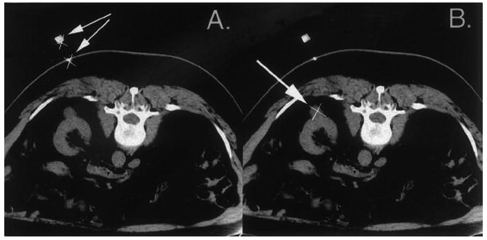
Figure 4.

Two images sent by the physician to the computer workstation. A, Image used to indicate the tip (×) and shaft (arrows) of the needle at the skin. B, Image used to indicate the target (×, arrow), an exophytic renal mass. The target is in the same image as is the needle tip, but it does not need to be.
