

Sir,
The genus Phaeoacremonium is intermediate between the genera Acremonium and Phialophora. It is distinguished from Acremonium by the presence of phaeoid, vegetative hyphae and conidiophores, and from Phialophora by its narrow, spine-like conidigenous cells. Phaeoacremonium species that cause human infections are Phaeoacremonium parasiticum, Phaeoacremonium infalitipes, and Phaeoacremonium rubrigenum.[1–3] We report here a subcutaneous, granulomatous infection caused by Phaeoacremonium infalitipes in the right foot of a man.
A 30 year-old male, a farmer by occupation, presented with pain in the right foot with a large lump which had been gradually enlarging over the past six months. There was a history of a minor injury due to a thorn prick one year ago. Examination of the left foot revealed a soft mass, which was slightly tender [Figure 1]. Biopsies from the sites were stained with Hematoxylin and Eosin stain and Gomori's Methenamine Silver stain, and revealed dense, proliferated, fibrous connective tissue with an inflammatory reaction and numerous, septate, hyphal fragments of various lengths. The biopsy material was cultured on Sabouraud's Dextrose Agar, with and without cycloheximide, and incubated at 25°C and 37°C. Colonies were initially glaborous, creamy to off-white, and later became brownish-black after two weeks of incubation. Slide cultures were performed on Potato Dextrose agar and incubated at 25°C. The Lactophenol Cotton Blue preparation from the slide culture showed phaeoid hyphae and subcylidrical phialides that were constricted at their bases, with larger conidia, some aggregated at the tips, some sliding down the sides of phialides [Figure 2].
Figure 1.
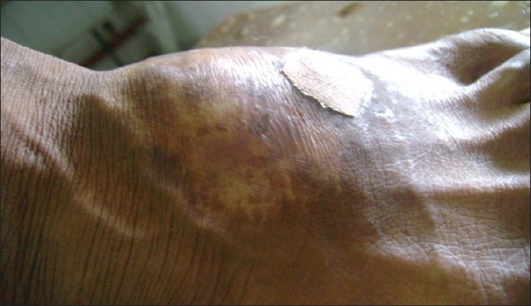
Swelling of the right foot
Figure 2.
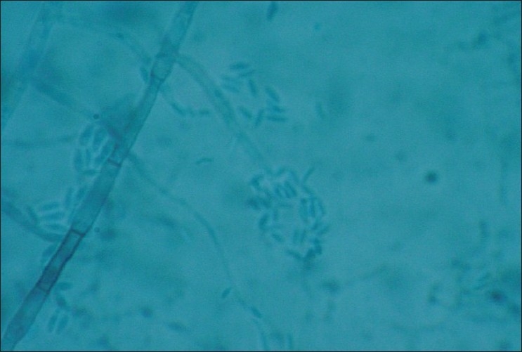
LPCB preparation from slide culture on PDA of Phaeoacremonium infalitipes showing subcylindrical phialides that are constricted at their bases and ellipsoid conidia (×1000)
Based on these findings, the isolate was identified as Phaeoacremonium infalitipes that could be distinguished from Phaeoacremonium parasiticum by its inflated phialides, which were constricted at the base. Surgical debridement was done and IV Amphotericin B was administered for two weeks. The debrided specimen also showed similar findings of hyphal forms on the GMS staining and Phaeoacremonium infalitipes was isolated from the cultures done from the debrided specimen.
The patient responded well to the treatment and was later discharged on oral Itraconazole for a period of two months. When last followed up, the patient showed no signs of recurrence and there was complete healing of the debrided wound.
The majority of the infections by Phaeoacremonium species are subcutaneous abscesses, cysts or chronic arthritis, synovitis, and mycetoma. Padhye et al.[3] reported similar presentation as in the present case caused by Phaeoacremonium infalitipes, and the patient responded to surgical debridement. The case is being reported here because this is the first such case from India.
References
- 1.Crous PW, Gams W, Wingfield MJ, Van Wyk PS. Phaeoacremonium gen.nov. associated with wilt and decline diseases of woody hosts and human infections. Mycologica. 1996;88:786–96. [Google Scholar]
- 2.Heath CH, Lendrum JL, Wetherall BL, Wesseling SL, Gordon DL. Phaeoacremonium parasiticum infective endocarditis following liver transplantataion. Clin Infect Dis. 1997;25:1251–2. doi: 10.1086/516963. [DOI] [PubMed] [Google Scholar]
- 3.Padhye AA, Davis M, Baer D, Reddick A, Sinha KK, Ott J. Phaehyphomycosis caused by Phaeoacremonium infalitipes. J Clin Microbiol. 1998;36:2763–5. doi: 10.1128/jcm.36.9.2763-2765.1998. [DOI] [PMC free article] [PubMed] [Google Scholar]