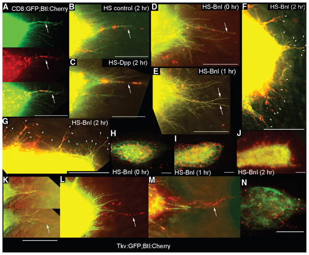
Fig. 3.
Dpp and FGF receptors segregate to separate locations in the ASP. (A to J) Expression of Btl:Cherry and CD8:GFP driven by btl-Gal4 mark ASP cytonemes (green) with Btl-containing puncta (red). [(A) and (B)] ASP tips from larvae lacking a HS transgene with (B) or without (A) heat shock. (C) Btl:Cherry puncta in ASP tip cytonemes after ectopic expression of Dpp. [(D) to (F) and (H) to (J)] Expression of Btl:Cherry and CD8:GFP released from Gal80ts repression by temperature elevation (3) and subjected to heat shock for the indicated times. (E) Image of ASP tip 1 hour after heat shock. (F) Image of ASP tip 2 hours after heat shock [also see table S4 and (3)]. (G) Image of one side of the ASP tube between the TC and tip of (F). Arrowheads in (F) and (G) indicate short cytonemes containing Btl:Cherry puncta. [(H) to (J)] Low-resolution (40×) images showing distribution of Btl:Cherry after overexpression of Bnl. (F), (G), and (J) are images of the same preparation. (K to N) Tip of ASPs expressing Tkv:GFP and Btl:Cherry driven by btl-Gal4. (K) Two focal planes from z-sections in which Tkv:GFP-containing cytonemes were in a focal plane (top) less proximal to the disc than those with Btl:Cherry (bottom). [(L) and (M)] Projection images show distinct Tkv:GFP and Btl:Cherry–containing cytonemes at ASP tip. (N) Image of mid-distal ASP. Arrows in (A) to (D) and (K) to (M) indicate long cytonemes containing Btl:Cherry puncta. All images oriented ASP tip to right. Scale bars, 30 μm.

