Summary
Recent discoveries of IgD in ancient vertebrates suggest that IgD has been preserved in evolution from fish to human for important immunological functions. A non-canonical form of class switching from IgM to IgD occurs in the human upper respiratory mucosa to generate IgD-secreting B cells highly reactive against respiratory pathogens and their products. In addition to enhancing mucosal immunity, IgD class-switched B cells enter the circulation to “arm” basophils and other innate immune cells with secreted IgD. Although the nature of the IgD receptor remains elusive, cross-linking of IgD on basophils stimulates release of immunoactivating, proinflammatory and antimicrobial mediators. This pathway is dysregulated in autoinflammatory disorders such as hyper-IgD syndrome, indicating that IgD orchestrates an ancestral surveillance system at the interface between immunity and inflammation.
Introduction
Five classes of antibodies named IgM, IgD, IgG, IgA and IgE exist in humans and mice. While the function of IgM, IgG, IgA and IgE is relatively well known, the function of IgD has remained obscure since the discovery of IgD in 1965 [1,2]. IgD is co-expressed with IgM on the surface of the majority of mature B cells prior to antigenic stimulation and functions as a transmembrane antigen receptor [3,4]. However, secreted IgD also exists and plays an elusive function in blood, mucosal secretions and on the surface of innate immune effector cells such as basophils [1,5]. In this article we review recent advances in our understanding of the regulation and function of IgD.
Evolutionary preservation of IgD
IgD was initially thought to be a recently evolved antibody class, because it was only detected in primates, mice, rats and dogs, but not guinea pigs, swine, cattle, sheep and frogs [6]. With the increasing availability of animal genome sequences and the rapid development of gene identification tools, the past 20 years have seen the discovery of IgD and its homologues and orthologues in more mammalian species as well as cartilaginous fishes, bony fishes, frogs and reptiles [7]. The most primitive of these species are cartilaginous fishes, which populated our planet about 500 million years ago, when jawed vertebrates first appeared and the adaptive immune system first evolved. This implies that IgD is an ancestral antibody class that has remained preserved in most jawed vertebrates throughout evolution [8]. Hence, IgD should exert some important immune functions that may confer a specific survival advantage to the host.
Structural diversity of IgD
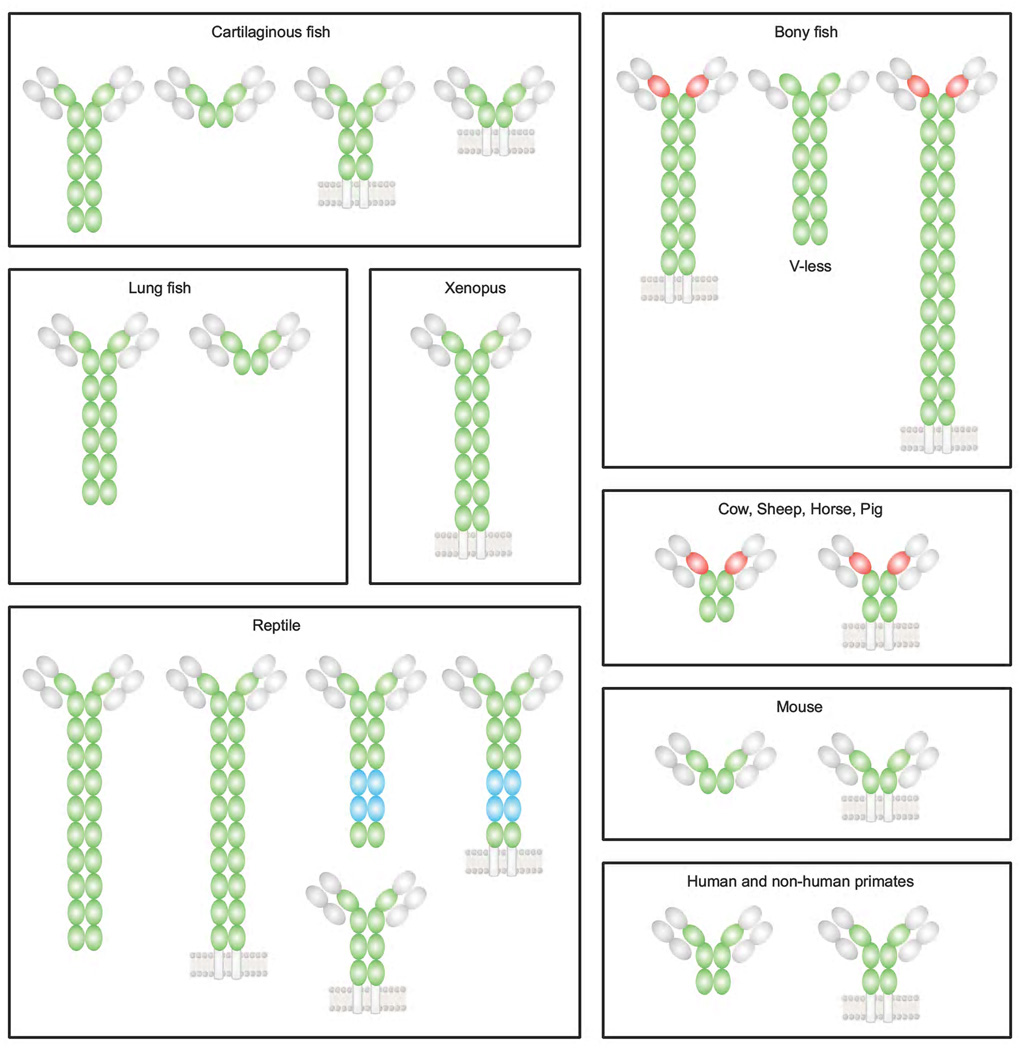
IgD exhibits much structural diversity throughout vertebrate evolution (Figure 1). B cells employ two strategies, including alternative RNA splicing and class switch recombination (CSR), to express IgD. Alternative splicing exists in all jawed vertebrates, including jawed fishes, while CSR is only found in higher vertebrates, from frogs to humans [9]. In fishes, the structure of the constant (C) region of IgD is highly diverse owing to various intragenic duplications of Cδ exons that can give rise to a large number of Cδ domains in the IgD molecule [6,7]. Alternative splicing further increases IgD diversity by creating different splice variants [8,10–12], perhaps to compensate for the lack of CSR. Interestingly, IgD molecules without antigen-binding variable (V) region have been detected in channel catfish, raising the possibility that Cδ exerts some form of “innate” immune function [13]. IgD also exhibits structural diversity in mammals. Indeed, IgD from both human and non-human primates has three Cδ domains (UniProtKB/Swiss-Prot Database; URL: http://www.uniprot.org/uniprot/P01880), while IgD from rodents only has two Cδ domains (UniProtKB/Swiss-Prot Database; URL: http://www.uniprot.org/uniprot/P018801). Interestingly, IgD from artiodactyls has three Cδ domains consisting of a Cμ1 domain that replaces a deleted Cδ1 domain and two additional Cδ domains [14,15]. This chimeric Cμ1-Cδ structure is typical of fish IgD and may be needed by the H chains of IgD to covalently bind to light (L) chains through Cμ1.
Figure 1.
Structural diversity of IgD. The heavy chain variable region and light chain of IgD are represented by gray ovals, whereas the Cδ domains of the heavy chain constant region of IgD are represented by colored ovals. Intragenic duplications of Cδ exons and alternative splicing generate structural diversity of IgD in fish. Transmembrane and secreted fish IgD molecules are shown to emphasize alternative splicing. No transmembrane forms have been described in lungfish. Xenopus has abundant transmembrane IgD as well as transcripts encoding secreted IgD. However, the structure of secreted IgD has not been clearly shown in xenopus. Presence of Ig light chain is predicted but not demonstrated in IgD from bony fish, xenopus, and lungfish. IgD of channel catfish and puffer fish, among other bony fishes, is shown. The red domain is encoded by a duplicated Cμ1 exon. IgD of leopard gecko and green anole lizard is shown. The blue domains denote Cα-like domains found in leopard gecko IgD. The red domains in cow, sheep, horse and pig IgD indicate the inclusion of a Cμ1 or Cμ1-like domain. Hinge regions of IgD are not shown.
The hinge (H) region of mammalian IgD is even more diverse in terms of length, amino acid composition and glycosylation. IgD from both human and non-human primates has a long H region consisting of an amino-terminal region rich in alanine and threonine residues and a carboxy-terminal region rich in charged lysine, glutamate and arginine residues, with up to seven O-linked glycans. The length of the H region renders human IgD capable of acquiring a flexible T shape rather than the traditional Y shape of other antibody isotypes, with two antigen-binding Fab arms swiveling at the two sides of the Fc region [16]. One possibility is that a flexible T shape may help IgD to bind epitopes that have a low density on the surface of particulate antigens. Of note, rodent IgD has a much shorter H region than human IgD, with a very different amino acid composition and only one N-linked glycan.
Structural differences in the H region may contribute to different functions of IgD in humans and mice. Human IgD seems to utilize O-linked glycans associated with the H region to bind a putative IgD receptor on the surface of activated T cells [17,18]. Human IgD could further utilize the highly charged segment of its H region to interact with heparin and/or heparan sulphate proteglycans expressed on the surface and in the granules of basophils and mast cells [7]. In all jawed vertebrates, an additional layer of structural diversity is generated by alternative splicing to create various transmembrane and secreted forms of IgD [7]. The reason underlying the structural diversity of IgD in evolution is that IgD may have been selected as a structurally flexible locus to complement the function of IgM [6,11]. One possibility is that the presence of IgD may ensure the preservation of essential immune functions in case of IgM defects, and the flexibility of IgD may provide additional immune functions in a species-specific manner.
IgD expression through alternative splicing
While IgM is first expressed by pre-B cells, IgD emerges later during B cell ontogeny, being mostly expressed at the transitional and mature B cell stage, at least in rodents and primates [6,7]. In most vertebrates, mature naive IgM+IgD+ B cells co-express IgM and IgD through alternative mRNA splicing, but the transcriptional ratio of Cμ and Cδ exons varies widely in different types of B cells [19]. The mechanisms regulating the ratio of Cμ to Cδ exon usage are poorly understood, but are likely to involve the post-translational modification of polymerase II and the induction of factors that regulate mRNA polyadenylation and splicing in response to antigenic stimulation and cellular differentiation. In plasma cells, the transcriptional elongation factor ELL2 associates with the carboxy-terminal portion of RNA polymerase II and with the polyadenylation factor CstF-64 to promote skipping of downstream exons through preferential usage of upstream mRNA cleavage and polyadenylation sites [20,21]. A similar transcriptional repression mechanism could explain the down-regulation of IgD expression that typically occurs in most antigen-experienced B cells, except IgM−IgD+ B cells.
IgD expression through class switching
In humans, a small subset of B cells express IgD but not IgM after undergoing an unconventional form of CSR [22,23]. These IgM−IgD+ B cells are found in the circulation as well as tonsils, nasal cavities, lachrymal glands and salivary glands, [7,24], but are rarely detected in non-respiratory mucosal districts. The specific topography of IgM−IgD+ B cells may result from the expression of tissue homing receptors that do not favor colonization of extra-respiratory mucosal sites such as the intestine [25]. Interestingly, IgM−IgD+ B cells are also found in channel catfish [13], but are not generated through IgM-to-IgD CSR. Indeed, although expressing a CSR-competent activation-induced cytidine deaminase (AID) molecule [26], catfish B cells seem to lack recognizable switch (S) regions [10,13], suggesting that IgM−IgD+ B cells originate from antigen-induced transcriptional inactivation of the IgM locus. Interestingly, the H chain of IgD from catfish IgM−IgD+ B cells is paired with an uncommon σ L chain [13]. Also human IgM−IgD+ B cells utilize a λ L chain instead of the more common κ L chain [22,23], which may hint to a derivation of human IgM−IgD+ B cells from a specific λ+ B cell lineage. In addition to specifically seeding the upper respiratory tract, λ+ B cell precursors of IgM−IgD+ B cells may be intrinsically committed to undergo IgM-to-IgD CSR. The mechanism of this unconventional form of CSR remains unclear.
S regions are highly repetitive intronic DNA sequences with G-rich non-template strands that precede each Cμ, Cγ, Cα and Cε gene and guide the process of CSR [27,28]. Upstream of each S region, there is a promoter associated with a short intronic (I) exon that mediates germline transcription [27,28]. While germline transcription of Cμ occurs in a constitutive manner, germline transcription of Cγ, Cα and Cε occurs after exposure of B cells to specific cytokines [27,28]. Germline transcription is crucial for CSR, as it renders the targeted S region substrate of AID, a DNA-editing enzyme essential for CSR [27–29]. Germline transcription of a given CX gene yields a primary IX-SX-CX transcript that is later spliced to form a secondary non-coding germline IX-CX transcript [27,28]. The primary transcript physically associates with the template strand of the S region DNA to form a stable DNA-RNA hybrid [27,28]. Such a structure generates R loops, in which the displaced non-template strand exists as a G-rich single-stranded DNA [27,28]. AID deaminates cytosine residues on both strands of S region DNA, thereby generating multiple DNA lesions that are ultimately processed into double-stranded DNA breaks [27,28]. Fusion of double-stranded DNA breaks at donor and acceptor SX regions through the non-homologous end-joining pathway induces looping-out deletion of the intervening DNA, thereby juxtaposing the recombined VHDJH exon encoding the antigen-binding VH region of the rearranging Ig molecule to a new CX gene [27,28].
Unlike Cμ, Cγ, Cα and Cε genes, the Cδ gene is not preceded by a canonical S region [27,28], which initially led to the idea that the genesis of IgM−IgD+ B cells does not involve CSR. However, a short, repetitive, G-rich intronic region called σδ lies between Cμ and Cδ genes in both human and cow IgH loci and functions as a cryptic acceptor site for the donor Sμ region to mediate non-homologous IgM-to-IgD CSR [7,22,23,30]. Additional studies found two identical, short intronic Iμ and Σμ regions upstream and downstream of Cμ in human and mouse IgH loci that might mediate homologous IgM-to-IgD CSR [31]. The involvement of CSR in the generation of IgM−IgD+ B cells is consistent with the recent observation that IgM−IgD+ B cells virtually disappear in patients with hyper-IgM syndrome type-2 (HIGM2) [22], which is characterized by a deficiency of AID [32]. Nonetheless, in HIGM2 patients, some IgD persists in the serum, but would derive from unusual IgM+IgD+ plasma cells secreting both IgM and IgD [22].
The mechanism by which AID targets the σδ S-like region remains obscure. Although constitutively transcribing both Cμ and Cδ loci and expressing AID in response to appropriate stimuli, only a minority of IgM+IgD+ B cells undergo AID-dependent IgM-to-IgD CSR [22]. One possible explanation is that AID does not target σδ in the absence of additional factors specifically expressed by λ+ precursors of IgM−IgD+ B cells. In addition, developmentally regulated epigenetic factors may render σδ impervious to AID in most B cells, except λ+ precursors of IgM+IgD− B cells. Clearly, more studies are needed to solve this intriguing puzzle.
Signals inducing IgD class switching
In addition to harboring unusual chromosomal Sμ-σδ and Iμ-Σμ rearrangements and biased λ L chain gene usage, most IgM−IgD+ B cells from the upper respiratory mucosa express highly hypermutated and clonally related V(D)J genes [23,33], suggesting massive oligoclonal expansion of IgM−IgD+ B cells in response to some respiratory antigen. Similar to mouse peritoneal B-1 cells, a large proportion of human tonsillar IgM−IgD+ B cells are highly polyreactive [34], which could enhance their ability to provide a rapid first line of humoral defense in the upper respiratory mucosa. Most of this polyreactivity may be a natural feature of unmutated λ+ precursors of IgM−IgD+ B cells, but additional polyreactivity may originate from SHM and antigen selection induced by local microbial determinants, including superantigens [34–36]. In this regard, human IgD can bind antigens from respiratory commensals and pathogens by utilizing the Cδ region instead of the V region [37,38]. The ensuing polyclonal activation of IgM+IgD+ and IgM−IgD+ B cells might compensate for the eventual accumulation of “crippling” mutations in the V region of at least some IgM−IgD+ B cells [39,40]. Interestingly, also the C region of catfish IgD might have innate antigen-binding properties, which may explain why in these animals the secreted form of IgD lacks an antigen-binding V region due to direct splicing of the Cδ1 domain to a leader exon [13].
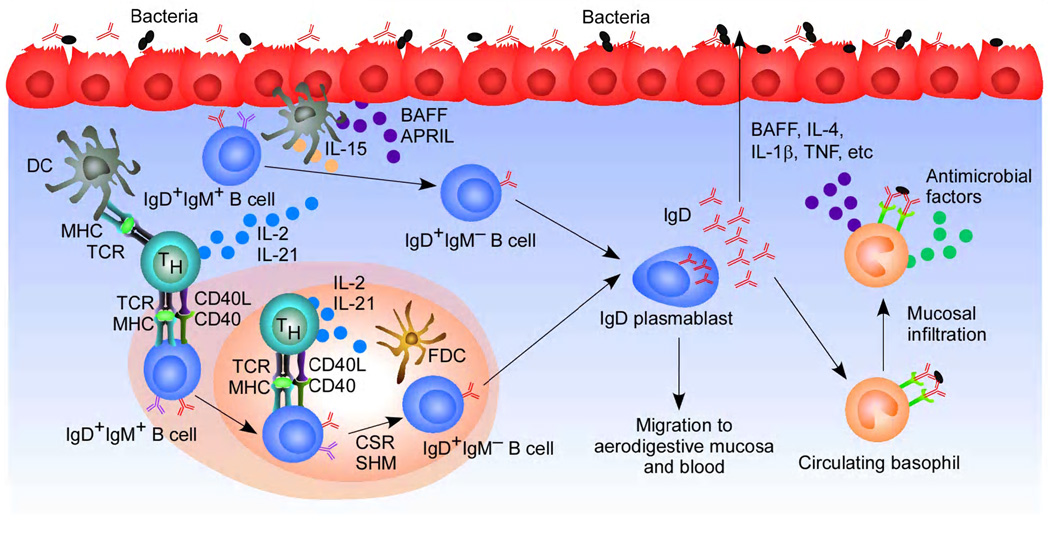
Signals capable of inducing IgM-to-IgD CSR include CD40 ligand (CD40L), a tumor necrosis factor (TNF) family ligand expressed by CD4+ T helper cells and required for B cell responses to T cell-dependent (TD) antigens, as well as B-cell activation factor of the TNF family (BAFF) and a proliferation-inducing ligand (APRIL) (Figure 2), two CD40L-related factors released by innate immune cells and involved in B cell responses to T cell-independent (TI) antigens [7,22]. Together with a combination of interleukin-15 (IL-15) and IL-21 or IL-2 and IL-21, CD40L, BAFF and APRIL not only induce Sμ-σδ CSR, but also promote the expression of a surface IgM−IgD+ phenotype typical of IgD class-switched B cells and the secretion of IgD [22]. This requirement for both TD and TI signals is further supported by the follicular and extrafollicular localization of IgM−IgD+ B cells, and by the fact that HIGM1 and HIGM3 patients with deleterious substitutions of CD40L and CD40 (OMIM Database: URLs: http://www.ncbi.nlm.nih.gov/omim/308230 and http://www.ncbi.nlm.nih.gov/omim/606843) as well as common variable immunodeficiency (CVID) patients with deleterious substitutions of calcium modulator and cyclophilin interactor (TACI, a CSR-inducing receptor for BAFF and APRIL) (OMIM Database; URL: http://www.ncbi.nlm.nih.gov/omim/240500) have less IgM−IgD+ B cells [22].
Figure 2.
Induction, regulation and function of mucosal IgD. Mucosal dendritic cells (DCs) present antigen to activate CD4+ T helper (TH) cells. These cells induce follicular IgM+IgD+ B cells to undergo IgM-to-IgD CSR through a TD pathway involving CD40L, IL-2 and IL-21. In addition, innate immune cells such as DCs, monocytes and epithelial cells produce BAFF, APRIL, IL-2 and IL-15 probably upon sensing microbial products. These mediators stimulate extrafollicular IgM+IgD+ B cells to undergo IgM-to-IgD CSR in a TI manner. The resulting IgD class-switched (IgM−IgD+) B cell differentiate into plasmablasts that secrete IgD molecules reactive against respiratory antigens. Secreted IgD also binds to an IgD receptor (IgDR) on circulating basophils. In the presence of IgD cross-linking antigens, basophils migrate to systemic or mucosal lymphoid tissues, where they enhance immunity by releasing immunoactivating, proinflammatory and antimicrobial factors such as BAFF, IL-4, IL-1β and TNF. These factors augment mucosal immune responses by promoting B and T cell activation, leukocyte recruitment and direct microbial killing.
Functions of membrane and secreted IgD
The reason why mature B cells express two IgM and IgD receptors remains unclear. One line of thought is that IgM and IgD deliver qualitatively different signals. Consistent with this possibility, IgM and IgD associate with distinct B cell receptor-associated proteins (BAPs) [6]. Additional evidence suggests a function of IgD in delivering tolerogenic or apoptotic signals. Mouse anergic B cells express more IgD than IgM [41–43]. Similarly, human B cells expressing more IgD than IgM show poor responsiveness to stimulation by antigen [34,36,44]. These B cells also express auto (poly) reactive IgD, which may lead to anergy through tolerogenic mechanisms [34,36,44]. Furthermore, transgenic mice ubiquitously expressing a cell surface superantigen that reacts with IgD show an arrest of B cell development at the immature stage [45], which clearly correlates with the commencement of membrane IgD expression at this stage. However, other seemingly contradicting findings show that IgD may actually protect B cells from tolerance [46,47]. Of note, the H chain of IgM is essential for the formation of the pre-B cell receptor, while the H chain of IgD is not [48], arguing against the old observation that IgD can substitute the function of IgM in B cell development [49]. These discrepancies may result from differences in the types of B cells and antigenic stimulations used, as the outcome of IgD signaling may be influenced by the maturation status of the B cell and the strength of the antigenic stimulus.
In general, the abundance of IgM−IgD+ B cells in the upper respiratory mucosa [22,24] and the fact that secreted IgD binds microbial virulence factors as well as pathogenic respiratory bacteria and viruses [7] support the notion that secreted IgD enhances mucosal immunity. Consistent with this possibility, patients suffering from selective IgA deficiency have markedly increased numbers of IgD-producing B cells in their respiratory mucosa [24]. In addition to binding antigen through both conventional V-mediated and unconventional Cδ-mediated mechanisms, secreted IgD activates an as yet unknown receptor on various innate immune cells. Early studies show that IgD binds to both myeloid cells and T cells [7]. More recent observations show that IgD binds to basophils, mast cells and, albeit to a lesser extent, monocytes, neutrophils and myeloid dendritic cells through a receptor distinct from IgG, IgA or IgE receptors [22,50–52]. The binding of IgD to basophils is evolutionarily conserved as IgD also binds a basophil-like subset of granulocytes in catfish [22]. Cross-linking of IgD induces basophil production of immunoactivating cytokines such as IL-4, IL-13 and BAFF, proinflammatory cytokines such as TNF and IL-1β, and chemokines such as IL-8 and CXC chemokine ligand 10 (CXCL10) [22]. Of note, production of BAFF (a mandatory B cell survival factor) and IL-4 (an IgG1- and IgE-inducing factor) by basophils in response to IgD cross-linking would be consistent with the development of peripheral B cell depletion, reduced serum IgE levels and impaired TD IgG1 production in mice lacking IgD [7,53,54]. Of note, IgD cross-linking triggers basophil release of antimicrobial factors such as cathelicidin [22], suggesting that IgD also prompts basophils to participate directly in antimicrobial immunity.
The ability of IgD to activate proinflammatory functions is supported by the observation that hyper-IgD syndrome (HIDS) caused by deleterious substitutions of mevalonate kinase (MvK) is associated with periodic fever, systemic antibiotic-resistant inflammation as well as elevated serum IgD, increased circulating IgM−IgD+ B cells [22], and abnormally activated macrophages [55]. The mechanism by which an enzyme of the cholesterol biosynthetic pathway such as MvK influences IgM−IgD+ B cells remains a mystery. One possibility is that mevalonate-derived products such as isoprenoids exert a negative control on the formation, survival and/or migration of IgM−IgD+ B cells. Alternatively, IgM−IgD+ B cells may increase as a result of the ongoing inflammatory reaction. Periodic fever-aphthous stomatitis-pharyngitis-adenitis (PFAPA) syndrome is another autoinflammatory disorder that causes periodic fever and aseptic mucosal inflammation together with elevated serum IgD, increased circulating and mucosal IgM−IgD+ B cells, and enhanced mucosal IgD-armed basophils [22]. The pathogenesis of PFAPA syndrome is unknown and therefore it is unclear whether dysregulated IgD production in this syndrome is a cause or rather an effect of the inflammatory reaction.
Conclusions
IgM−IgD+ B cells originate in the human upper respiratory from both TD and TI pathways involving CD40L, BAFF and APRIL [7,22]. These mediators are not specific to the respiratory tract, suggesting the involvement of additional factors in the topography of IgM−IgD+ B cells. One possibility is that naturally polyreactive and λ L chain-expressing precursors of IgM−IgD+ B cells preferentially home to the respiratory mucosa from the bone marrow. Such precursors may have an IgH locus “geared” to undergo IgM-to-IgD CSR and further increase their polyreactivity by undergoing SHM in mucosal follicles. SHM may also generate IgD molecules with more specific reactivity against respiratory commensals and pathogens [7]. Secretion of IgD by plasmacytoid IgM−IgD+ B cells would then lead to the binding of IgD to an as yet unknown IgD receptor on mucosal and circulating myeloid cells, including basophils [22]. In this manner, IgD may educate the innate immune system as to the antigenic composition of the upper respiratory tract, thereby enhancing local and systemic surveillance against airborne pathogens. The seemingly conserved nature of this and other immune functions of IgD from fish to humans further supports the notion that IgD is part of an ancestral surveillance system involving microbial sensing and immune activation [13,22]. A dysregulation of this system may contribute to the pathogenesis of inflammation as seen in autoinflammatory disorders associated with hyper-IgD production. Further immunological characterization of these disorders as well as IgD–knockout mice [53,54] and MvK–knockout mice [56] should yield more insights into the function of IgD in immunity and homeostasis.
Acknowledgements
Supported by US National Institutes of Health grants R01 AI-074378, ARRA AI-61093, funds from The Hemsley Foundation for IBD research, Ministerio de Ciencia e Innovación grant SAF 2008-02725, and funds from Fundacio’ IMIM (to A.C.).
Footnotes
Publisher's Disclaimer: This is a PDF file of an unedited manuscript that has been accepted for publication. As a service to our customers we are providing this early version of the manuscript. The manuscript will undergo copyediting, typesetting, and review of the resulting proof before it is published in its final citable form. Please note that during the production process errors may be discovered which could affect the content, and all legal disclaimers that apply to the journal pertain.
Contributor Information
Kang Chen, Email: kang.chen@mssm.edu.
Andrea Cerutti, Email: acerutti@imim.es, andrea.cerutti@mssm.edu.
References and recommended reading
- 1.Rowe DS, Fahey JL. A new class of human immunoglobulins. Ii. Normal serum IgD. J. Exp. Med. 1965;121:185–199. doi: 10.1084/jem.121.1.185. [DOI] [PMC free article] [PubMed] [Google Scholar]
- 2.Rowe DS, Fahey JL. A new class of human immunoglobulins. I. A unique myeloma protein. J. Exp. Med. 1965;121:171–184. doi: 10.1084/jem.121.1.171. [DOI] [PMC free article] [PubMed] [Google Scholar]
- 3.Finkelman FD, van Boxel JA, Asofsky R, Paul WE. Cell membrane IgD: demonstration of IgD on human lymphocytes by enzyme-catalyzed iodination and comparison with cell surface Ig of mouse, guinea pig, and rabbit. J. Immunol. 1976;116:1173–1181. [PubMed] [Google Scholar]
- 4.Ruddick JH, Leslie GA. Structure and biologic functions of human IgD. XI. Identification and ontogeny of a rat lymphocyte immunoglobulin having antigenic cross-reactivity with human IgD. J. Immunol. 1977;118:1025–1031. [PubMed] [Google Scholar]
- 5.Finkelman FD, Woods VL, Berning A, Scher I. Demonstration of mouse serum IgD. J. Immunol. 1979;123:1253–1259. [PubMed] [Google Scholar]
- 6.Preud'homme JL, Petit I, Barra A, Morel F, Lecron JC, Lelievre E. Structural and functional properties of membrane and secreted IgD. Mol. Immunol. 2000;37:871–887. doi: 10.1016/s0161-5890(01)00006-2. [DOI] [PubMed] [Google Scholar]
- 7.Chen K, Cerutti A. New insights into the enigma of immunoglobulin D. Immunol Rev. 2010;237:160–179. doi: 10.1111/j.1600-065X.2010.00929.x. [DOI] [PMC free article] [PubMed] [Google Scholar]
- 8. Ohta Y, Flajnik M. IgD, like IgM, is a primordial immunoglobulin class perpetuated in most jawed vertebrates. Proc. Natl. Acad. Sci. USA. 2006;103:10723–10728. doi: 10.1073/pnas.0601407103. By discovering the IgD gene in xenopus, the authors presented the concept that IgD is an evolutionarily preserved antibody class with structural flexibility in most jawed vertebrates and functions to complement the function of IgM.
- 9.Stavnezer J, Amemiya CT. Evolution of isotype switching. Semin. Immunol. 2004;16:257–275. doi: 10.1016/j.smim.2004.08.005. [DOI] [PubMed] [Google Scholar]
- 10.Bengten E, Clem LW, Miller NW, Warr GW, Wilson M. Channel catfish immunoglobulins: repertoire and expression. Dev. Comp. Immunol. 2006;30:77–92. doi: 10.1016/j.dci.2005.06.016. [DOI] [PubMed] [Google Scholar]
- 11.Bengten E, Quiniou SM, Stuge TB, Katagiri T, Miller NW, Clem LW, Warr GW, Wilson M. The IgH locus of the channel catfish, Ictalurus punctatus, contains multiple constant region gene sequences: different genes encode heavy chains of membrane and secreted IgD. J. Immunol. 2002;169:2488–2497. doi: 10.4049/jimmunol.169.5.2488. [DOI] [PubMed] [Google Scholar]
- 12.Wilson M, Bengten E, Miller NW, Clem LW, Du Pasquier L, Warr GW. A novel chimeric Ig heavy chain from a teleost fish shares similarities to IgD. Proc. Natl. Acad. Sci. USA. 1997;94:4593–4597. doi: 10.1073/pnas.94.9.4593. [DOI] [PMC free article] [PubMed] [Google Scholar]
- 13. Edholm ES, Bengten E, Stafford JL, Sahoo M, Taylor EB, Miller NW, Wilson M. Identification of two IgD+ B cell populations in channel catfish, Ictalurus punctatus. J. Immunol. 2010;185:4082–4094. doi: 10.4049/jimmunol.1000631. This article demonstrated the existence of IgM−IgD+ B cells in catfish that preferentially express σ light chain, analogous to the biased usage of λ light chain by human IgM−IgD+ B cells. Secreted catfish IgD heavy chain contained no variable region, suggesting that IgD may function as a pattern recognition molecule like human IgD.
- 14.Zhao Y, Kacskovics I, Pan Q, Liberles DA, Geli J, Davis SK, Rabbani H, Hammarstrom L. Artiodactyl IgD: the missing link. J. Immunol. 2002;169:4408–4416. doi: 10.4049/jimmunol.169.8.4408. [DOI] [PubMed] [Google Scholar]
- 15.Zhao Y, Kacskovics I, Rabbani H, Hammarstrom L. Physical mapping of the bovine immunoglobulin heavy chain constant region gene locus. J Biol Chem. 2003;278:35024–35032. doi: 10.1074/jbc.M301337200. [DOI] [PubMed] [Google Scholar]
- 16.Sun Z, Almogren A, Furtado PB, Chowdhury B, Kerr MA, Perkins SJ. Semi-extended solution structure of human myeloma immunoglobulin D determined by constrained X-ray scattering. J. Mol. Biol. 2005;353:155–173. doi: 10.1016/j.jmb.2005.07.072. [DOI] [PubMed] [Google Scholar]
- 17.Rudd PM, Fortune F, Patel T, Parekh RB, Dwek RA, Lehner T. A human T-cell receptor recognizes 'O'-linked sugars from the hinge region of human IgA1 and IgD. Immunology. 1994;83:99–106. [PMC free article] [PubMed] [Google Scholar]
- 18.Swenson CD, Patel T, Parekh RB, Tamma SM, Coico RF, Thorbecke GJ, Amin AR. Human T cell IgD receptors react with O-glycans on both human IgD and IgA1. Eur. J. Immunol. 1998;28:2366–2372. doi: 10.1002/(SICI)1521-4141(199808)28:08<2366::AID-IMMU2366>3.0.CO;2-D. [DOI] [PubMed] [Google Scholar]
- 19.Kerr WG, Hendershot LM, Burrows PD. Regulation of IgM and IgD expression in human B-lineage cells. J. Immunol. 1991;146:3314–3321. [PubMed] [Google Scholar]
- 20. Martincic K, Alkan SA, Cheatle A, Borghesi L, Milcarek C. Transcription elongation factor ELL2 directs immunoglobulin secretion in plasma cells by stimulating altered RNA processing. Nat. Immunol. 2009;10:1102–1109. doi: 10.1038/ni.1786. This study revealed the regulation of immunoglobulin mRNA splicing mediated by cell stage-specific expression and binding of elongation and polyadenylation factors to RNA polymerase II. Similar mechanisms may operate in the regulation of alternative RNA splicing in IgD expression.
- 21.Takagaki Y, Seipelt RL, Peterson ML, Manley JL. The polyadenylation factor CstF-64 regulates alternative processing of IgM heavy chain pre-mRNA during B cell differentiation. Cell. 1996;87:941–952. doi: 10.1016/s0092-8674(00)82000-0. [DOI] [PubMed] [Google Scholar]
- 22. Chen K, Xu W, Wilson M, He B, Miller NW, Bengten E, Edholm ES, Santini PA, Rath P, Chiu A, et al. Immunoglobulin D enhances immune surveillance by activating antimicrobial, proinflammatory and B cell-stimulating programs in basophils. Nat. Immunol. 2009;10:889–898. doi: 10.1038/ni.1748. This study identified the cytokine signals that promote the CSR and production of human IgD, and the function of IgD in inducing immuno-activating, pro-inflammatory and antimicrobial functions of basophils. Findings also suggest that such functions are evolutionarily conserved from fish to human.
- 23.Arpin C, de Bouteiller O, Razanajaona D, Fugier-Vivier I, Briere F, Banchereau J, Lebecque S, Liu YJ. The normal counterpart of IgD myeloma cells in germinal center displays extensively mutated IgVH gene, Cmu-Cdelta switch, and lambda light chain expression. J. Exp. Med. 1998;187:1169–1178. doi: 10.1084/jem.187.8.1169. [DOI] [PMC free article] [PubMed] [Google Scholar]
- 24.Brandtzaeg P, Carlsen HS, Farstad IN. The human mucosal B-cell system. In: Mestecky J, Lamm ME, Strober W, Bienenstock J, McGhee JR, Mayer L, editors. Mucosal Immunology. Elsevier Academic Press; 2005. [Google Scholar]
- 25.Johansen FE, Baekkevold ES, Carlsen HS, Farstad IN, Soler D, Brandtzaeg P. Regional induction of adhesion molecules and chemokine receptors explains disparate homing of human B cells to systemic and mucosal effector sites: dispersion from tonsils. Blood. 2005;106:593–600. doi: 10.1182/blood-2004-12-4630. [DOI] [PubMed] [Google Scholar]
- 26.Wakae K, Magor BG, Saunders H, Nagaoka H, Kawamura A, Kinoshita K, Honjo T, Muramatsu M. Evolution of class switch recombination function in fish activation-induced cytidine deaminase, AID. Int. Immunol. 2006;18:41–47. doi: 10.1093/intimm/dxh347. [DOI] [PubMed] [Google Scholar]
- 27.Chaudhuri J, Alt FW. Class-switch recombination: interplay of transcription, DNA deamination and DNA repair. Nat. Rev. Immunol. 2004;4:541–552. doi: 10.1038/nri1395. [DOI] [PubMed] [Google Scholar]
- 28.Stavnezer J, Guikema JE, Schrader CE. Mechanism and regulation of class switch recombination. Annu. Rev. Immunol. 2008;26:261–292. doi: 10.1146/annurev.immunol.26.021607.090248. [DOI] [PMC free article] [PubMed] [Google Scholar]
- 29.Muramatsu M, Kinoshita K, Fagarasan S, Yamada S, Shinkai Y, Honjo T. Class switch recombination and hypermutation require activation-induced cytidine deaminase (AID), a potential RNA editing enzyme. Cell. 2000;102:553–563. doi: 10.1016/s0092-8674(00)00078-7. [DOI] [PubMed] [Google Scholar]
- 30.Kluin PM, Kayano H, Zani VJ, Kluin-Nelemans HC, Tucker PW, Satterwhite E, Dyer MJ. IgD class switching: identification of a novel recombination site in neoplastic and normal B cells. Eur. J. Immunol. 1995;25:3504–3508. doi: 10.1002/eji.1830251244. [DOI] [PubMed] [Google Scholar]
- 31.White MB, Word CJ, Humphries CG, Blattner FR, Tucker PW. Immunoglobulin D switching can occur through homologous recombination in human B cells. Mol. Cell Biol. 1990;10:3690–3699. doi: 10.1128/mcb.10.7.3690. [DOI] [PMC free article] [PubMed] [Google Scholar]
- 32.Revy P, Muto T, Levy Y, Geissmann F, Plebani A, Sanal O, Catalan N, Forveille M, Dufourcq-Labelouse R, Gennery A, et al. Activation-induced cytidine deaminase (AID) deficiency causes the autosomal recessive form of the Hyper-IgM syndrome (HIGM2) Cell. 2000;102:565–575. doi: 10.1016/s0092-8674(00)00079-9. [DOI] [PubMed] [Google Scholar]
- 33.Liu YJ, de Bouteiller O, Arpin C, Briere F, Galibert L, Ho S, Martinez-Valdez H, Banchereau J, Lebecque S. Normal human IgD+IgM− germinal center B cells can express up to 80 mutations in the variable region of their IgD transcripts. Immunity. 1996;4:603–613. doi: 10.1016/s1074-7613(00)80486-0. [DOI] [PubMed] [Google Scholar]
- 34.Koelsch K, Zheng NY, Zhang Q, Duty A, Helms C, Mathias MD, Jared M, Smith K, Capra JD, Wilson PC. Mature B cells class switched to IgD are autoreactive in healthy individuals. J. Clin. Invest. 2007;117:1558–1565. doi: 10.1172/JCI27628. [DOI] [PMC free article] [PubMed] [Google Scholar]
- 35.Seifert M, Steimle-Grauer SA, Goossens T, Hansmann ML, Brauninger A, Kuppers R. A model for the development of human IgD-only B cells: Genotypic analyses suggest their generation in superantigen driven immune responses. Mol. Immunol. 2009;46:630–639. doi: 10.1016/j.molimm.2008.07.032. [DOI] [PubMed] [Google Scholar]
- 36.Zheng NY, Wilson K, Wang X, Boston A, Kolar G, Jackson SM, Liu YJ, Pascual V, Capra JD, Wilson PC. Human immunoglobulin selection associated with class switch and possible tolerogenic origins for C delta class-switched B cells. J. Clin. Invest. 2004;113:1188–1201. doi: 10.1172/JCI20255. [DOI] [PMC free article] [PubMed] [Google Scholar]
- 37.Forsgren A, Brant M, Mollenkvist A, Muyombwe A, Janson H, Woin N, Riesbeck K. Isolation and characterization of a novel IgD-binding protein from Moraxella catarrhalis. J. Immunol. 2001;167:2112–2120. doi: 10.4049/jimmunol.167.4.2112. [DOI] [PubMed] [Google Scholar]
- 38.Samuelsson M, Hallstrom T, Forsgren A, Riesbeck K. Characterization of the IgD binding site of encapsulated Haemophilus influenzae serotype b. J. Immunol. 2007;178:6316–6319. doi: 10.4049/jimmunol.178.10.6316. [DOI] [PubMed] [Google Scholar]
- 39.Jendholm J, Morgelin M, Perez Vidakovics ML, Carlsson M, Leffler H, Cardell LO, Riesbeck K. Superantigen- and TLR-dependent activation of tonsillar B cells after receptor-mediated endocytosis. J. Immunol. 2009;182:4713–4720. doi: 10.4049/jimmunol.0803032. [DOI] [PubMed] [Google Scholar]
- 40.Vidakovics ML, Jendholm J, Morgelin M, Mansson A, Larsson C, Cardell LO, Riesbeck K. B cell activation by outer membrane vesicles--a novel virulence mechanism. PLoS Pathog. 2010;6:e1000724. doi: 10.1371/journal.ppat.1000724. [DOI] [PMC free article] [PubMed] [Google Scholar]
- 41.Cyster JG, Hartley SB, Goodnow CC. Competition for follicular niches excludes self-reactive cells from the recirculating B-cell repertoire. Nature. 1994;371:389–395. doi: 10.1038/371389a0. [DOI] [PubMed] [Google Scholar]
- 42.Fulcher DA, Basten A. Reduced life span of anergic self-reactive B cells in a double-transgenic model. J. Exp. Med. 1994;179:125–134. doi: 10.1084/jem.179.1.125. [DOI] [PMC free article] [PubMed] [Google Scholar]
- 43.Goodnow CC, Crosbie J, Adelstein S, Lavoie TB, Smith-Gill SJ, Brink RA, Pritchard-Briscoe H, Wotherspoon JS, Loblay RH, Raphael K, et al. Altered immunoglobulin expression and functional silencing of self-reactive B lymphocytes in transgenic mice. Nature. 1988;334:676–682. doi: 10.1038/334676a0. [DOI] [PubMed] [Google Scholar]
- 44.Duty JA, Szodoray P, Zheng NY, Koelsch KA, Zhang Q, Swiatkowski M, Mathias M, Garman L, Helms C, Nakken B, et al. Functional anergy in a subpopulation of naive B cells from healthy humans that express autoreactive immunoglobulin receptors. J. Exp. Med. 2009;206:139–151. doi: 10.1084/jem.20080611. [DOI] [PMC free article] [PubMed] [Google Scholar]
- 45.Duong BH, Ota T, Ait-Azzouzene D, Aoki-Ota M, Vela JL, Huber C, Walsh K, Gavin AL, Nemazee D. Peripheral B cell tolerance and function in transgenic mice expressing an IgD superantigen. J. Immunol. 2010;184:4143–4158. doi: 10.4049/jimmunol.0903564. [DOI] [PMC free article] [PubMed] [Google Scholar]
- 46.Carsetti R, Kohler G, Lamers MC. Transitional B cells are the target of negative selection in the B cell compartment. J. Exp. Med. 1995;181:2129–2140. doi: 10.1084/jem.181.6.2129. [DOI] [PMC free article] [PubMed] [Google Scholar]
- 47.Carsetti R, Kohler G, Lamers MC. A role for immunoglobulin D: interference with tolerance induction. Eur. J. Immunol. 1993;23:168–178. doi: 10.1002/eji.1830230127. [DOI] [PubMed] [Google Scholar]
- 48. Ubelhart R, Bach MP, Eschbach C, Wossning T, Reth M, Jumaa H. N-linked glycosylation selectively regulates autonomous precursor BCR function. Nat. Immunol. 2010;11:759–765. doi: 10.1038/ni.1903. This articles showed that the heavy chain of IgM but not IgD was capable of forming the pre-B cell receptor and direct early B cell differentiation, owing to the presence of a conserved N-linked glycan in the Cμ1 domain unique to IgM, highlighting the role of distinct glycosylation in conferring differential functions of IgM and IgD.
- 49.Lutz C, Ledermann B, Kosco-Vilbois MH, Ochsenbein AF, Zinkernagel RM, Kohler G, Brombacher F. IgD can largely substitute for loss of IgM function in B cells. Nature. 1998;393:797–801. doi: 10.1038/31716. [DOI] [PubMed] [Google Scholar]
- 50.Sechet B, Meseri-Delwail A, Arock M, Wijdenes J, Lecron JC, Sarrouilhe D. Immunoglobulin D enhances interleukin-6 release from the KU812 human prebasophil cell line. Gen. Physiol. Biophys. 2003;22:255–263. [PubMed] [Google Scholar]
- 51.Nguyen TG, Little CB, Yenson VM, Jackson CJ, McCracken SA, Warning J, Stevens V, Gallery EG, Morris JM. Anti-IgD antibody attenuates collagen-induced arthritis by selectively depleting mature B-cells and promoting immune tolerance. J. Autoimmun. 2010;35:86–97. doi: 10.1016/j.jaut.2010.03.003. [DOI] [PubMed] [Google Scholar]
- 52.Drenth JP, Goertz J, Daha MR, van der Meer JW. Immunoglobulin D enhances the release of tumor necrosis factor-alpha, and interleukin-1 beta as well as interleukin-1 receptor antagonist from human mononuclear cells. Immunology. 1996;88:355–362. doi: 10.1046/j.1365-2567.1996.d01-672.x. [DOI] [PMC free article] [PubMed] [Google Scholar]
- 53.Nitschke L, Kosco MH, Kohler G, Lamers MC. Immunoglobulin D-deficient mice can mount normal immune responses to thymus-independent and -dependent antigens. Proc. Natl. Acad. Sci. USA. 1993;90:1887–1891. doi: 10.1073/pnas.90.5.1887. [DOI] [PMC free article] [PubMed] [Google Scholar]
- 54.Roes J, Rajewsky K. Immunoglobulin D (IgD)-deficient mice reveal an auxiliary receptor function for IgD in antigen-mediated recruitment of B cells. J. Exp. Med. 1993;177:45–55. doi: 10.1084/jem.177.1.45. [DOI] [PMC free article] [PubMed] [Google Scholar]
- 55.Rigante D, Capoluongo E, Bertoni B, Ansuini V, Chiaretti A, Piastra M, Pulitano S, Genovese O, Compagnone A, Stabile A. First report of macrophage activation syndrome in hyperimmunoglobulinemia D with periodic fever syndrome. Arthritis Rheum. 2007;56:658–661. doi: 10.1002/art.22409. [DOI] [PubMed] [Google Scholar]
- 56. Hager EJ, Tse HM, Piganelli JD, Gupta M, Baetscher M, Tse TE, Pappu AS, Steiner RD, Hoffmann GF, Gibson KM. Deletion of a single mevalonate kinase (Mvk) allele yields a murine model of hyper-IgD syndrome. J. Inherit. Metab. Dis. 2007;30:888–895. doi: 10.1007/s10545-007-0776-7. By disrupting a single copy of the MvK gene, the authors created a mouse model of hyper-IgD syndrome (HIDS), enabling the study of the production and function of IgD in vivo and the pathogenesis of HIDS.


