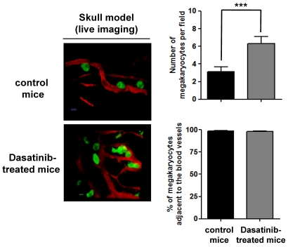
Figure 7.
Localization of MKs in BM of dasatinib-treated mice. Localization of MKs in vivo was visualized by 2-photon intravital microscopy using mouse skull BM. The MKs (green) were identified using CD41-YFPki/+ mice in which enhanced YFP was expressed as a targeted transgene from the endogenous gene locus for CD41, a MK- and platelet-specific integrin. BM vasculature (red) was visualized by injection of TRITC-dextran (2 MDa) immediately before imaging. The average number of MKs per field and the percentage of MKs adjacent to the BM vasculature were determined in control CD41-YFPki/+ mice compared with dasatinib-treated mice (scale bar = 20 μm); ***P < .005.

