Abstract
Purpose/Background:
Hip abduction strengthening exercises may be critical in the prevention and rehabilitation of both overuse and traumatic injuries where knee frontal plane alignment is considered to be important. The purpose of the current investigation was to examine the muscular activation of the gluteus maximus and gluteus medius during the double-leg squat (DLS), single-leg squat (SLS), or front step-up (FSU), and the same exercises when an added load was used to pull the knee medially.
Methods:
Eighteen healthy females (ages 18-26) performed six exercises: DLS, DLS with load, FSU, FSU with load, SLS, and SLS with load. Integrated and peak surface electromyography of gluteus maximus and gluteus medius of the dominant leg were recorded and normalized. Motion analysis was used to measure knee abduction angle during each exercise.
Results:
SLS had the highest integrated and peak activation for both muscles, regardless of load. Adding load, only increased DLS integrated gluteus maximus activation (p=0.019). Load did not increase integrated gluteus medius or peak gluteus maximus activation. Adding load decreased SLS peak gluteus medius activation (p=0.003). Adding load increased peak knee abduction angle during DLS (p=0.013), FSU (p=0.000), and SLS (p=0.011).
Conclusions:
Overall, the SLS was most effective exercise for activating the gluteus maximus and gluteus medius. Applied knee load does not appear to increase muscle activation during SLS and FSU. DLS with an applied load may be more beneficial in activating the gluteus maximus. Overall, the use of applied loads appears to promote poorer musculoskeletal alignment in terms of peak knee valgus angle.
Level of Evidence: 3
Keywords: Electromyography, injury, lower extremity, muscle activation
INTRODUCTION
Maintaining frontal plane knee alignment during strengthening and neuromuscular training exercises may be important in the prevention and rehabilitation of both overuse and traumatic injuries. Hip abduction strengthening exercises are often utilized in lower extremity rehabilitation programs since they have functional implications in activities of daily living1 and may facilitate positive outcomes for hip injuries and in knee dysfunction.2,3 Hewett et al.4 reported that a six week strength, flexibility, and neuromuscular training program emphasizing the hip musculature resulted in a decreased incidence of knee injuries in female athletes over a single playing season.
During weight bearing activities, the combination of ground reaction forces, ligamentous forces, and muscular/tendon forces throughout all lower extremity (LE) joints are interrelated; therefore, abnormal/excessive stress, inefficient neuromuscular patterns, or muscular weakness at one joint may have an effect on the entire LE kinetic chain. With these combined effects influencing the LE kinetic chain, hip and ankle kinetics and kinematics during weight bearing activities may have a direct impact on knee biomechanics and thus, relate to knee injury risk.5–8 Heinert et al.9 reported that females with decreased hip abductor strength demonstrated a greater peak knee abduction angle during stance phase of treadmill running when compared to females with greater hip abductor strength. Hip strength also influences landing mechanics. Lawrence et al.10 reported that female subjects with greater strength in the hip external rotators demonstrated lower vertical ground reaction forces during single-leg landings when compared to females with weak hip external rotators. These authors also reported a decrease in external knee valgus moments during landing. Leetun et al11 reported that athletes who sustained lower extremity injury had weaker hip abduction and external rotation strength than athletes who were uninjured, when strength was measured before the injury occurred. Hip external rotation strength was significantly reduced by 15% for athletes who sustained a lower extremity injury in their investigation. In addition, Jaramillo et al.12 reported greater weakness in both peak and endurance force in the ipsilateral flexors/extensors and abductors/adductors of the hip musculature of the injured extremity following knee surgery.
Hip abduction strengthening exercises used in exercise include both weight bearing (closed kinetic chain) and non weight bearing (open kinetic chain) activities. These may include sidelying or standing hip abduction and other single-leg standing (SLS) exercises, such as squats, lateral step downs, proprioceptive training, and plyometric activities. SLS activities require hip abductor muscle activation of the weight-bearing side in order to control pelvic positioning in the frontal plane during the exercise.1,13,14 Other weight bearing exercises include double-leg and single-leg squats, leg press, forward and lateral lunges, and step-ups. Performing these exercises with proper biomechanics may improve hip muscle recruitment and force production of the extensors and abductors during functional and sports specific movements.7 Weight bearing exercises are generally favored because they better replicate functional and athletic movements as compared to non weight bearing exercises.7,15,16
Hip abductor strengthening has been described as an important intervention in individuals with knee pain.1,13 Several authors describe the activation of gluteus maximus and gluteus minimus during specific exercises1,13,14,17 as well as gluteus maximus and gluteus medius activation during stepping, squatting activities, and lunges.13,14 Presently, the authors of this paper are not aware of research that has identified how the activity of these muscles is altered when a frontal plane load is applied to the knee during stepping and squatting exercises. Therefore, the primary purpose of this study was to determine which of three exercises (double-leg squat, single-leg squat, or front step-up) was most effective in facilitating hip muscle activation while maintaining a more neutral varus/valgus knee frontal plane alignment in healthy female subjects. A secondary objective was to determine how added medial pull on the knee during these exercises altered the magnitude of gluteus maximus and gluteus medius activation while enabling the maintenance of neutral varus/valgus knee alignment during the task.
METHODS
A repeated measures study design was used. The independent variables included the exercise (double-leg squat, single-leg squat, or front step-up) and the medial or unloaded condition. The dependent variables were the integrated percentage of the maximal voluntary isometric contraction (% MVIC*s), peak %MVIC, and peak knee abduction angle during exercise.
Eighteen healthy females between the ages 18-26 years (average age 22.3 years, SD 2.3) with an average height 166.82 cm (SD 9.2) and average weight 61.1 kg (SD 7.1) were recruited to participate in this study. Inclusion criteria were that participants had no current injuries or pain in their lower extremities. Injury was defined as an event that occurred during athletic participation that required treatment or required refraining from participation for at least one day. Participants also reported no history of surgical intervention of the spine or lower extremities that may have influenced the data. The dominant leg was determined by asking the subject which leg she would use to kick a soccer ball.16 Each subject's height, weight, leg length were recorded. Before participating, subjects were informed of possible risks and signed an informed consent form approved by the University institutional review board.
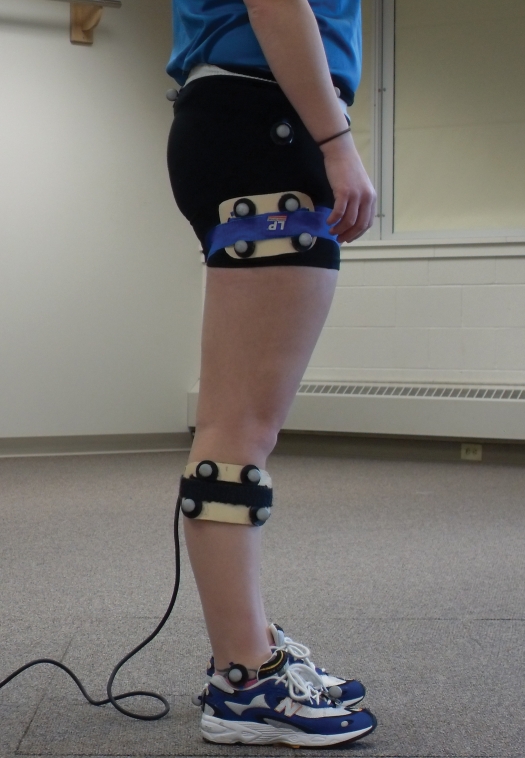
Eight Eagle high speed video cameras (Motion Analysis Corp., Santa Rosa California) were positioned around the laboratory such that at least two cameras could identify and track each retroreflective marker placed on the subject. Three markers placed on rigid shells were strapped to the dominant thigh and shank (Figure 1). A static neutral standing trial was used to identify joint centers of the knee and ankle by anatomical markers on the medial and lateral condyles and malleoli. Ankle and knee centers were determined by taking half the distance between the medial and lateral markers. The hip joint center was determined based on markers placed on the greater trochanters of the femur, and 25% of the horizontal distance from the tested leg was used to calculate this location.18 All video from the cameras and analog data from the force platform measures were collected (120 Hz for video and 1200 Hz for analog) using Eva 6.4 (Motion Analysis Corp.) and stored on a personal computer. All marker data were identified and exported to the Motion Monitor Software (Version 7.0, Innovative Sports Training, Chicago, IL) to create the rigid bodies of the thigh and shank. All data were smoothed using a low pass Butterworth recursive (fourth order) at eight Hz based on the frequency content of the data. Knee abduction angles were determined from the thigh and shank coordinate data.10
Figure 1.
Participant shown with marker set used for motion capture. Rigid shells with retroreflective markers were placed on the thigh and shank to measure peak movement of the varus/valgus knee motion during each exercise.
Surface electromyography (EMG) recordings were collected from two muscles of the dominant leg of each subject, the gluteus medius and gluteus maximus, using the landmarks described by Cram et al.19 Before placement of the surface electrodes on the subject, the skin was prepared by abrading the skin with sandpaper and cleansing the skin with alcohol to reduce skin impedance. EMG data were captured at 960 Hz using the Data Pac 2K2 acquisition software (Run Technologies, Version 3, Mission Viejo, CA, USA). Delsys DE2.1 surface electrodes and a Bagnoli 8 amplifier (Delsys, Inc, Boston, MA, USA) interfaced with a second PC. The system bandwidth was 20-450 Hz with a gain of 1000. Electrode placements were based on those used by Cram et al.19 Data were high pass filtered using a 4th order Butterworth recursive filter at 30 Hz, rectified and low pass filtered with a 4th order Butterworth recursive filter at 6 Hz.20 EMG data were collected using DataPac 2k2 (Run Technologies, Mission Viejo, CA) and integrated over the duration of each exercise trial using a customized Matlab program (The Mathworks, Inc., Natick, Massachusetts). Integrating the individual EMG signals serves to determine the net activation required by the muscles involved in the task by determining the area under the processed curves. The data were normalized to the % MVIC to allow for comparison between subjects.
During these exercises, EMG data was synchronized with video data by using a force platform interfaced with both the EMG and the motion analysis system on each computer. The subject was instructed to perform each exercise stepping onto and off of the force platform. Data based on a portion of the participant's body weight based on the vertical ground reaction force (increase of greater than 5%) was used to determine when movement began on both recording systems. The force platform loading and unloading events served as a point of synchronization between systems.
After the surface electrodes were attached to the subject, the subject performed three maximal voluntary isometric contractions (MVIC) for each muscle.21 An investigator applied manual resistance for the MVIC which were held for five seconds. To obtain the MVIC for the gluteus maximus, the subject was placed in a prone position with the knee flexed to approximately 90 degrees and asked to maximally extend the hip. To obtain the MVIC for the gluteus medius, the subject was placed in a side-lying position with the hip in a neutral position and asked to maximally abduct the hip while maintaining the lower extremity in the frontal plane. All subsequent EMG recordings were normalized to the MVIC and were reported as a percent of MVIC.
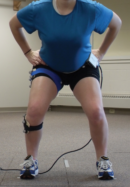
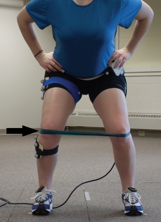
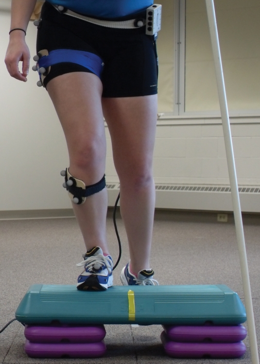
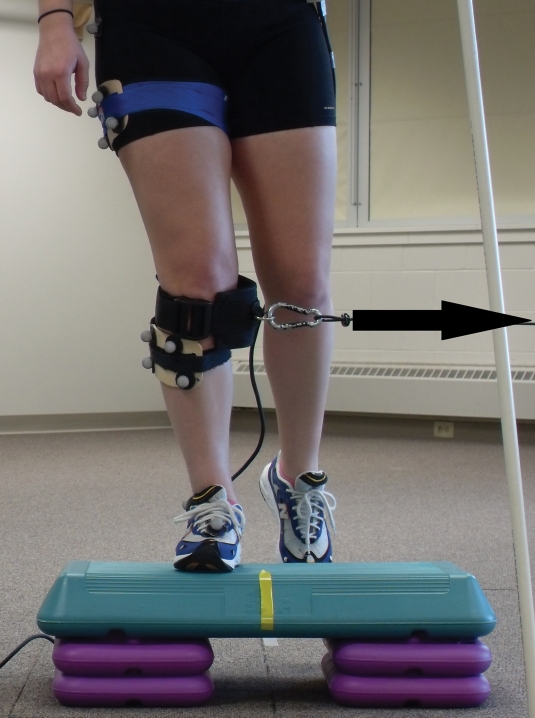
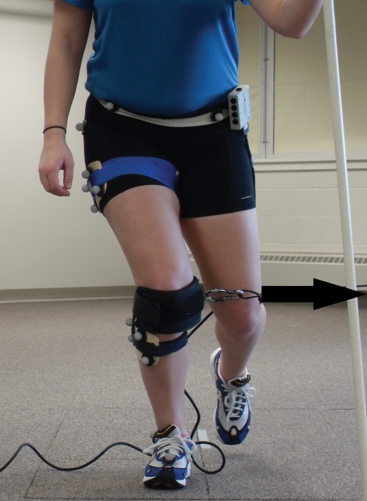
After the MVIC data were collected, the retroreflective markers were placed on the subject. After collection of the static, anatomical neutral motion capture file to identify the location of the joint centers for the ankle and hip, the investigator then provided instruction for how each exercise would be performed. The subject performed the following six exercise activities in a randomized order: double-leg squat (Figure 2), double-leg squat with lateral resistance via a green resistance band applied at the knee (Theraband, Hygenic Corp, Akron, Ohio) tied in a 30 cm loop and applied around both LE's (Figure 3), front step-up (Figure 4), front step-up with an applied resistance via a cable column pulling horizontally in a medial direction (Figure 5), single-leg squat (Figure 6), and single-leg squat with resistance via a cable column pulling horizontally in a medial direction (Figure 7). The subject was allowed to practice each exercise several times before data were collected. Five repetitions of each exercise were performed and used for simultaneous EMG and motion analysis data collection. Between each repetition was a 10-15 second rest interval and between each exercise was a 45-60 second rest interval. The level of applied resistance for the front step-up and single-leg squat was set to 15% of the subject's body weight. This weight was established after a pilot investigation of 5 subjects not involved in this study. It was determined that this weight would provide ample force without disturbing the subjects ability to perform the desired maneuver. The cable with the weight stack resistance was created specifically for this investigation such that the pull of the cable could be raised or lowered such that the line of pull was perpendicular to the limb. Each exercise was performed to the beat of a metronome set at 40 beats per minute where beat 1 initiated the repetition, beat two marked the mid-point between the concentric and eccentric phases, and beat three marked the end of the repetition. One full repetition lasted three-seconds in duration. Each subject was allowed to use a vertical pole to maintain balance on the side being tested. The subjects were monitored by visually observing for excessive trunk lean toward the pole to ensure that significant weightbearing did not occur through the upper extremity holding onto the pole. Other authors have utilized a similar protocol during their data collection to maintain balance.13 The subjects were encouraged to maintain their knee over their ankle during each exercise. They were also given verbal cues to maintain an upright and vertical position of the head and trunk. The average normalized integrated EMG, peak EMG activity and peak knee valgus angle from five trials were used for statistical analysis.
Figure 2.
Participant performing a double-leg squat exercise.
Figure 3.
Participant performing a double-leg squat exercise with medial knee resistance via a resistance band. Arrow depicts direction of pull on the knee.
Figure 4.
Participant performing a front step-up exercise.
Figure 5.
Participant performing a front step up exercise with an applied resistance to the knee via a cable column. Arrow depicts direction of pull on the knee.
Figure 6.
Participant performing a single-leg squat exercise.
Figure 7.
Participant performing a single-leg squat exercise with an applied resistance to the knee via a cable attached to a weight stack. Arrow depicts direction of pull on the knee.
STATISTICAL ANALYSIS
A two way analysis of variance with repeated measures using two within subject factors (exercise: double-leg squat, single-leg squat, front step-up; and with or without an applied load to the knee) was utilized. These were run separately on the average normalized integrated gluteus maximus and gluteus medius activity, average peak normalized gluteus maximus and gluteus medius activity and the peak knee valgus angle. Alpha for all statistical tests was chosen, apriori to be 0.05. Post-hoc comparisons were performed as warranted by running pairwise t-tests using the Bonferoni procedure.
RESULTS
Muscle activation of gluteus maximus and gluteus medius were monitored during three exercises: double-leg squat, single-leg squat, and step-up for two conditions (with and without an applied load to the knee). Table 1 depicts the integrated gluteus maximus activation during these exercises with and without an applied load to the knee. There was a statistically significant difference in muscle activation between the three exercises (double-leg squat, single-leg squat, and step-up) for gluteus maximus (p=0.000). The application of an applied load did not change the amount of integrated activation across all exercises for the gluteus maximus (p=0.550). There was an exercise by applied load interaction for the gluteus maximus (p=0.017) indicating that the presence of the load did influence the integrated muscle activation among the three exercises. When an applied load was added to the double-leg squat, gluteus maximus activation increased by 6% MVIC*sec (p=0.019). When an applied load was added to the step-up and single-leg squat gluteus maximus activation decreased by 2% MVIC*sec and 7.4% MVIC*sec, respectively. However, this change in muscle activation was not large enough to be considered significant for either the step-up (p=0.598) or the single-leg squat (p=0.053).
Table 1.
Two-way analysis of variance results and means (SD) for integrated gluteus maximus electromyography (EMG) during exercises with and without an applied load to the knee.
| Exercise Type | No Applied Load Mean (SD) | Applied Knee Load Mean (SD) | Exercise Main Effect Mean (SD) |
|---|---|---|---|
| Double Leg Squat | 21.7 (15.8) | 27.7 (20.3) | 24.7 (18.1) |
| Step-Up | 36.4 (18.6) | 34.4 (11.6) | 35.4 (15.1) |
| Single Leg Squat | 47.4 (21.2) | 40.0 (17.6) | 43.7 (19.4) |
| Load Main Effect Mean (SD) | 35.2 (18.5) | 34.0 (16.5) |
Main Effect of Exercise (Double-Leg Squat, Step-Up, Single-Leg Squat) p = 0.000
Main Effect of Load (No Applied Load, Applied Load) p = 0.019
Interaction Effect of Exercise and Load p = 0.001
*Note: Data are expressed as a percentage of maximum voluntary isometric contraction (% MVIC * sec).
The integrated gluteus medius activation during the exercises with and without an applied load to the knee is depicted in Table 2. There was also a statistically significant difference in normalized integrated muscle activation across the three exercises (double-leg squat, single-leg squat, and step-up) for gluteus medius (p=0.000) as well as a change in the normalized integrated activation across all exercises with the application of a valgus load (p=0.019). There was an exercise by applied load interaction indicating that the presence of load did influence normalized integrated muscle activation among the three exercises for gluteus medius (p=0.001). When an applied load was added to the double-leg squat, the normalized integrated gluteus medius activation increased non-significantly, or by only 2.9% MVIC*sec (p=0.112). The application of an applied load to the step-up decreased the amount of integrated gluteus medius activation non-significantly by 3.0% MVIC*sec (p=0.223). When an applied load was added to the single-leg squat, there was a significant 11.9% MVIC*sec decrease in the integrated gluteus medius activation (p=0.002).
Table 2.
Two-way analysis of variance results and means (SD) for integrated gluteus medius electromyography (EMG) during exercises with and without an applied load to the knee.
| Exercise Type | No Applied Load Mean (SD) | Applied Knee Load Mean (SD) | Exercise Main Effect Mean (SD) |
|---|---|---|---|
| Double Leg Squat | 20.8 (14.7) | 23.7 (16.3) | 22.5 (15.5) |
| Step-Up | 48.2 (20.4) | 45.2 (21.7) | 46.7 (21.1) |
| Single Leg Squat | 65.6 (23.8) | 53.7 (27.6) | 59.7 (25.7) |
| Load Main Effect Mean (SD) | 44.9 (19.6) | 40.9 (21.9) |
Main Effect of Exercise (Double-Leg Squat, Step-Up, Single-Leg Squat) p = 0.000
Main Effect of Load (No Applied Load, Applied Load) p = 0.019
Interaction Effect of Exercise and Load p = 0.001
*Note: Data are expressed as a percentage of maximum voluntary isometric contraction (% MVIC * sec).
Table 3 depicts the peak gluteus maximus activity during the exercises with and without an applied load. When examining average peak normalized muscle activation, there was a significant difference in activation across the three exercises for gluteus maximus (p=0.000). There was also a change in the average peak normalized gluteus maximus activation across all exercises with the application of the load (p=0.042). Follow up pairwise comparisons tests showed there was no difference between the means for any of the three exercises with or without load (p>0.05). There was no exercise by load interaction for the average peak normalized gluteus maximus activation (p>0.05).
Table 3.
Two-way analysis of variance results and means (SD) for peak gluteus maximus electromyography (EMG) during three different exercises with and without an applied load to the knee.
| Exercise Type | No Applied Load Mean (SD) | Applied Knee Load Mean (SD) | Exercise Main Effect Mean (SD) |
|---|---|---|---|
| Double Leg Squat | 22.0 (16.6) | 25.8 (18.7) | 23.9 (17.7) |
| Step-Up | 33.5 (13.4) | 37.8 (11.4) | 35.7 (12.4) |
| Single Leg Squat | 40.5 (16.8) | 36.7 (11.2) | 38.6 (14.0) |
| Load Main Effect Mean (SD) | 32.0 (15.6) | 33.4 (13.8) |
Main Effect of Exercise (Double-Leg Squat, Step-Up, Single-Leg Squat) p = 0.000
Main Effect of Load (No Applied Load, Applied Load) p = 0.042
Interaction Effect of Exercise and Load p = 0.087
*Note: Data are expressed as a percentage of maximum voluntary isometric contraction (% MVIC).
The average normalized peak gluteus medius activation during the exercises with and without a load is provided in Table 4. Examining average peak normalized muscle activation, there was a significant difference across the three exercises for gluteus medius (p=0.000). There was also exercise by applied load interaction in the average peak normalized gluteus maximus activation (p=0.019) indicating that the load influenced the peak activation among the three exercises. There was an exercise by load interaction for average peak gluteus medius activation (p=0.026). With the addition of the load to the single-leg squat, there was a 7.5% MVIC decrease in peak activation for the gluteus medius (p=0.003). There was no significant change in the average normalized peak activation of the gluteus medius during the front step-up (p=0.924) or double–leg squat (p=0.560) with the addition of a load.
Table 4.
Two-way analysis of variance results and means (SD) for peak gluteus medius electromyography (EMG) during exercises with and without an applied load to the knee.
| Exercise Type | No Applied Load Mean (SD) | Applied Knee Load Mean (SD) | Exercise Main Effect Mean (SD) |
|---|---|---|---|
| Double Leg Squat | 17.6 (10.4) | 18.7 (11.1) | 18.2 (10.8) |
| Step-Up | 43.5 (14.4) | 43.8 (20.1) | 43.7 (17.3) |
| Single Leg Squat | 47.5 (13.2) | 40.0 (13.8) | 43.8 (13.5) |
| Load Main Effect Mean (SD) | 36.2 (12.7) | 34.2 (15.0) |
Main Effect of Exercise (Double-Leg Squat, Step-Up, Single-Leg Squat) p = 0.000
Main Effect of Load (No Applied Load, Applied Load) p = 0.019
Interaction Effect of Exercise and Load p = 0.026
*Note: Data are expressed as a percentage of maximum voluntary isometric contraction (% MVIC).
Table 5 depicts the significant difference in peak knee abduction angles between the three exercises (p=0.011) which occurred between conditions with or without a medial pull applied to the knee (p=0.000). There was an interaction between the exercise and applied load on the amount of knee abduction (p=0.016). When the load was added, there was an increased knee abduction angle demonstrated across all three exercises. With the load applied, the peak knee abduction angle increased by 1.9° during the double-leg squat (p=0.013), 6° during the step-up (p=0.000), and 3.9° during the single-leg squat (p=0.011), each of which was a significant increase.
Table 5.
Two-way analysis of variance results and means (SD) for peak knee abduction angles (degrees) during exercises with and without an applied load to the knee.
| Exercise Type | No Applied Load Mean (SD) | Applied Knee Load Mean (SD) | Exercise Main Effect Mean (SD) |
|---|---|---|---|
| Double Leg Squat | −4.1 (3.5) | −6.0 (2.6) | −5.1 (3.1) |
| Step-Up | −4.2 (3.8) | −10.2 (2.9) | −7.2 (3.4) |
| Single Leg Squat | −5.6 (4.0) | −9.2 (3.5) | −7.4 (3.8) |
| Load Main Effect Mean (SD) | −4.6 (3.8) | −8.5 (3.0) |
Main Effect of Exercise (Double-Leg Squat, Step-Up, Single-Leg Squat) p = 0.011
Main Effect of Load (No Applied Load, Applied Load) p = 0.000
Interaction Effect of Exercise and Load p = 0.016
*Note: Data are expressed in degrees. Negative numbers depict knee abduction or valgus angles and positive numbers represent knee adduction or varus angles.
DISCUSSION
The researchers examined the average normalized peak and integrated EMG activation of the gluteus maximus and gluteus medius during three different LE exercises (double-leg squat, single-leg squat, and step-up) under two separate conditions (with and without a load pulling the knee in a medial direction). The single-leg squat had the highest integrated activation for both gluteus maximus (47.4% MVIC*s) and gluteus medius (65.6 %MVIC*s), regardless of whether a load was applied. Therefore, the results of this study suggest that the single-leg squat may be the most effective of the three exercises for activating gluteus maximus and gluteus medius.
Normalized average peak activation was also assessed to determine which of the three exercises would be most beneficial for strengthening gluteus maximus and gluteus medius. Ekstrom et al.22 reported that a peak activation of 45%-50% of MVIC was required to strengthen trunk, hip, and thigh musculature. Andersen et al.23 indicated that an adaptive threshold of 40% to 60% of maximal effort is needed for strengthening the lower extremity musculature during therapeutic exercise. When viewed in the context of the current investigation, these suggestions indicate that the single-leg squat without the medial pull may be the only LE exercise to achieve this activation percentage in gluteus maximus (40.5% MVIC). For the gluteus medius, the step-up (43.5% MVIC), step-up with applied load (43.8% MVIC), single-leg squat (47.5% MVIC), and the single-leg squat with applied load (40.0% MVIC) all reached this adaptive threshold for muscle strengthening. Overall, the single-leg squat reached the adaptive threshold for strengthening in both muscles, with the highest normalized activation percentage of all the exercises tested.
Although the front step-up exercise for the gluteus maximus and the double-leg squat for both gluteus maximus and medius did not reach the adaptive threshold for strengthening, they may still be considered for use in muscle endurance training. Souza and Powers24 reported that women with patellofemoral pain (PFP) had significantly greater hip internal rotation during running (p < 0.001), decreased hip muscle strength in eight of ten hip strength measurements (p < 0.05), and decreased hip extension isotonic endurance (p <0.05) when compared to a control group. Of the hip muscle performance tests, isotonic hip extension endurance was the best predictor (p = 0.004, r = –0.451) of average hip internal rotation motion during running. These findings may support the use of muscular endurance training in rehabilitation of those with PFP. As the gluteus maximus is the primary extensor and external rotator of the hip, poor gluteus maximus endurance may result in decreased ability to control transverse plane motions of the lower extremity during sustained weight bearing activities. Therefore, exercises that target the gluteus maximus may be useful in endurance training protocols for the hip in rehabilitation and prevention of injury.
A secondary goal of this study was to determine if an added load increased the activation of the tested hip muscles. Although the double-leg squat achieved the lowest integrated activation, using a resistance band to add apply a medial pull to the knees increased integrated muscle activation by 6% MVIC*sec. The valgus load increased knee abduction by 1.9 degrees due to the applied medial pull, which the authors suspect is not large enough to be deleterious for those who perform this exercise. This medial pull was greater than the standing reference angle based on the static neutral trial obtained during testing prior to the dynamic movement trials. Certainly, if the addition of a medial pull during a double-leg squat is provocative of pain, professionals should consider removing or reducing this load. Although the double-leg squat resulted in lower muscle activation than single-leg exercises, the addition of load while completing squats may be indicated in a population with reduced strength of the hip extensors and abductors.
While the addition of an applied load increased the amount of integrated muscle activation during the double-leg squat, this was not the case in the single leg stance exercises. Both the single-leg squat and the step-up exercise showed a decreased integrated muscle activation and an increased knee abduction angle when the load was applied. In the single-leg squat, integrated muscle activation of the gluteus medius decreased by 11.9% MVIC*sec indicating that adding the load from the cable column did not provide cueing to prompt greater integrated muscle activation in order to resist knee abduction. This may indicate that the subjects compensated for the applied load with means other than increasing their integrated gluteus maximus and gluteus medius activation such as trunk movement and weight shifting over their stance leg. The results during single leg squat activities with added load may also indicate that the 15% of the participant's body weight applied by the cable column may have been too large to resist during the single-leg squat and front step-up. It is possible that a smaller applied load may be sufficient for eliciting enhanced muscle activation while maintaining appropriate knee abduction during the exercise.
The data from this investigation may also be used to identify those exercises that activate the gluteus maximus and gluteus medius and promote knee abduction alignment. Greater muscle activation at the hip may help in achieving safer knee mechanics during functional and athletic movements. Once exercises that are effective in increasing muscle activation and strength at the hip are identified, they may be employed to improve training programs for reducing knee injury risks caused by athletic movements that involve large amounts of deceleration such as landing, and cutting.7,25,26
Research suggests that the combination of hip adduction and internal rotation25 along with inadequate or abnormal neuromuscular control of lower extremity biomechanics are factors related to the ACL-injury mechanism.2,8,25–28 Other studies have also established that increased knee abduction, caused by hip adduction and internal rotation, increases ACL strain.29–33 Weakness in the gluteus maximus and gluteus medius may predispose an individual to increased knee valgus since the gluteus maximus functions as a powerful hip extensor, external rotator and abductor.34 The gluteus medius also functions as a hip abductor and external rotator.2
Other knee overuse pathologies may also be related to decreased hip abductor strength. Fredericson et al2 reported that runners with iliotibial band syndrome (ITBS) exhibited weaker hip abductor strength on their affected side compared to their unaffected side. They reported that improving hip abductor strength resulted in a decrease in ITBS symptoms. Females with PFP have also been reported to have weaker hip abductors, extensors, and external rotators as compared to age-matched controls.34,35 Strengthening these muscles resulted in a significant improvement of PFP, lower extremity kinematics, and a return to activity.
As the gluteus maximus and gluteus medius are both involved in frontal plane knee alignment and have been shown to be connected to ACL injuries, ITBS, and PFP, strengthening of these muscles becomes a primary goal in both the prevention and rehabilitation of these injuries. The results of the current study indicate that the single leg squat may be the most effective exercise, of those tested in this investigation, for activating the hip abductors and extensors.
There are several limitations present in this study. Researchers chose to utilize surface EMG rather than the use of fine wire techniques to quantify the activation of the tested muscles. Surface EMG was chosen since it may be able to better reflect the gross muscle activation of these muscles for a study of their function during exercise.36 The use of young, healthy volunteers as subjects limits the ability to apply these results to patients recovering from injury; however, using healthy subjects supports the use of these exercises for the prevention of ACL injuries. While subjects were allowed to use a pole for balance during single leg stance activities, this may not have been sufficient to prevent changes in trunk position, therfore allowing participants to lean or shift their weight within their base of support. The pole was used on the side of weight bearing LE in this investigation. This use could have artificially changed the activation state of the hip musculature due to the additional stabilizing moment added to the hip.37 Also, trunk lean and pelvis positioning could have influenced the magnitude of muscle activation. Bolga and Uhl1 described how trunk lean and resultant pelvis positioning toward the weight bearing side could influence the external moment due to the upper body reducing the demands of the hip abductors during exercise. Better experimental control for trunk and pelvis position may be warranted in future investigations on this topic. This control, however, limits the applicability of the results in a clinical setting as there would likely be less control over trunk position.
Another limitation was the use of a metronome. Using a metronome requires the subjects to move at a speed that may not be typical during daily activities. At a non-controlled speed, the frontal plane angle and gluteus maximus and medius activation may have been different, which may limit the application to a clinical setting.
Further research is needed to validate these findings and to analyze the effect of other strengthening exercises in activating the gluteus maximus and gluteus medius used in exercise programs and clinical practice. Research in the use of lower resistance loads applied at the knee may also be beneficial to determine if lower loads result in higher hip muscle activation levels with less trunk compensation. An alternative method may be to add a load to the contralateral side to increase muscle activation in the weight bearing leg. This approach was demonstrated by Neumann et al.37 who reported an increase in EMG activation of the hip abductors during the stance phase of gait when carrying a load in the contralateral hand.
The results of the current study demonstrate that the use of exercises with applied load to the knee as supplied in this study may not increase the integrated or peak muscle activation of the gluteus maximus and gluteus medius during single-leg squat and step-up. However, the double-leg squat exercise with an applied load provided by a resistance band may be more beneficial than the same exercise performed without the band. The single leg squat achieved the highest integrated and peak activation of the gluteus maximus and gluteus medius of the three exercises examined.
ACKNOWLEDGEMENTS
Partial funding was received from the Gundersen Lutheran Medical Foundation in support of this work.
REFERENCES
- 1. Bolgla LA, Uhl TL. Electromyographic analysis of hip rehabilitation exercises in a group of healthy subjects. J Orthop Sports Phys Ther. 2005;35:487–494 [DOI] [PubMed] [Google Scholar]
- 2. Fredericson M, Cookingham CL, Chaudhari AM, Dowdell BC, Oestreicher N, Sahrmann SA. Hip abductor weakness in distance runners with iliotibial band syndrome. Clin Sports Med. 2000;10:169–175 [DOI] [PubMed] [Google Scholar]
- 3. Mascal CL, Landel R, Powers C. Management of patellofemoral pain targeting hip, pelvis, and trunk muscle function: 2 case reports. J Orthop Sports Phys Ther. 2003;33:647–660 [DOI] [PubMed] [Google Scholar]
- 4. Hewett T.E., Lindenfeld T.N., Riccobene J.V., Noyes F.R. The effect of neuromuscular training on the incidence of knee injury in female athletes. A prospective study. [see comment]. Am J Sports Med. 1999;27:699–706 [DOI] [PubMed] [Google Scholar]
- 5. Griffin LY, Agel J, Albohm MJ, et al. Noncontact anterior cruciate ligament injuries: risk factors and prevention strategies. J Am Acad Orthop Surg. 2000;8:141–150 [DOI] [PubMed] [Google Scholar]
- 6. Ireland ML. The female ACL: Why is it more prone to injury? Orthop Clin North Am. 2002;33:637–651 [DOI] [PubMed] [Google Scholar]
- 7. Hall CM, Brody LT. Therapeutic exercise: Moving toward function. Philadelphia, PA: Lippincott Williams & Wilkins; 2005: 488 [Google Scholar]
- 8. McLean SG, Lipfert SW, van den Bogert AJ. Effect of gender and defensive opponent on the biomechanics of sidestep cutting. Med Sci Sports Exerc. 2004;36:1008–1016 [DOI] [PubMed] [Google Scholar]
- 9. Heinert BL, Kernozek TW, Greany JF, Fater DC. Hip abductor weakness and lower extremity kinematics during running. J Sport Rehab. 2008;17:243–256 [DOI] [PubMed] [Google Scholar]
- 10. Lawrence RK, Kernozek TW, Miller EJ, Torry MR, Reuteman P. (2008). Influences of hip external rotation strength on knee mechanics during single-leg drop landings in females. Clin Biomech. 2008;23:806–13 [DOI] [PubMed] [Google Scholar]
- 11. Leetun DT, Ireland ML, Willson JD, Ballantyne BT, Davis IM. Core stability measures as risk factors for lower extremity injury in athletes. Med Sci Sports Exerc. 2004;36:926–934 [DOI] [PubMed] [Google Scholar]
- 12. Jaramillo J, Worrell TW, Ingersoll CD. Hip isometric strength following knee surgery. J Orthop Sports Phys Ther. 1994;20,160–165 [DOI] [PubMed] [Google Scholar]
- 13. Ayotte NW, Stetts DM, Keenan G, Greenway EH. Electromyographical analysis of selected lower extremity muscles during 5 unilateral weight-bearing exercises. J Orthop Sports Phys Ther. 2007;37:48–55 [DOI] [PubMed] [Google Scholar]
- 14. Boudreu SN, Dwyer MK, Mattacola CG, Lattermann C, Uhl TL, McKeon JM. Hip-muscle activation during the lunge, single leg squat and stepup and over exercises. J Sport Rehab. 2009;18:91–103 [DOI] [PubMed] [Google Scholar]
- 15. Shields RK, Madhavan S, Gregg E, et al. Neuromuscular control of the knee during a resisted single-limb squat exercise. Am J Sports Med. 2005;33:1520–1526 [DOI] [PMC free article] [PubMed] [Google Scholar]
- 16. Zeller BL, McCrory JL, Kibler W., Uhl TL. Differences in kinematics and electromyographic activity between men and women during the single-legged squat. Am J Sports Med. 2003;31:449–456 [DOI] [PubMed] [Google Scholar]
- 17. Jacobs CA, Lewis M, Bolgla LA, Christensen CP, Nitz AJ, Uhl TL. Electromyographic analysis of hip abductor exercises performed by a sample of total hip arthroplasty patients. J Arthroplasty, 2008;00:1–7 [DOI] [PubMed] [Google Scholar]
- 18. Robertson DG, Caldwell GE, Hamill J, Kamen J, Whittlesey SN. Research Methods in Biomechanics. Champaign, IL: Human Kinetics, 2004:309 [Google Scholar]
- 19. Cram JR, Kasman GS, Holtz J. Introduction to Surface Electromyography. Gaithersburg, MD: Aspen Publishers, Inc. 1998:408 [Google Scholar]
- 20. Lloyd DG, Besier TF. An EMG-driven musculoskeletal model to estimate muscle forces and knee joint moments in vivo. J Biomech. 2003;36:765–776 [DOI] [PubMed] [Google Scholar]
- 21. Kendall FP, McCreary EK, Provance PG, Rodgers MI, Romani WI. Muscle testing and function with posture and pain (5th Edition). Baltimore: Lippincott Williams and Wilkins; 2005:433–436 [Google Scholar]
- 22. Ekstrom RA, Donatelli RA, Carp KC. Electromyographic analysis of core trunk, hip, and thigh muscles during 9 rehabilitation exercises. J Orthop Sports Phys Ther. 2007;37:754–762 [DOI] [PubMed] [Google Scholar]
- 23. Andersen LL, Magnusson SP, Nielson M, Haleem J, Poulsen K, Aagaard P. Neuromuscular activation in conventional therapeutic exercise and heavy resistance training: implications for rehabilitation. Phys Ther. 2006;86:683–697 [PubMed] [Google Scholar]
- 24. Souza RB, Powers CM. Predictors of hip internal rotation during running. Am J Sports Med. 2009;37:579–587 [DOI] [PubMed] [Google Scholar]
- 25. Ireland ML. Anterior cruciate ligament injury in female athletes: epidemiology. J Athl Train. 1999;34:150–154 [PMC free article] [PubMed] [Google Scholar]
- 26. Hewett TE. Neuromuscular and hormonal factors associated with knee injuries in female athletes: Strategies for intervention. Sports Medicine. 2000;29:313–327 [DOI] [PubMed] [Google Scholar]
- 27. Ford KR, Myer GD, Hewett TE. Valgus knee motion during landing in high school female and male basketball players. Med Sci Sports Exerc. 2003;35:1745–1750 [DOI] [PubMed] [Google Scholar]
- 28. Hewett TE, Myer GD, Ford KR, et al. Biomechanical measures of neuromuscular control and valgus loading of the knee predict anterior cruciate ligament injury risk in female athletes: a prospective study. Am J Sports Med. 2005;33:492–501 [DOI] [PubMed] [Google Scholar]
- 29. Markolf KL, Burchfield DM, Shapiro MM, Shepard MF, Finerman GAM, Slauterbeck JL. Combined knee loading states that generate high anterior cruciate ligament forces. J Orthop Res. 1995;13:930–935 [DOI] [PubMed] [Google Scholar]
- 30. Lloyd DG, Buchanan TS. Strategies of muscular support of varus and valgus isometric loads at the human knee. J Biomech. 2001;34:1257–1267 [DOI] [PubMed] [Google Scholar]
- 31. Fukuda Y, Woo SL, Loh JC, et al. A quantitative analysis of valgus torque on the ACL: a human cadaveric study. J OrthopRes. 2003;21:1107–1112 [DOI] [PubMed] [Google Scholar]
- 32. Kanamori A, Woo SL, Ma CB, et al. The forces in the anterior cruciate ligament and knee kinematics during a simulated pivot shift test: A human cadaveric study using robotic technology. Arthroscopy. 2000;16:633–639 [DOI] [PubMed] [Google Scholar]
- 33. McLean SG, Huang X, Su A, van den Bogert AJ. Sagittal plane biomechanics cannot injure the ACL during sidestep cutting. Clin Biomech. 2004;19:828–838 [DOI] [PubMed] [Google Scholar]
- 34. Ireland ML, Willson JD, Ballantyne BT, Davis IM. Hip strength in females with and without patellofemoral pain. J Orthop Sports Phys Ther. 2003;33:671–676 [DOI] [PubMed] [Google Scholar]
- 35. Robinson RL, Nee RJ. Analysis of hip strength in females seeking physical therapy treatment for unilateral patellofemoral pain syndrome. J Orthop Sports Phys Ther. 2007;37:232–238 [DOI] [PubMed] [Google Scholar]
- 36. Soderberg GL, Cook TM. Electromyography in biomechanics. Phys Ther. 1984;64:1813–1820 [DOI] [PubMed] [Google Scholar]
- 37. Neumann DA, Cook TM, Sholty RL, Sobush DC. An electromyographic analysis of the hip abductor muscle activity when subjects are carrying loads in one or both hands. Phys Ther. 1992;72:207–217 [DOI] [PubMed] [Google Scholar]


