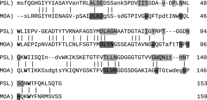
Fig. 3.
Secondary structure-based sequence alignment and manual motif identification of the β-trefoil fold (residues 1–153) of PSL1a and MOA with bound Galα(1,3)[Fucα(1,2)]Gal (PDBID 3EF2) using Sequoia (Bruns et al. 1999) and Coot (Emsley and Cowtan 2004) . RMSD: 2.34 Å. Light grey: rPSL residues within 4 Å of the central Gal of the ligand from the MOA structure. Dark Grey: residues within 4 Å of the central Gal of the ligand in MOA. Bold: conserved tryptophans from the ricin B-type (Q/N-x-W) motifs. In capital letters: residues whose atomic coordinates are structurally equivalent in the alignment.

