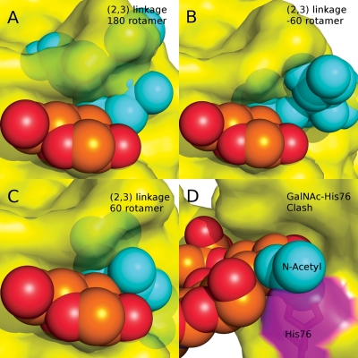
Fig. 6.
(A–C) Models of the Neu5Acα2-3Galβ-1-4GlcNAc trisaccharide bound to PSL, indicating that no rotamers of the 2-3 linkage can be adopted that avoid collisions with the PSL surface. (D) Orientation of a GalNAc-containing trisaccharide in the binding site of PSL, indicating a collision with His76 (magenta).

